
D. Adam Algren and Michael R. Christian
Chemicals have been used to impede, incapacitate, or kill the opposition throughout history. Although international treaties (e.g., 1925 Geneva Protocol, 1997 Chemical Weapons Convention) ban the development, production, stockpiling, and use of chemical weapons, Iraq used both nerve and blistering agents in its war with Iran and against Kurdish civilians in the 1980s. The Russian military used a gas thought to contain a fentanyl derivative and halothane to subdue Chechen rebels in a Moscow theater in 2002 and there is reported use of nerve agents in 2013 against rebels by the Syrian National Army (1).
Of greater concern is that chemical warfare agents (CWAs) may be used by terrorists. CWAs are fairly inexpensive and easy to acquire, synthesize, deploy, and hide. On three occasions, the terrorist group Aum Shinrikyo released nerve agents (sarin and VX) in Japan (2). The Tokyo subway incident in 1995 resulted in 12 deaths and more than 5,000 civilian casualties (2). The 1996 Defense against Weapons of Mass Destruction Act, directed the US Department of Defense to establish preparedness programs of local, state, and federal agencies to respond to chemical and biologic incidents (3) and these became better supported following the September 11, 2001 terrorist attacks.
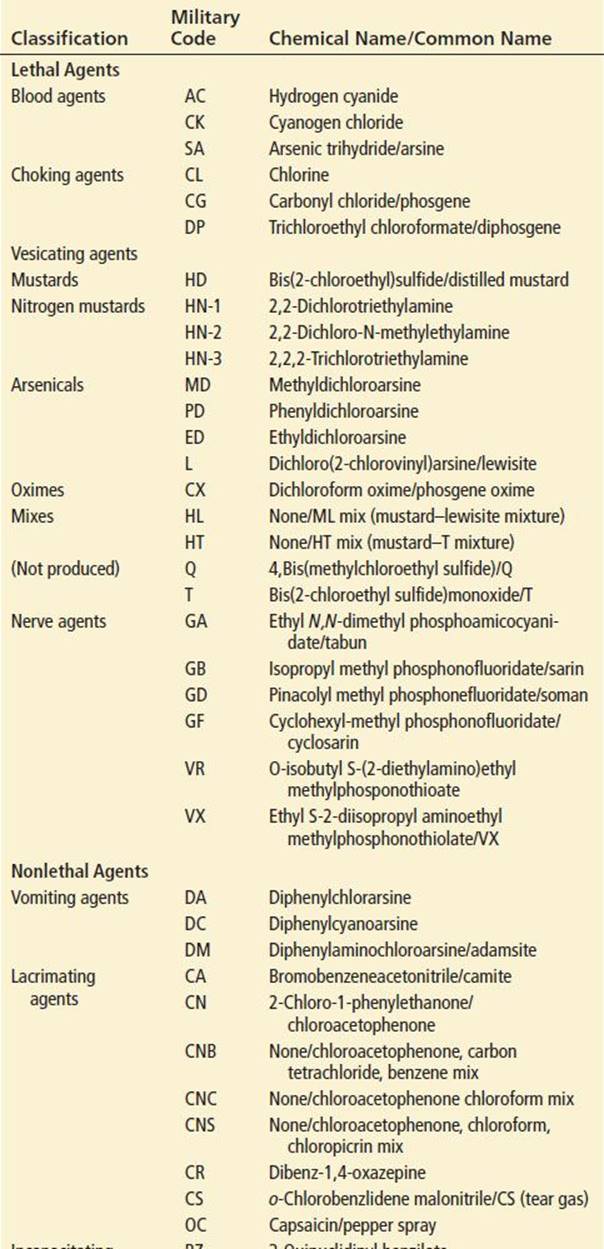
CWAs can be lethal or nonlethal and fall into one of seven major categories: (a) chemical choking agents, (b) vesicating or blister agents, (c) lacrimating agents, (d) nerve agents, (e) vomiting agents, (f) systemic asphyxiants, and (g) incapacitating agents (Table 350.1) (4). Lacrimating agents are also used for personal protection and by police for crowd dispersal and incapacitating criminal suspects.
TABLE 350.1
Chemical Warfare Agents
CWAs can be aerosolized or vaporized and delivered in multiple ways. They may be dispersed from handheld or vehicle-mounted devices, low-flying aircraft, crop dusters, cruise missiles, and artillery. Sarin was dispersed by evaporation from punctured plastic bags left on Tokyo subway cars (2). Although CWAs could be added to water or food supplies, aerosolization is favored by terrorists and military groups as it allows for the greatest exposure with the goal of inflicting the maximum number of casualties (4).
Systemic asphyxiants incorrectly labeled as “blood agents” by the military include the gases cyanogen chloride, hydrogen cyanide (see Chapter 329), and hydrogen sulfide (see Chapter 332), and the hemolytic gas arsine (see Chapter 336) (4). Hydrogen cyanide (prussic acid) and cyanogen chloride are clear liquids with a bitter almond odor that readily volatilize at temperatures >26°F (1). As warfare agents, their efficacy is limited by physical properties that result in ineffective dispersal (4).
The choking agents are pulmonary irritants introduced in World War I and include chlorine, phosgene, and diphosgene (see Chapter 336). Chlorine, a dense, water-soluble, yellow-green gas that smells like concentrated bleach, reacts with tissue water to form hydrochloric acid and oxygen-free radicals, which produce corrosive and cytotoxic effects. Toxicity is proportional to the gas concentration, duration of contact, and water content of exposed mucous membranes.
Phosgene (carbonyl chloride) was probably responsible for more than 80% of all chemical-agent fatalities in World War I (4) and can be formed by decomposition of chlorinated hydrocarbons. It is a colorless gas that is heavier than air. Its propensity to settle into low-lying areas, such as trenches, led to its tactical success. Phosgene smells like freshly mown hay, but odor is not a reliable indicator of exposure. Because of its low water solubility it is only mildly irritating to the eyes and upper respiratory tract and the initial exposure may go unnoticed (4). Phosgene hydrolyzes with terminal bronchial and alveolar mucosal water to produce hydrochloric acid. This and other toxic by-products of phosgene (e.g., oxygen-free radicals) result in pulmonary edema that can be delayed up to 24 hours. The latency period is shorter with higher concentrations and longer durations of exposure.
Vesicating agents, which cause blistering of exposed surfaces, include mustard gases and organic arsenicals. Sulfur mustard was used in World War I and in the Iran–Iraq war (4). Most injuries were disabling but nonfatal; mortality during World War I was <2%, and rates of 3% to 4% were reported in the Iran–Iraq war (4,5). Street clothing offers no protection, but military protective garments (chemical-resistant suit) can provide up to 6 hours of protection following exposure (6). Low volatility of the liquid results in persistence for several days in temperate climates, thereby restricting enemy use of contaminated equipment and terrain (4).
Sulfur mustard is an oily amber liquid that smells like burning garlic or mustard. Upon tissue contact, it spontaneously forms highly reactive and unstable episulfonium compounds, which irreversibly alkylate nucleic acids and cellular proteins within minutes of exposure (6). This permanently damages DNA and alters or inhibits cell replication, protein synthesis, and enzyme function often leading to cell death. Blisters form after destruction of anchoring filaments in the epidermal–dermal junction (6).
Lewisite, the best known organic arsenical, was developed in 1918 to complement sulfur mustard as a semipersistent blistering agent. Its vapor is about seven times heavier than air and will flow into lower terrain (4). It has low water solubility and persists in the environment for up to 24 hours under temperate conditions. Many countries have large stockpiles of lewisite, but its actual use in combat is not well documented (4).
Lewisite is an amber, oily liquid that smells like geraniums. It is rapidly absorbed after vapor inhalation and, as a result of high lipid solubility, penetrates intact skin within 15 minutes, more rapidly than mustard agents (4). In the dermis, it hydrolyzes to hydrochloric acid and chlorovinylarsenious oxide, the vesicating agent. The organic arsenical moiety binds to the dithiol component of pyruvate dehydrogenase, prevents formation of acetyl coenzyme A from pyruvate, and produces systemic toxicity similar to other arsenicals (see Chapter 326). Increased capillary permeability and extensive interstitial fluid losses may result in hypovolemia or “lewisite shock” (4).
Nerve agents are similar in structure and function to organophosphorus pesticides (see Chapter 318) but are more potent and typically have a more rapid onset and shorter duration of action. The “G” (German) agents tabun (GA), sarin (GB), and soman (GD) were developed in Germany between 1936 and 1944 (4,7). A fourth, VX, with the “V” standing for “viper,” was synthesized in 1952 in England (4,5). Although commonly referred to as gases, these agents are colorless, odorless liquids that under temperate conditions form vapors are heavier than air and concentrate close to the ground (4,7). Lethality correlates directly to the product of vapor concentration and time of exposure (LCt) (4,7). The G agents are moderately volatile, evaporate within several hours, and are environmentally nonpersistent (7,4). VX, an oily liquid, is much less volatile and persists for several weeks after dispersion (4). It is the most potent nerve agent, with an LCt50 (vapor concentration–time product necessary to kill 50% of an unprotected population) that is 5, 10, 40, and 500 times more potent than soman, sarin, tabun, and hydrogen cyanide, respectively (4,7). VX also has a slower rate of elimination than G agents and is expected to have a longer duration of action. In addition to inhalational exposures nerve agents can also be absorbed through the skin due to their high lipid solubility. Time of onset and severity are determined by the dose, agent, and skin integrity/location (4,7).
Toxicity of nerve agents results primarily from phosphorylation and inactivation of acetylcholinesterase (AChE) at its hydroxy group in its serine active site (see Chapter 318). AChE accumulates at nerve terminals, initially stimulating and then paralyzing cholinergic neurotransmission at both central and peripheral nicotinic and muscarinic receptors. Once inactivated, the phosphorylated AChE enzyme may be permanently inhibited or it may be reactivated by nucleophilic oximes to end the nerve gas effect. Reactivation is impossible once dealkylation (“aging”) of the phosphorylated cholinesterase occurs. The rate of aging varies among the nerve agents. It occurs within 2 minutes after soman exposure making it difficult to treat, 5 to 8 hours after sarin exposure, and more than 40 hours after tabun or VX exposure (4,7).
Other mechanisms likely contribute to effects on central nervous system (CNS) neurotransmission that result in seizures and CNS neuropathology. This likely explains why some compounds that are neuroprotective do not reactivate AChE and why CNS-active oximes (e.g., pralidoxime) are not more effective than oximes that do not reach the brain.
Vomiting agents are arsenic-based compounds used for riot control (4). They are normally solids that are vaporized by heating and then condense to form aerosols that produce rapid irritation of exposed tissues.
Lacrimating, or riot-control, agents, include o-chlorobenzylidenemalonitrile (CS or tear gas), ω-chloroacetophenone (CN or mace), dibenz-1,4-oxazepine (CR), and oleoresin capsaicin (OC or pepper spray). Except for capsaicin, toxicity results from alkylation and inhibition of sulfhydryl-containing enzymes. Generation of bradykinin also contributes to their toxicity. Capsaicin is a lipid-soluble alkaloid that first stimulates release of substance P from nociceptive neurons and then blocks its synthesis and transport. Substance P depolarizes neurons to produce dilatation of blood vessels, stimulation of smooth muscle, and activation of sensory nerve endings that mediate pain. With repeated application, nociceptive fiber response and number are decreased (desensitization).
CN and CS are usually delivered as a vapor or cloud of suspended solid particles (smoke) produced by a thermal grenade. CS is the riot-control agent of choice in the United States. It is about 10 times more potent as a lacrimator than CN but is much less corrosive to tissues (6,7). Newer CS formulations consist of an unheated micropulverized powder that can remain active in the environment for several weeks. OC is used frequently by civilians for personal protection and by law-enforcement personnel for immobilization of combative subjects. “Pepper mace” can contain CS, oleoresin of capsicum, or both.
Incapacitating agents include stimulants (e.g., amphetamines), depressants (e.g., opioids), psychedelics (e.g., d-lysergic acid diethylamide, LSD), and deliriants (e.g., anticholinergics) (4). Historically, the anticholinergic agent 3-quinuclidinyl benzilate (BZ) was the only incapacitating agent considered feasible for use by the US military (4). BZ is a potent, glycolate, anticholinergic agent that is a crystalline solid under normal atmospheric conditions but most likely dispersed as a vapor or liquid aerosol by military personnel or terrorists (4). Although ultrapotent fentanyl derivatives (alfentanil, carfentanil, remifentanil, and sufentanil) show some promise, these agents have unpredictable effects as a result of high lipid solubilities and large volumes of distribution (1). The unpredictable nature of these agents is evidenced by the tragic end to the October 2002 Moscow hostage crisis in which 127 of 800 hostages died from a fentanyl-halothane gas intended to incapacitate its victims (1).
CLINICAL PRESENTATION
Choking Agents
Clinical symptoms begin within minutes after significant chlorine exposure and include lacrimation, conjunctival irritation, rhinorrhea, cough, sore throat, chest burning, dyspnea, sputum production, nausea, headache, and respiratory failure. Corneal abrasions and cutaneous burns may result from eye and skin exposure, respectively. Following significant chlorine exposures, pulmonary edema develops within 2 to 4 hours and peaks at 12 to 24 hours (4). The chest x-ray may be normal or may show noncardiogenic pulmonary edema. Patients with significant phosgene exposure have a typical latency period of 4 to 6 hours (range, 1 to 24 hours) and then present with dyspnea, chest tightness, cyanosis, hemoptysis, hypotension, and pulmonary edema. Chest x-ray findings of pulmonary edema are rather late and nonspecific, occurring 6 to 8 hours after exposure. Following significant pulmonary agent exposure (e.g., chlorine gas), patients may subsequently develop reactive airways dysfunction syndrome, a chronic asthma-like condition.
Vesicating Agents
After mustard exposure, there is typically a latency period of 4 to 12 hours before the onset of symptoms. The latency period is shorter with high concentrations and long exposures, with increased ambient temperature and humidity, and in victims previously exposed to or innately susceptible to mustard (4,6). Sites of injury principally involve the skin, eye, and respiratory tract and may follow vapor or liquid exposure. Heavy exposure may lead to systemic effects such as bone marrow depression and sloughing of intestinal mucosa.
Cutaneous injury resulting from mustard exposure ranges from erythema to blisters and skin necrosis. The moist, thinner skin of the neck, axilla, and groin is more severely affected. After of 4 to 12 hours, erythema and edema develop. Vesication typically starts within 24 hours and evolves over several days. Vesicles coalesce into blisters, and skin necrosis occurs over 24 to 72 hours (4). Blister fluid does not contain active mustard and is not toxic (4). Skin denudation occurs over 6 to 9 days, and healing may take 4 to 10 weeks.
The eye is the organ most sensitive to sulfur mustard. Tissue injury occurs rapidly, but symptoms develop gradually over 4 to 8 hours and include eye pain, lacrimation, photophobia, and blurred vision (4,6). Physical findings include blepharospasm, eyelid edema, conjunctival injection and edema, chemosis, anterior chamber cellular infiltrates, and decreased vision. Corneal edema begins within 1 hour after exposure, and the corneal epithelium vesicates and sloughs within 4 to 36 hours (4). Resolution of injury depends on the severity of exposure and typically takes 1 to 2 weeks. About 90% of victims are visually disabled for 10 days with conjunctivitis, photophobia, and corneal swelling. The remaining 10% are severely affected and are at risk for permanent blindness from corneal opacification, scarring, and ulceration (4).
Respiratory epithelial damage occurs several hours after exposure. Victims may develop rhinitis, nasal bleeding, sinus discomfort, hoarseness, sore throat, cough, sputum production, and dyspnea of increasing severity. Hemorrhagic inflammation and erosions of the upper airway mucosa are followed by fibrinous pseudomembrane formation and sloughing of necrotic, ulcerated mucosa. Bronchospasm and partial airway obstruction result. After high-dose exposures, severe bronchitis, secondary bronchopneumonia, and respiratory failure may develop within 24 to 48 hours (4,6). Pulmonary edema is uncommon, as mustard does not typically affect the pulmonary parenchyma. The need for mechanical ventilation is a poor prognostic sign, and the majority of these patients subsequently die from progressive respiratory failure.
Bone marrow and gastrointestinal (GI) mucosal toxicity may occur with high concentration or prolonged exposure. Leukopenia develops within 5 to 7 days, with the white cell nadir occurring 10 days after exposure. Thrombocytopenia and anemia typically follow. The development of hematologic effects from mustard poisoning is a grave prognostic sign (4). Respiratory failure, bone marrow suppression, or superimposed bacterial infection are the most common causes of death from sulfur mustard poisoning (5,6).
The clinical manifestations of lewisite poisoning are similar to those of sulfur mustard, although lewisite causes immediate severe pain on contact with the skin, eyes, and nasal mucosa. Vapor condensing on the skin causes erythema within 30 minutes and blister formation within 2 to 3 hours (4). The eyes are very sensitive to lewisite, and permanent blindness from corneal destruction may result if decontamination is not initiated within 1 minute. Inhalation of lewisite vapor can result in death within 10 minutes from rapid respiratory mucosal sloughing and bleeding, leading to asphyxiation. Pulmonary edema may also develop.
Nerve Agents
Toxicity may occur after inhalation of vapor, skin contact with vapor or liquid, or ingestion. The rate of onset and severity of effects are determined by the route of exposure and dose (4). At high ambient temperature, skin absorption is rapid and increasing amounts of the agent are volatilized, leading to increased inhalation (4). Small amounts of vapor cause miosis, rhinorrhea, mild dyspnea, cough, and wheezing (4). This can occur within seconds to minutes. Symptoms include eye pain, blurred and dim vision, headache, and dizziness (2). As the dose of vapor increases, nausea and vomiting, increased respiratory difficulty, progressive muscular weakness, and agitation develop. A high vapor concentration causes rapid loss of consciousness, seizures, flaccid paralysis, and respiratory arrest within seconds to minutes. Following sarin vapor exposure in the Tokyo incident, the most common manifestations were miosis (99%), headache (75%), dyspnea (63%), nausea (60%), eye pain (45%), blurred vision (40%), and vomiting (37%) (2). After a mass-casualty incident due to a nerve agent, miosis may serve as a useful marker of exposure. Tachycardia and hypertension were common, but bradycardia and bronchorrhea were not. Electrocardiographic manifestations include supraventricular and ventricular dysrhythmias, ischemic ST–T changes, atrioventricular conduction disturbances, and QTc interval prolongation (7). Seizures may be single and brief or multiple and persistent; status epilepticus will often occur in severely poisoned patients (2,7). Death is primarily a result of depression of the central respiratory drive (4,7). Respirations cease before significant neuromuscular blockade or bronchoconstriction occurs.
Victims of vapor exposure are unlikely to deteriorate once removed from exposure (2). In contrast, those with dermal exposure may subsequently worsen. Skin absorption of a lethal dose may occur within 1 to 2 minutes, yet symptoms are commonly delayed and develop after a latency period of 30 minutes to 18 hours (4,6,7). Skin exposure may produce local sweating and twitching or fasciculations before systemic toxicity (4,7). Conversely, serious effects may also occur abruptly, without antecedent local or mild symptoms after dermal exposure (7). In contrast to vapor exposure, miosis is absent early following skin exposure. Miosis may be present for weeks after exposure and does not readily respond to systemic treatment (7).
Lacrimating Agents
Within seconds to minutes of exposure, lacrimating agents cause intense eye discomfort, blepharospasm, lacrimation, stinging and burning of the mouth and nose, salivation, rhinorrhea, irritation of the respiratory tract and stomach with coughing and vomiting, and skin irritation leading to burning pain and erythema (4). Because they are miscible in sweat, skin irritation is amplified in areas of increased sweat, such as the axilla, buttocks, and popliteal, antecubital, and inguinal regions. Skin effects increase with concentrated liquid spray or aerosol exposures, in warm, moist environments, and in those with pre-existing skin conditions. Toxic effects following exposure to tear agents typically resolve within 20 to 30 minutes (4). High-concentration, enclosed-space exposures, however, may result in significant respiratory effects that include acute laryngotracheobronchitis, pulmonary edema, and death. Pulmonary edema can be delayed 4 to 8 hours; peak effects should be evident by 12 hours.
Prolonged or repeated exposures to lacrimating agents may produce malaise, skin blistering, chest tightness, coughing, wheezing, hemoptysis, shortness of breath, and a feeling of suffocation. Reactive airways dysfunction (RADS) and allergic contact dermatitis have been described in sensitized individuals. A severe dermatitis, labeled “Hunan hand syndrome,” has been described after prolonged skin exposure to capsaicin containing chili peppers.
Incapacitating Agents
Manifestations of BZ and fentanyl derivative poisoning are described in detail in Chapter 344 and Chapter 305, respectively. BZ poisoning may occur after inhalation of vapor or liquid aerosol, intravenous or intramuscular injection, or, to a small degree, skin contact with liquid. Following exposure by all routes (except skin), the onset of effects takes 1 hour, peaks at 8 hours, and gradually subsides over 24 to 72 hours (4). Effects may be delayed up to 24 hours after dermal exposure. Doubling the dose produces incapacitation within 1 hour and prolongs recovery an additional 48 hours. Wide variability in the onset, severity, and duration of effects should occur after BZ deployment by aerosol or vapor (4). The rate of onset and duration of clinical effects after inhalation of aerosolized fentanyl derivatives has not been fully described (1). The gas mixture utilized in the Moscow hostage crisis produced effects within 15 minutes of exposure; toxicity lasted days in some survivors (1).
DIFFERENTIAL DIAGNOSIS
Deliberate release of chemicals by terrorists is likely to involve substances that cannot be immediately identified. Thus, the most likely diagnosis is likely to be made by history, physical examination, and initial diagnostic testing (8,9). Diagnosis is also strongly suggested by the complete reversal of toxic effects following empiric antidotal therapy (1,2,10).
CWA poisoning should be suspected when a group of patients present with the abrupt onset of a similar constellation of signs or symptoms, particularly if they involve the exposed mucosal or skin surfaces, the respiratory tract, or sudden systemic effects (8,10). Following a chemical attack, clinical effects will occur within minutes to hours, and large numbers of symptomatic patients from the same location will present to medical facilities within a short period of time (3,8,10). There may be an associated explosion and/or reported vapor cloud with or without a characteristic odor. In contrast, clinical effects after a biologic agent attack are delayed as a result of the incubation period of the illness and patients will present insidiously, often with nonspecific signs and symptoms. It will be difficult to identify the release site of the weapon, and the geographic distribution of patients may be wide by the time symptomatic disease develops (see Chapter 349) (8,10).
Irritant or corrosive gas inhalation, hydrocarbon aspiration, and vesicating agent and high-dose lacrimating agent exposures may produce pulmonary signs and symptoms similar to those caused by choking agents. Exacerbations of asthma, chronic obstructive pulmonary disease, and allergic or infectious pneumonitis may present similarly. The vesiculobullous skin lesions that result from vesicating agents are most pronounced on exposed skin but may mimic those resulting from Stevens–Johnson syndrome, toxic epidermal necrolysis, pemphigus vulgaris, bullous pemphigoid, scalded skin syndrome, thermal and chemical burns, and hypersensitivity reactions. Nerve-agent poisoning is nearly identical to organophosphate and carbamate pesticide poisoning. Other agents and conditions that can produce similar clinical effects are botulism; nicotine; cholinergic drugs such as bethanechol, carbachol, edrophonium, methacholine, neostigmine, pilocarpine, physostigmine, pyridostigmine, and succinylcholine; and pyrethroids.
Nerve-agent, cyanide, or fentanyl-derivative exposure should be suspected if victims become comatose within minutes of exposure (4,8,10). Although the presence of coma, miosis, and respiratory depression is likely following exposure to both opioid and nerve agents, the additional presence of a seizure, fasciculations, and/or cholinergic findings strongly suggest intoxication with a nerve agent (10). Although seizures may be present following exposure to both cyanide and nerve agents, the presence of intractable hypotension and acidemia despite adequate oxygenation suggests severe cyanide toxicity (4,8,10). BZ will produce effects identical to other anticholinergic agents (4). In the absence of history, unintentional atropine poisoning following use of a nerve-agent autoinjector cannot be differentiated from BZ toxicity. Lacrimating agents cause effects similar to those of irritant gas and lewisite exposure. Anxiety and hysterical reactions must also be considered in the differential diagnosis of patients exposed to warfare agents.
ED EVALUATION
Despite the probable confusion and panic and conflicting reports, an attempt should be made to document the amount, time, nature, and duration of exposure. The color and odor of the toxic agent may provide clues to its identity. The time of onset, nature, progression, severity of symptoms, and prehospital treatment should be noted.
Physical examination should first focus on vital signs and an assessment of neuromuscular and cardiopulmonary function and then on the eyes, skin, and GI tract. Following stabilization, patients with eye symptoms should have fluorescein and eventual slit-lamp examinations. If signs and symptoms of cyanide, choking, vesicating, or lacrimating agent toxicity are present, ancillary studies should include a chest radiograph, electrocardiogram (ECG), and arterial blood gas analysis, depending on the severity of symptoms. Additional laboratory evaluation after exposure to cyanide or a vesicating agent should include baseline complete blood count and serum electrolyte, blood urea nitrogen, creatinine, and glucose levels. Measurements of whole blood or plasma cyanide confirm cyanide poisoning but are rarely readily available. High blood lactate concentrations, a large anion gap metabolic acidosis, and high venous blood oxygen content suggest cyanide poisoning.
Patients with signs and symptoms of nerve agent toxicity should have a chest radiograph, ECG, arterial blood gas analysis, routine admission laboratory studies, and measurement of peak expiratory flow rate and plasma or red blood cell cholinesterase activity (see Chapter 318). Cholinesterase activity, however, will not be available in time to be useful in making treatment decisions, and it does not always correlate with the severity of disease (4,10). In the Tokyo sarin attack, 27% of patients with clinical manifestations of moderate poisoning had plasma cholinesterase levels in the normal range (2).
Routine toxicology testing will not identify CWAs. The diagnosis of CWA poisoning is confirmed by detecting chemical agents and their degradation products or cellular macromolecule adducts in environmental or body fluid samples. Formal identification of CWAs is likely to be performed at state and federal law enforcement laboratories with the assistance of federal public health authorities (Centers for Disease Control laboratory). Rapid-detection kits are used by law enforcement in the prehospital setting to quickly identify the agent involved.
One of the objectives during the evaluation is to separate the truly exposed from the worried-well or anxious patients to focus the resources on patients who are in the greatest need.
KEY TESTING
• Obtain a chest x-ray in those with respiratory symptoms following exposure to choking, vesicating, nerve, or lacrimating agents.
• Obtain an arterial blood gas, serum chemistries, lactate level, and whole blood or serum cyanide level in those with findings suggestive of cyanide poisoning.
• Consider obtaining RBC and plasma cholinesterase levels in those with symptoms suggestive of nerve agent exposure.
ED MANAGEMENT
In the event of a chemical weapon attack, emergency field personnel will be responsible for performing on-scene triage, decontamination, and initial treatment prior to patient transport. Appropriate scene and decontamination control zones should be established. Emergency field personnel should wear appropriate personal protective equipment (PPE). If the hazardous substance is unknown, this consists of an encapsulated, vapor-impermeable, and chemical-resistant suit; chemical-resistant gloves and boots; and a positive-pressure, self-contained breathing apparatus (level A PPE) (3,5,8,10). The importance of first-responder personal protection was illustrated in the Matsumoto and Tokyo sarin gas attacks in which 10% to 35% of rescuers developed mild toxicity (2). In each incident, patients were not decontaminated, and rescuers wore standard work clothing without respiratory protection. Mouth-to-mouth rescue breathing should be avoided as 10% of inhaled nerve agents are expired and could result in rescuer toxicity. Hospitals should be prepared to activate their disaster plans and be able to decontaminate those are inadequately decontaminated or those that arrive by self-transport (3,10). Details of disaster planning and management are discussed in Chapter 378. ED personnel treating a patient exposed to an unknown chemical agent should wear a chemical-resistant suit, gloves, and boots and an external self-contained breathing apparatus (SCBA) or positive-pressure supplied air respirator (level B PPE) until the threat of secondary exposure is cleared (3,5,10). Removal of dry substances along with removal of clothing eliminates up to 90% of the hazard and is the most effective step in decontamination (7–10). Clothing should be discarded in impervious plastic bags, particularly leather items (e.g., shoes, watchbands), which can absorb chemicals and result in reexposure. Dermal decontamination involves irrigation with tepid water, followed by 0.5% hypochlorite solution (household bleach diluted 1:10 with water) or alkaline soap, and then repeated, thorough water rinsing (3,4,10). The wastewater should be collected and isolated, if possible. If exposed, the eyes should be irrigated with copious amounts of water or saline. Rapid dermal decontamination is critical following exposure to the liquid or aerosolized formulations but less important following isolated vapor exposure (3).
After decontamination, management of victims includes establishing an airway and supporting breathing and circulation. Ideally, all patients should have oxygen provided and receive continuous cardiac and oxygen saturation monitoring, but in mass-casualty situations priority must be given to the most severely affected patients.
Other workup is dictated by clinical severity including management of traumatic injuries. For inhalation exposure initial treatment includes administration of 100% humidified oxygen, bronchodilators, and assisted ventilation, as necessary.
Choking Agents
Treatment of choking agent poisoning consists of providing humidified oxygen, β-adrenergic agonist bronchodilators, ventilatory support, intravenous crystalloids for hypotension (4). Nebulized 2% sodium bicarbonate may provide symptomatic relief following acute chlorine gas exposure (8,10). Although frequently recommended for chlorine and phosgene exposure, corticosteroids have no proven benefit.
Vesicating Agents
Because of rapid and irreversible binding to tissues, immediate decontamination is the best form of treatment for mustard agent exposure (4,6,10). Water alone is ineffective, as mustard is relatively water-insoluble. Dilute bleach is recommended as it produces “free” chlorine, which inactivates the mustard compound. Copious water or saline irrigation is the favored method for ocular decontamination. Exposure to tissue or vesicle fluid will not produce secondary injury to healthcare personnel (5).
Treatment is otherwise supportive. Blisters and necrotic skin should be debrided. Skin burns should be treated with topical antibiotics. Ocular injury requires urgent ophthalmologic consultation. Treatment may include topical anesthetics, antibiotic ointment to prevent infection, and mydriatic or cycloplegic medication to prevent adhesions between iris and cornea (4,6,10). Respiratory care is the same as for choking agents.
Potential antidotes for sulfur mustard poisoning include mustard scavengers (glutathione, N-acetylcysteine, thiosulfate), antioxidants (vitamin E), NAD+-level stabilizers (niacin and nicotinamide), anti-inflammatory drugs (corticosteroids, indomethacin), and nitric oxide synthase inhibitors (L-nitroarginine methyl ester) (4). Bone-marrow suppression may respond to granulocyte-colony–stimulating factor.
Treatment of lewisite exposure involves washing the skin with water, soap, solutions of dilute chlorine bleach (0.5%), or baking soda (sodium bicarbonate) (4). Neither scrubbing nor hot water is appropriate, because both enhance lewisite absorption and toxicity. Blisters, shown to contain arsenic, should be opened and drained of fluid. Other treatment is identical to that of thermal burns.
Historically, systemic lewisite poisoning has been treated by intramuscular British antilewisite (BAL, 2,3-dimercapto-propanol, or dimercaprol), an arsenic chelator antidote, along with supportive care (see Chapter 326). BAL has also been used topically as a 5% ointment for skin lesions and as a 5% to 10% oil solution for ocular symptoms. Newer, less toxic dithiol analogs of BAL, meso-dimercaptosuccinic acid (DMSA) or 2,3-dimercapto-1-propan-sulfonic acid (DMPS) can be administered and have equal efficacy (see Chapters 326 and 334).
Nerve Agents
Intubation, mechanical ventilation, and the use of muscarinic antagonists (e.g., atropine), oximes (e.g., pralidoxime), and benzodiazepines (e.g., diazepam) may be necessary. If endotracheal intubation is necessary, a nondepolarizing neuromuscular blocker such as rocuronium (1.0 mg/kg IV) is recommended to avoid prolonged paralysis from succinylcholine. Antidotal therapy is discussed in Chapter 318. Initially, atropine should be administered intravenously at a dose of 2 mg every 3 to 5 minutes (0.02 to 0.05 mg/kg in children) until respiratory secretions clear, bronchospasm resolves, and ventilation is normal (5,7,9). Heart rate and pupil size are poor endpoints for adequate atropinization (2,4,7,9). Cumulative atropine doses of 10 to 20 mg are usually adequate over the first 2 to 3 hours, with little or no therapy required thereafter (2,4). This differs from organophosphate pesticide poisoning, which may require significantly greater amounts of atropine and a longer duration of therapy. In the Tokyo sarin attacks, only 19% of poisoned patients required more than 2 mg of atropine; severely poisoned patients required 1.5 to 15 mg of atropine (mean, 6 mg) (2). Topical mydriatic cycloplegic eye drops such as tropicamide (Mydriacyl) may be used for intractable eye pain as a result of ciliary spasm. No treatment is needed for miosis as an isolated ocular finding.
Oxime therapy is recommended for all cases of nerve-agent poisoning. Because unlike with insecticides, nerve agents age extremely rapidly, pralidoxime must be given as soon as possible after exposure to be effective. Initially, 1 to 2 g of pralidoxime should be administered as an intravenous bolus over 10 minutes (20 to 50 mg/kg in children), followed by a continuous infusion of 500 mg/hr (10 to 20 mg/kg/hr in children) (5,7,10). Side effects of pralidoxime include hypertension and tachycardia. Pralidoxime is given until signs and symptoms of intoxication have resolved (7). In the Tokyo sarin attacks, severely poisoned patients required 1 to 36 g pralidoxime (mean, 11 g) (2). Conventional oximes (pralidoxime preparations, obidoxime, trimedoxime) are not clinically useful against soman. Newer H-series (Hagedorn) oximes are superior in their ability to reactivate unaged soman-inhibited AChE. They also have direct antimuscarinic and antinicotinic (ganglia-blocking and nondepolarizing neuromuscular-blocking) actions but do not protect from nerve agent–induced seizures (4). HI-6, a bisquaternary pyridinum oxide, is currently the most promising AChE reactivator for the treatment of nerve-agent poisoning; it is effective against all nerve agents and does not result in significant side effects in human studies (4,10). Currently, other oximes are not commonly available in the United States.
Pretreating those at risk with a reversible AChE inhibitor, such as the carbamate pyridostigmine (Mestinon), is also effective against soman, in which enzyme aging is rapid and pralidoxime alone is unlikely to be effective (4). The intent is to carbamylate or bind 20% to 40% of AChE and limit subsequent nerve-agent binding. Carbamylated cholinesterase then spontaneously reactivates or is regenerated with oxime therapy, leaving the victim with enough AChE to function normally. Pyridostigmine pretreatment does not protect against sarin and VX, in which aging is slower and pralidoxime is effective. During the Persian Gulf War, US soldiers were given pyridostigmine bromide in blister packs containing 21 30-mg tablets (4,7). Pyridostigmine was taken orally every 8 hours without causing impaired performance. Side effects were minimal, but they can mimic mild nerve gas poisoning.
Seizure control can protect against sudden death and brain injury (1,4,10). Seizures should be aggressively treated with high-dose benzodiazepines, such as intravenous diazepam 10 mg (0.2 to 0.4 mg/kg) or midazolam (Versed) (0.1 to 0.3 mg/kg), in addition to standard therapy with an antimuscarinic and oxime agents (7,9,10). Both antimuscarinic agents and benzodiazepines are effective in treating nerve-agent seizures in experimental animals. Trihexyphenidyl (Artane) and midazolam are the most potent and rapidly acting agents. Diazepam should be administered to all severely exposed victims (4).
Lacrimating Agents
Treatment of lacrimating agent exposure is frequently unnecessary because effects resolve quickly, even without irrigation. Symptomatic patients may require supportive care and decontamination with soap and water. Contact dermatitis may respond to topical corticosteroids and antipruritics. Tearing usually irrigates the eyes adequately, but topical anesthetics and saline irrigation may be helpful. Chemical conjunctivitis may benefit from symptomatic treatment with topical vasoconstrictors (see Chapter 56). Corneal injuries are treated with cycloplegics, topical antibiotics, and ophthalmologic follow-up (see Chapter 57). Patients exposed to high concentrations of aerosols, as in enclosed spaces or near exploding tear-gas canisters, should be observed for 12 to 24 hours because of delayed pulmonary effects (4). Treatment of pulmonary toxicity consists of humidified oxygen, bronchodilators, and assisted ventilation, as needed.
Incapacitating Agents
Treatment of incapacitating-agent toxicity includes supportive care and the administration of antidotes. Standard doses of physostigmine (1 to 2 mg or 0.02 mg/kg IV) are usually effective in reversing the anticholinergic effects of BZ (see Chapter 344) (4,10). For unclear reasons, however, physostigmine is ineffective if administered during the first 4 to 6 hours after the onset of clinical effects (4). The treatment of fentanyl-derivative poisoning is similar to that for other opioids (see Chapter 305). Because these agents bind with high affinity to opiate receptors, large doses of naloxone may be required for complete reversal of toxic effects. As a result of high lipophilicity, fentanyl derivatives redistribute from tissue stores to the CNS and are more likely to produce recurrent CNS depression (1). Based on animal studies, naloxone antagonist redosing is likely to be necessary for a period of 2 to 24 hours (1).
CRITICAL INTERVENTIONS
• Decontaminate victims of CWA exposure by removing, bagging, and discarding all clothing, and washing the skin with copious amounts of soap and water.
• Wear level B PPE (e.g., chemical-resistant suit, gloves, boots, SCBA) when the CWA is unknown and protect caregivers from exposure.
• Notify local and federal law-enforcement authorities along with local or state health departments when CWA poisoning is suspected.
• Activate the hospital disaster plan and establish a decontamination area outside the ED for mass-casualty incidents.
• Administer oxygen, bronchodilators, and artificial ventilation to patients with respiratory symptoms following choking, vesicating, and lacrimating agent exposure.
• Administer antidotal therapy to victims with cyanide, nerve agent, anticholinergic, and opioid poisoning and titrate to clinical improvement.
DISPOSITION
Asymptomatic patients with choking agent exposure should be observed and monitored for at least 6 hours. Mild to moderately symptomatic patients may be discharged safely after several hours of observation if symptoms have improved or resolved with treatment. Victims of lacrimating agents may be treated and released if there is no significant pulmonary toxicity. Patients with phosgene exposures should be admitted and closely observed because of phosgene’s varying latency period and high potential morbidity. Asymptomatic patients with vesicating agent exposure should be admitted and monitored for 12 to 24 hours. Following exposure to nerve agent vapor, patients with signs and symptoms confined to the eyes may be discharged safely after a short period of observation (2). Those with skin exposure to nerve agents (liquid or aerosolized droplets) should be observed for 24 hours. All patients with systemic symptoms, pulmonary toxicity, and severe or extensive dermal injury should be hospitalized. Patients with corneal or skin burns should have follow-up with an ophthalmologist or plastic surgeon. As a result of the long duration and recrudescent nature of clinical effects from incapacitating agents, all symptomatic victims should be observed for a minimum of 24 hours. The appropriate level of care will depend on clinical severity.
ACKNOWLEDGMENTS
The authors acknowledge the contributions of Michael Joseph Burns and Christopher H. Linden to previous editions of this chapter.
Common Pitfalls
• Failure to protect rescuers and medical personnel from secondary contamination.
• Failure to decontaminate victims of chemical agent exposure in a timely fashion.
• Failure to appreciate that effects can be delayed and progressive and to observe asymptomatic victims for appropriate periods of time.
• Failure to administer antidotes in a timely manner.
REFERENCES
1. Wax PM, Becker CE, Curry SE. Unexpected “gas” casualties in Moscow: A medical toxicology perspective. Ann Emerg Med. 2003;41:700–705.
2. Okumura T, Takasu N, Ishimatsu S, et al. Report on 640 victims of the Tokyo subway sarin attack. Ann Emerg Med. 1996;28:129–135.
3. Brennan RJ, Waeckerle JF, Sharp TW, et al. Chemical warfare agents: Emergency medical and emergency public health issues. Ann Emerg Med. 1999;34:191–204.
4. Zajtchuk R, Bellamy RD, eds. Textbook of Military Medicine: Medical Aspects of Chemical and Biological Warfare, Part I. Washington, DC: Office of the Surgeon General, US. Department of the Army; 1997.
5. Borak J, Sidell FR. Agents of chemical warfare: Sulfur mustard. Ann Emerg Med. 1992;21:303–308.
6. Bozeman WP, Dilbero D, Schauben JL. Biologic and chemical weapons of mass destruction. Emerg Med Clin North Am. 2002;20:975–993.
7. Leikin JB, Thomas TGH, Walter R, et al. A review of nerve agent exposure for the critical care physician. Crit Care Med. 2002;30:2346–2354.
8. Kales SN, Christiani DC. Acute chemical emergencies. N Engl J Med. 2004;350:800–808.
9. Shih T-M, Duniho SM, McDonough JH. Control of nerve agent-induced seizures is critical for neuroprotection and survival. Toxicol Appl Pharmacol. 2003;188:69–80.
10. Zilker T. Medical management of incidents with chemical warfare agents. Toxicology. 2005;214:221–231.