
Joe Suyama
Radiation injuries can arise from a range of potential scenarios ranging from accidental exposure to nuclear power or medical devices, a radiologic dispersion device (RDD), or “dirty bomb,” to the intentional poisoning of an individual.
Radiation is defined as the transfer of energy in the form of waves, rays, or particles. Ionizing radiation originates from the decay of inherently unstable radionuclides and is emitted in the form of α- and β-particles, γ- and x-rays, and neutrons. Positively charged α-particles consist of two neutrons and two protons and do not penetrate further than 0.05 to 0.1 mm into tissue. However, they cause high linear energy transfer (LET) to tissues. Simplistically, the amount of subsequent cellular damage increases with increasing LET. Internalization of α-particles is associated with the potential for significant radiologic damage. Polonium-210 is an example of an α-particle emitter, which must either be inhaled or ingested to be radiotoxic (1). After entering the blood stream, it is deposited in the liver, spleen, bone marrow, and other soft tissues of the reticuloendothelial system, causing death at high enough doses. Negatively charged β-particles are low LET particles with a penetration depth of 5 to 30 mm. γ- and x-rays lack both charge and mass and are low LET rays, but they are highly penetrating. Neutrons lack a charge, are deeply penetrating, and are classified as high LET particles. Neutrons do not ionize tissues directly but rather transfer energy to atoms in the tissues, which then create α- and β-particles, γ- and x-rays, and subsequent tissue effects. Tissue effects are either deterministic, immediate and clinically apparent tissue damage, or stochastic, which largely refers to the teratogenic effects of radiation (2).
The amount of energy transferred to tissues per unit mass of the target is quantified by the radiation absorbed dose (rad). One hundred rads equals one Gray (Gy). Potential for tissue injury is dependent on both the dose and radiation quality, which refers to varying biologic effects based on LETs. The radiation equivalent in man (rem) reflects the biologic impact of the absorbed radiation dose. The rem is expressed as sieverts (Sv) in SI nomenclature, with 100 rem = 1 Sv, which is now the more widely accepted unit for expressing the health effects of radiation.
Ionizing radiation can produce adverse health effects from both internal and external exposure. Ingestion or inhalation of contaminated materials can cause significant local effects. Incorporation occurs when the body replaces regular components with its radioactive counterpart or similar molecule. Bone and thyroid are particularly prone to the replacement of elements with radionuclides. Radium (Ra-226) and uranium (U-235) accumulate in bone, radioactive iodine (I-131) can replace its nonradioactive isotope in thyroid, and from a more therapeutic and diagnostic perspective, radioactive phosphorus (P-32) can replace phosphorus in a multitude of biochemical processes and structures. Radionuclides that are incorporated are difficult to eliminate and have the potential to produce long-term health effects in addition to acute symptoms. External radiation sources produce local effects from direct skin or clothing contamination or by dosing the entire body with irradiation.
Ionizing radiation initiates a cascade of detrimental chemical reactions by generating free radicals as tissue is energized. Tissue is energized directly by α- and β-particles but γ- and x-rays excite atoms in the tissue by transferring energy to an electron, which subsequently ionizes other molecules in proximity. Although subcellular damage can occur by structural protein or enzyme degradation, the majority of cellular damage and death occurs through disruption of DNA and the mitotic apparatus. The most radiosensitive cells and tissues are those with high turnover rates, such as hematopoietic, reproductive, gastrointestinal (GI), and epithelial cells. Relatively radiation-resistant tissues include bone, solid organs, muscle, and neuronal tissues.
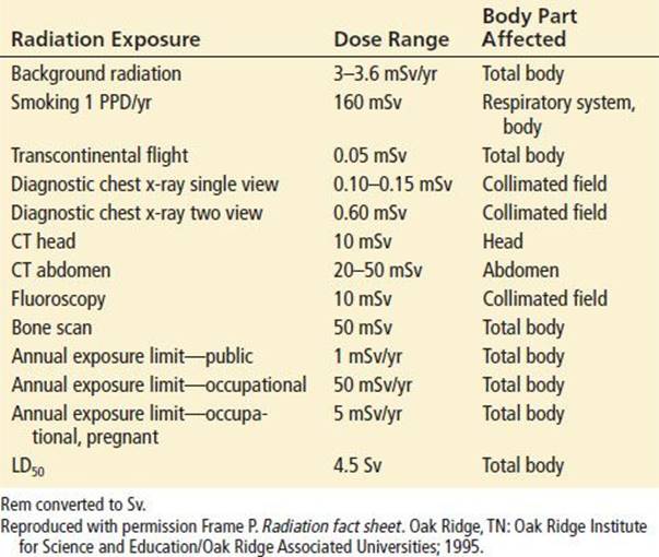
Radioactive substances can be found in many settings and forms. Radioactive materials are found in medical settings, research facilities, industrial sites, and nuclear power plants (3). Naturally occurring background radiation exposure accounts for 3 to 3.6 mSv per year; this and other radiation doses from medical procedures are listed in Table 366.1. According to the Department of Energy–Radiation Emergency Assistance Center/Training Site (REAC/TS) Radiation Accidents Registry, the majority of radiation accidents occur in the medical, energy production, or industrial settings (4). Current occupational exposure limits for individuals working with radiation sources are 0.05 Sv per year and 5 mSv per year for pregnant women. For the general public, current limits are 1 mSv per year (5).
TABLE 366.1
Dose Ranges from Background Radiation Exposures and Selected Medical Procedures
Radiation accidents are typically secondary to the improper use of radiation devices and improper use of radioisotopes. Between 1944 and 2002, a total of 428 radiation accidents have been documented worldwide, resulting in 133,815 persons affected and 134 fatalities. Nearly 90% of these victims were the result of the 1986 Chernobyl nuclear reactor accident. More recently, the near-critical event and leakage of radioactive materials from the Fukushima Daiichi plant after the 2011 Great East earthquake and tsunami in Japan reinforced the need to be locally prepared to respond to the immediate and long-term medical needs of the people living in proximity to nuclear facilities and of the emergency responders to radiologic incidents. Other historically important mass-casualty radiation accidents include the rupture of an iridium source in Venezuela; a cobalt-therapy unit accident in Ciudad Juarez, Mexico; and the theft and subsequent large exposure of a cesium source in Goiania, Brazil (4). A comprehensive planning guide to these and other types of radiation events has been produced by the International Commission on Radiological Protection to assist local medical planning and response and recovery efforts (6).
Individuals and groups may intentionally use radiologic materials for terrorist purposes. Such use may introduce new combinations of radiologic injury, or radiation injury may be misdiagnosed as other routine medical conditions. For example, an RDD may combine blast and radiation injuries and may disperse radioactive substances, creating a contamination hazard for the environment and both real and psychological casualties (7). Initial fatalities from an RDD are anticipated to be minimal and related to blast injuries (8), but the greater threat from the unrecognized radioactivity of RDD detonation is the contamination of receiving facilities. The mysterious death of Alexander Litvinenko in November 2006 from multisystem organ failure was initially difficult to recognize as Polonium-210 ingestion without further specialized diagnostic testing. Finally, direct attacks on nuclear power plants would not yield a nuclear explosion, but intentional meltdown of the core may cause similar environmental and health effects as seen at Chernobyl (9). Such events may not create a large number of immediate casualties, but the public health risk related to carcinogenesis may be significant over time. The greatest impact may be the fear and uncertainty that follows these events.
CLINICAL PRESENTATION
Acute radiation injury to the skin and underlying structures causes changes that clinically resemble thermal burns, with desquamation and blistering. Unlike thermal burns, these signs develop after a period of at least 48 to 72 hours after the contact. The clinical effects of dermal radiation exposure are listed in Table 366.2. Delayed skin effects include carcinogenesis, vascular insufficiency, and chronic nonhealing ulcers.
TABLE 366.2
Cutaneous Manifestations of Radiation Exposure

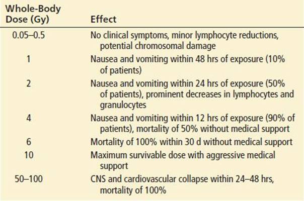
Whole-body radiation exposure clinically manifests as acute radiation sickness (ARS), a progression of systemic effects from derangements of the hematopoietic, GI, and neurologic systems. ARS has four general phases: (1) prodromal, (2) latent, (3) illness, and (4) recovery or death. Depending on the dose rate and total absorbed dose, ARS may progress rapidly to death or be completely asymptomatic (Table 366.3). Larger total-body doses correlate with increasing severity of symptoms and decreasing time interval to onset (10). The prodromal phase usually lasts 48 to 72 hours and manifests as nausea, vomiting, diarrhea, and nonspecific constitutional symptoms. The latent phase, which may last 3 weeks, is characterized by the lack of clinical symptoms and apparent recovery from the prodromal phase (7). The latent phase is followed by the illness or hematopoietic syndrome, in which initial damage to the bone marrow becomes clinically apparent. Pancytopenia and the subsequent sequelae of immunosuppression, bleeding diathesis, and anemia often lead to death from sepsis unless adequately supported. If the patient survives the illness phase, which lasts up to 30 days postexposure, recovery occurs 3 to 6 weeks after initial exposure.
TABLE 366.3
Acute Radiation Syndrome and Dose-Related Effects
Patients receiving total body doses in excess of 7 Sv manifest a distinct entity referred to as the GI syndrome, in addition to the prodromal symptoms (Table 366.4). The GI syndrome results in significant fluid and electrolyte losses, loss of bowel integrity to enteric bacteria, and subsequent death in more than 80% of patients. A pulmonary syndrome is noted at doses of 5 to 10 Sv, manifesting as pneumonitis, respiratory failure, and subsequent pulmonary fibrosis and cor pulmonale. Patients with total-body doses of 9 to 17 Sv develop a cerebrovascular syndrome, and patients receiving more than 50 Sv develop acute encephalopathy, with rapid (hours to days) progression of cerebral edema, cardiovascular collapse, and 100% mortality.
TABLE 366.4
Acute Radiation Syndrome: Symptoms and Radiation Dose

DIFFERENTIAL DIAGNOSIS
Early symptoms of nausea and vomiting are nonspecific, resembling gastroenteritis or food poisoning. Lymphopenia, immunosuppression, and anemia are not only characteristics of radiation sickness but also can be manifestations of HIV and other viral infections, leukemia, use of chemotherapeutic agents, toxic ingestions, and blood dyscrasias.
The differential diagnosis of radiation-induced dermal injury includes chemical, ultraviolet and thermal burns. In contrast to these entities, radiation-associated dermal injury is typically delayed up to 2 weeks. Other dermal lesions to consider include bullous pemphigoid, necrotic arachnidism, and stasis ulcers.
Concomitant radiation contamination should be suspected in any potential terrorist event. Although extraordinarily large doses of radiation can cause rapidly progressive shock, radiation typically does not cause sudden death. It is also unlikely to cause the immediate incapacitation of large numbers of people resulting from weapons of mass destruction incident. Other agents should be suspected should such scenarios arise.
ED EVALUATION
Because of the extreme rarity and potential calamity of an unexpected radiation exposure, the Joint Commission requires that every hospital develop a formal plan to deal with such an event. Prudence also dictates that every regional emergency management system includes radiation accidents in its disaster management protocol. Specific prehospital management guidelines have been developed by REAC/TS, and are available online at http://www.orau.gov/reacts/manage.htm.
When notified that a radiation accident may have occurred in which many people were exposed to high levels of radiation, the physician should invoke the hospital and regional plans, following them as closely as the circumstances of the particular incident allow. The hospital’s radiation safety officer, and the radiation safety officer employed at the site of the accident (or at the company whose truck has crashed, for instance), must be contacted immediately. Police and fire authorities and a public relations official should be called. The physician should communicate with the scene while the area designated in the hospital to receive patients is prepared. Specialized radiation detectors can be used in emergency departments (EDs) to identify patients who are emitting radiation from nonmedical radionuclides that should not be present in a healthcare environment. Such devices help to filter out the noise of the background medicinal and diagnostic radionuclides typically present in hospitals, and can provide an early warning of the presence of inappropriate radiation contamination (11).
It must be determined exactly what sort of accident has happened: an exposure to external radiation, contamination by radioactive matter, or possible incorporation of ionizing substances via open wounds or the GI or respiratory tracts. A history of trauma or other toxic exposures and the nature, severity, and time of onset of any symptoms should be noted. The duration of exposure (partial or whole body), the nature of and distance from the source, and shielding and dosimeters used, if any, all constitute important data.
Patients require careful evaluation by a designated radiologic emergency response team to minimize contamination of the ED (12). When possible, arriving patients should be undressed in the ambulance, and clothes should be collected, bagged, and stored at a point distant from the patient care areas. A designated receiving area large enough to manage the anticipated number of casualties should be established. The decontamination area should be prepared and ready for use for both ambulatory and nonambulatory patients. Areas that are likely to be traversed by contaminated patients and workers should be cordoned off. Paper or plastic sheets should be placed on the floor. Entry points to the radiation treatment areas need to be staffed by radiation officers trained to scan for contamination as workers exit the radiation treatment area, and large numbers of gloves, boots, gowns, and masks need to be available for workers entering the area. Strict isolation and double-bagging procedures should be maintained, and a buffer zone should be established for additional contamination control. No special ventilation controls are currently recommended.
All care providers must wear barrier protection, masks, and eye protection to prevent internal and external contamination. Furthermore, four ALARA (As Low As Reasonably Achievable) principles provide the foundation for radiation protection: timing, distance, shielding, and quantity. For timing, all personnel must wear a radiation detector to determine the whole body dose accumulated during patient contact. Personal dosimeters with real-time displays are preferred over radiation badges to limit any unnecessary exposure and to determine when to rotate teams of care providers. For routine procedures and care, teams should be rotated once 5 rem have been accumulated. The upper limit to provide lifesaving interventions must be predetermined by the radiation safety officer. For those performing lifesaving interventions, the recommended limits for total effective dose range from 0.25 to 1.5 Sv, or a rate of exposure ≤0.1 mGy/hr, depending on the radiation organization cited (13). For simplicity, a total-body exposure of 0.5 Sv is the National Council of Radiation Protection and Measurements/International Council of Radiation Protection and Measurements (NCRP/ICRP) recommended dose acceptable for preserving life (5). Workers in this setting should be volunteers chosen on the basis of age (older) and lowest accumulated lifetime effective doses.
Distance from a radiation source should be maximized when feasible, because the exposure dose decreases in proportion to the square of the distance from the source. Shielding high-density materials such as lead or concrete provides greater relative levels of protection from ionizing radiation than do less dense materials. Most structural elements (e.g., walls, floors, etc.) and personal protective equipment (PPE) will not block high-energy γ-rays. PPE will, however, provide a physical barrier to contamination from radioactive fallout and debris from RDDs. PPE is also effective in blocking the effects of α-particles and a proportion of β-particles as well. The quantity of ionizing radiation can also be limited by removing the radioactive source material from the area.
If no life-threatening illness is present, a rapid survey of patients using a “pancake” detector or a GM counter should be performed. Ideally, survey meters capable of detecting both high-range (up to 5 Sv/hr) and low-range (up to 0.2 Sv/hr) exposures and also common (γ and β) or uncommon (α and neutron) sources should be available. The initial scan should progress rapidly from head to toe, focusing on any open wounds or obvious debris. For the ambulatory patient, the survey should focus on areas, such as the hair and shoulders, where fallout generally accumulates. If the patient is unstable, critical interventions should be performed in conjunction with the radiation survey (14).
Anatomic charts should be used to document the location of all radiation emission and the radiation counts. Example charts and forms can be downloaded from http://www.afrri.usuhs.mil/www/outreach/pdf/afrriform331.pdf. Samples with cotton swabs should be taken of all orifices if external contamination is suspected. The physical examination should include evaluation for concomitant trauma. In all cases of radiation injury, a complete blood count and differential, a type and screen, and serum electrolytes and urinalysis should be obtained to determine kidney function. Subsequent investigations are driven by the patient’s clinical presentation.
KEY TESTING
• A CBC with differential count should be obtained for the purposes of identifying the absolute lymphocyte count and should be interpreted in the context of the temporal relationship to the radiation injury.
• Trauma laboratory work and imaging should be performed as warranted, and should be performed in conjunction with decontamination.
• Radiation dosimetry should be attempted, and initial and persistent contamination severity and the affected anatomical areas should be documented with a portable GM counter.
• Radiation spectrometry can be performed to determine if radiation sources are typical hospital radioisotopes or are nonmedicinal in nature.
• Biologic samples and fluids should be obtained and stored for analysis by radiation safety and health physics consultants.
ED MANAGEMENT
The organized delivery of care to radiation-injured patients can increase the survival of those with whole-body radiation injury (4). A tremendous amount of information and relevant protocols for the ED management of radiation injuries can be found on the REMM website: http://www.remm.nlm.gov. ED management of radiation injuries is summarized here, starting with initial contact and decontamination procedures (6,12,15).
Decontamination procedures begin with the removal of all clothing. This eliminates 80% to 90% of external contamination. Because radioactive debris may remain in the hair after removal of grossly contaminated clothing, surgical caps should be placed on the unstable contaminated patient to provide some limited shielding and contamination containment to protect staff while performing advanced life-support procedures. All clothes, debris, and bandages must be properly stored. The hospital radiation safety officer must properly dispose of the waste once surveyed. When contaminated debris or shrapnel are removed from patients, they should be placed in appropriate lead-shielded containers and removed from patient-care areas.
Wounds should be decontaminated aggressively, then treated as usual and covered with plastic and tape prior to decontamination of intact skin. External decontamination should proceed from the area with the most debris to the area with least. Uncontaminated areas should also be covered and taped. Using tepid water and mild soap or nonionic detergent, very gentle scrubbing should be done for 3 minutes with the patient lying either on a special decontamination table (similar to an autopsy table) or on a makeshift plastic trough placed on a stretcher in slight reverse Trendelenburg. Hot water should be avoided because it causes capillary vasodilation and raises the potential for increased radionuclide absorption. Aggressive scrubbing should likewise be avoided because of the potential for abrasions that can increase dermal absorption. This procedure should be repeated with serial radioactive screening and continued as long as counts are decreasing. Gentle brushing under nails, in hard to clean areas such as between fingers, axilla, groin, of skin folds, hair and clipping of hair, including body hair (not shaving, which might leave small open wounds) may be required. All fluids should be collected if possible. If the patient is stable, decontamination attempts can stop when the radioactivity level cannot be further reduced with serial GM counter testing.
Most isolated radiation injuries are not medical emergencies and only require supportive care, baseline laboratory testing, and follow-up for specialty care such as burns, infectious disease, and hematology/oncology. Specific antidotes and chelation agents are available that may assist in mitigating the effects of the radionuclide burden from internally contaminated patients. These agents do so by decreasing the overall radionuclide burden by dilution, displacement, blockage, or chelation. However, it is difficult to differentiate between radionuclides without scintillators or other advanced laboratory equipment that is found only in select laboratory settings. If identification is possible or a likely radionuclide is suspected from the type of radiation accident, antidotes must be administered promptly or the effectiveness will be limited. There are several tools, such as the Biological Assessment Tool (available at the Armed Forces Radiobiology Research Institute website: http://www.afrri.usuhs.mil) and the Radiation Injury Severity Classification (RISC) system, that can help the clinician determine the total whole-body radiation delivered based upon clinical symptoms (biologic dosimetry) alone (16).
Treatment of ingestions should begin with attempts to decrease the chance of internal irradiation or incorporation. Whole-bowel irrigation, using a polyethylene glycol bowel prep solution (e.g., GoLYTELY, Colyte) at 0.5 to 2 L/hr until the rectal effluent is clear, may be beneficial. Activated charcoal is also recommended. Washings should be saved, surveyed, and disposed of by the radiation safety officer. Bronchoalveolar lavage may be helpful if stained or radioactive sputum confirm that radioactive dust has been inhaled.
Specific therapy for radiation injury includes medical countermeasures, prophylaxis, and supportive care. There are specific medical countermeasures for certain radionuclides, such as Prussian blue for thallium, cesium, or rubidium; and barium sulfate or aluminum-based antacids for radium or strontium. Prussian blue (Radiogardase) works by increasing fecal elimination of the radionuclide. Prussian blue is taken orally and will not compete with other treatment agents such as oral potassium iodide (KI). Clinical side effects include constipation and GI upset. Prussian blue is most effective when administered soon after exposure but should be administered as soon as cesium or thallium has been identified as the radionuclide in question. In the Goiania incident involving cesium, Prussian blue at doses ranging from 3 to 10 g/day resulted in a 71% reduction in mean dose (3).
Potassium iodide (KI) should be administered to those in whom exposure to iodine-131 (I-131) is likely (18). Such events include an RDD contaminated with medical I-131, release of radioisotopes from a nuclear reactor, and nuclear detonation. Children may benefit most from the protective effects of KI AsKI works by competition to prevent the incorporation of radioactive iodine by the thyroid gland, the maximum benefit is conferred when KI is administered before exposure to I-131. It will protect the thyroid from increased risk for cancer but will not mitigate any other forms of acute or long-term radiation effects. The dose of KI is approximately 2 mg/kg. The protective effects of KI last approximately 24 hours, and daily dosing for up to 2 weeks, is usually at the discretion of the local health department. Short-term treatment of KI at thyroid-blocking doses is safe, although side effects of sialadenitis, GI upset, rashes, and allergic reactions are most common. As internal contamination of radioiodine will partition into breast milk, lactating mothers should not breastfeed their infants until public health authorities have deemed it safe to consume the milk.
Other countermeasures include pentetate calcium trisodium injection (Ca-DTPA) and pentetate zinc trisodium injection (Zn-DTPA). Both agents are now approved by the US Food and Drug Administration (FDA) to treat internal contamination with plutonium, americium, or curium (17). For specific conditions relating to ARS, such as the hematopoietic syndrome, use of hematopoietic colony–stimulating factors may enhance neutrophilic recovery by increasing the absolute numbers of white blood cells and decreasing the period of neutropenia (20).
Supportive care is needed for ARS, for medical and surgical comorbidities, and for psychological trauma. The initial management of concomitant trauma is the same as for other patients. Wound healing is adversely affected by radiation, and the likelihood of infection is increased due to fibroblast damage, bleeding diathesis, and loss of bone marrow that occurs over time. In light of this, definitive surgery should be performed within the first 48 hours of exposure. Otherwise, nonemergent procedures such as internal fixation of fractures is best delayed at least 3 to 60 days until bone marrow production returns to normal (19). Prophylaxis against bacterial infection in neutropenic patients may require empiric antibiotic therapy, specifically with antipseudomonal agents.
Children may have greater health effects with regard to radiation injury. Children are more metabolically active, and have potential greater risks for both deterministic and stochastic effects. They are also more likely to incorporate radioisotopes by ingestion or inhalation. Although psychological support is necessary for all patients, the pediatric population may be particularly susceptible to long-term psychological sequelae from real or suspected radiation injury (21).
During the UK Health Protection Agency investigation of the death of Alexander Litvinenko, approximately 596 individuals tested positive for polonium-210 (22), although only 13 were found to have had significant exposures. However, fears of polonium-210 contamination led thousands to seek medical attention. Physical manifestations of acute stress reaction, including nausea, vomiting, and vague constitutional complaints, can mimic those of acute radiation syndrome and can complicate the appropriate disposition of these patients (20,23).
CRITICAL INTERVENTIONS
• Notify the hospital radiation safety officer and activate the hospital radiation exposure plan for patients with possible radionuclide contamination.
• Use time, distance, shielding, and quantity-reduction techniques to minimize exposure.
• Wear barrier protection, masks, eye protection, and dosimeter, if available.
• Perform resuscitation and stabilization as necessary.
• Perform external and internal decontamination and isolate and remove contaminated clothes, debris, and fluids.
DISPOSITION
After initial ED management, patients may be discharged for outpatient management depending on extent of prodromal symptoms and absolute lymphocyte count at presentation. Onset of prodromal ARS symptoms <30 minutes after exposure indicates a total-body dose >6 Sv, although onset >2 hours after exposure indicates a dose <2 Sv. Onset of prodromal symptoms more than 24 hours after exposure indicates a dose <0.7 Sv (Table 366.4) (15). Severe nausea and vomiting indicate a substantial whole-body dose, (i.e., 2 to 10 Gy) that will require inpatient monitoring for supportive care.
The lymphocyte count is routinely used to aid in disposition and management of radiation injuries. If the patient is asymptomatic and has a normal lymphocyte count, early follow-up and frequent complete blood counts may be appropriate. Prognosis is directly tied to the 48-hour lymphocyte count, which is dose-dependent (Table 366.5). Lymphocyte counts >1,200/μL indicate a good prognosis, and counts <300/μL indicate a poor prognosis (24). Individuals with total-body doses >10 Sv will likely need admission, and those with doses >30 Sv may require reverse isolation precautions.
TABLE 366.5
Relationship Between Absolute Lymphocyte Count and Outcome After Total-Body Radiation Exposure

Hematology consultation will be required to administer growth-factor therapy and colony-stimulating factors, treat anemia and thrombocytopenia, and provide advanced cytokine therapy or cord/placental-blood progenitor cells. Burn, infectious disease, and bone marrow transplant consultation may also be necessary (25).
Questions regarding radiation accidents and exposures can be referred to the US Department of Energy through the REAC/TS, Oak Ridge, TN, by the web, http://www.orau.gov/reacts, or by phone, 865-576-1005. Other online references include the Radiation Event Medical Management website (http://www.remm.nlm.gov) hosted by the Department of Health and Human Services, which provides guidance in the diagnosis and treatment of radiation injuries, and the Centers for Disease Control website (http://www.bt.cdc.gov/radiation/clinicians.asp), related to radiation emergencies.
Common Pitfalls
• Failure to consider and identify radioactive contamination or irradiation injury.
• Failure to conduct disaster training, or performing such drills without addressing decontamination, use of PPE, and radiation surveys.
• Failure to manage concomitant injuries appropriately and to accomplish this prior to decontamination in critically ill patients.
• Failure to obtain an initial lymphocyte count for comparison to a follow-up 48-hour count.
• Failure to request appropriate consultation or to provide indicated chelation or antidote therapy.
• Failure to address psychological components of radiation exposure with patients and staff.
REFERENCES
1. Chambers JA, Purdue GF. Radiation injury and the surgeon. J Am Coll Surg. 2007;204:128–139.
2. Wolbarst AB, Wiley AL, Nemhauser JB, et al. Medical response to a major radiological emergency: A primer for medical and public health practitioners. Radiology. 2010;254:660–677.
3. U.S. Department of Health, Center for Drug Evaluation and Research. Guidance: Potassium Iodide as a Thyroid Blocking Agent in Radiation Emergencies: Use in Radiation Emergencies: Treatment Recommendations. Food and Drug Administration website. Available at http://www.fda.gov/cder/guidance/4825fnl.htm#KI. Accessed December 9, 2003.
4. Anonymous. DOE-REAC/TS Radiation Accident Registry System (REAC/TS Course Material). Oak Ridge, TN: Oak Ridge Institute for Science and Education; 2003.
5. Anonymous. Limitations of Exposure to Ionizing Radiation (NCRP report no. 116). Bethesda, MD: National Council on Radiation Protection and Measurements; 1993.
6. Waselenko JK, MacVittie TJ, Blakely WF, et al. Medical management of the acute radiation syndrome: Recommendations of the Strategic National Stockpile Radiation Working Group. Ann Intern Med.2004;140:1037–1051.
7. Jarrett DG, Sedlak RG, Dickerson WE, et al. Medical treatment of radiation injuries—current US status. Radiat Meas. 2007;42(6–7):1063–1074.
8. Gusev I, Guskova AK, Mettler FA Jr. Medical Management of Radiation Accidents. 2nd ed. Boca Raton, FL: CRC Press; 2001.
9. Weisdorf D, Chao N, Waselenko JK, et al. Acute radiation injury: Contingency planning for triage, supportive care, and transplantation. Biol Blood Marrow Transplant. 2006;12:672–682.
10. Goans RE, Waselenko JK. Medical management of radiological casualties. Health Phys. 2005;89(5):505–512.
11. Dickson R, Kim JO, Huq MS, et al. Interceptor and phantom trials of EDNS at UPMC. Health Phys. 2013;105(5 suppl 3):S199–S208.
12. Centers for Disease Control. Guidance for Public Health Departments and Clinicians Caring for Individuals Who may have been Recently Exposed to Polonium 210 (Po-210). Available online at http://www.bt.cdc.gov/radiation. Accessed January 27, 2014.
13. Jarrett DG. Medical Management of Radiologic Casualties Handbook. 1st ed. Bethesda, MD: Armed Forces Radiobiology Research Institute; 1999.
14. Koenig KL, Goans RE, Hatchett RJ, et al. Medical treatment of radiological casualties: Current concepts. Ann Emerg Med. 2005;45:643–652.
15. Mettler FA Jr, Voelz GL. Current concepts: Major radiation exposure—what to expect and how to respond. New Engl J Med. 2002;346(20):1554–1561.
16. Kuniak M, Azizova T, Day R, et al. The Radiation Injury Severity Classification (RISC) system: An early injury assessment tool for the frontline health-care provider. Br J Radiol. 2008;81:232–243.
17. Food and Drug Administration press release. FDA Approves Drugs to Treat Internal Contamination from Radioactive Elements. Food and Drug Administration website. Available online at Accessed January 27, 2014.
18. Bushberg JT, Kroger LA, Hartman MB, et al. Nuclear/radiological terrorism: Emergency department management of radiation casualties. J Emerg Med. 2007;32(1):71–85.
19. Flynn DF, Goans RE. Nuclear terrorism: Triage and medical management of radiation and combined-injury casualties. Surg Clin N Am. 2006;86:601–636.
20. Weinberg AD, Kripalani S, McCarthy, PL, et al. Caring for survivors of the Chernobyl disaster: What the clinician should know. JAMA. 1995;274:408–412.
21. American Academy of Pediatrics, Committee on Environmental Health.. Policy statement, radiation disasters and children. Pediatrics. 2003;111(6 Pt 1):1455–1466.
22. International Commission on Radiological Protection. 1990 recommendations of the International Commission on Radiological Protection. In Annals of the ICRP, vol. 21, nos. 1–3. Oxford: Pergamon Press; 1991:60.
23. Valentin J. Protecting people against radiation exposure in the event of a radiological attack: A report of the International Commission on Radiological Protection. Ann ICRP. 2005;35(1):1–110.
24. Collins DL. Human responses to the threat of or exposure to ionizing radiation at Three Mile Island, Pennsylvania, and Goiania, Brazil. Mil Med. 2002;167(2 suppl):137–138.
25. Berger ME, Christensen DM, Lowry PC, et al. Medical Management of radiation injuries: Current approaches. Occup Med (Lond). 2006;56:162–172.