
Andrew D. Perron, Christopher T. Bowe, and Christine Irish
Acute compartment syndrome is a serious life- and limb-threate-ning complication of extremity trauma. Fractures, crush injuries, burns, and arterial injuries all can result in an acute compartment syndrome. Compartment syndrome is defined as the presence of pathologically elevated intramuscular pressures within confined osseofascial compartments resulting first in tissue ischemia and ultimately infarction and subsequent contractures if not treated in a timely and appropriate manner (1). Dr. Richard Von Volkmann first described this entity in 1881; he noted the complication of muscle ischemia, paralysis, and ultimate contracture in a child with a supracondylar fracture (2,3). The various compartments in the extremities are composed of muscle groups bound by dense fascial tissue planes that result in limited ability to accommodate increased pressure. In addition to muscle, these compartments contain various nerves and blood vessels. Elevated pressure in these spaces, above normal physiologic ranges, can compromise capillary blood flow and produce predictable clinical signs and symptoms. Delays in diagnosis and treatment can result in myonecrosis, permanent nerve damage, distal ischemic injury, and muscle contracture. The exact incidence of compartment syndrome is unknown, although it is thought to be relatively rare. Three-quarters of cases are associated with fractures. Fractures of the tibia are the most likely to result in compartment syndrome; the incidence in patients with tibial fractures is reported to range from 1% to 29% (2). The most frequently involved compartments are the anterior compartment of the lower leg and the flexor compartment in the forearm (2). Table 48.1 lists the common locations of compartment syndromes.
TABLE 48.1
Common Locations of Compartment Syndromes

The pathophysiology of compartment syndrome involves local hydrostatic and osmotic pressure conditions within the myofascial compartment. When the intracompartmental pressure increases above a specific level due to the above factors, perfusion to the compartment is impaired, resulting in disruption of skeletal muscle metabolic processes. Cell wall membrane integrity is compromised, leading to cytolysis with the release of osmotically active cellular contents into the extracellular compartment. This draws additional fluid from plasma into the interstitial space. The net effect is increased intracompartmental pressure and further impairment of perfusion to the closed space of the myofascial compartment as well as to distal structures in that vascular distribution. This progressively leads to compromise of circulation and nerve conduction and ultimately to irreversible damage to the contents of the compartment (4).
Normal compartment pressure is below 10 to 12 mm Hg (2). Capillary blood flow depends on the pressure gradient between the arterial and venous systems. With elevated compartmental pressures, this gradient is interrupted, compromising the blood flow across the capillary bed. When the pressure within a compartment rises sufficiently to interrupt this arteriovenous gradient, blood flow will be reduced to the point at which the metabolic demands cannot be met. Then functional abnormalities and compartment syndrome occur (2). Two general mechanisms can result in elevated compartment pressures: Either an extrinsic compressive force constricts the compartment (e.g., a cast) or there is an intrinsic increase in the size of the fascia-bound compartment owing to edema or hemorrhage.
Clinically significant reduction of blood flow and blood oxygenation can occur at tissue pressures above 20 mm Hg (5,6). Cellular metabolic derangements are closely associated with the difference between mean arterial blood pressure and compartment pressure. This difference is known as the delta P. In one study, the lowest delta P that was compatible with a normal metabolic state was 30 mm Hg in normal muscle (7). The length of time that tissue can tolerate elevated pressures before pathologic changes occur is less clear. Sensory changes including paresthesias and hypesthesia can develop within 30 minutes of ischemia, whereas irreversible nerve damage begins as soon as 6 hours and almost universally after 12 to 24 hours (2).
Other factors should also be considered when assessing potential compartment syndrome. Externally applied pressure (i.e., casting) tends to produce less elevation in compartment pressures as compared to similar levels of internally developed pressures (i.e., edema or hemorrhage) (8). Postischemic compartment syndrome is caused by revascularization after a prolonged period of ischemia (2), leading to the release of vasoactive factors that result in fluid extravasation and elevated pressures. Concurrent hypotension, hypoxia, and associated premorbid conditions such as peripheral vascular disease adversely affect the ability of ischemic tissue to tolerate even mildly elevated tissue pressures.
CLINICAL PRESENTATION
Pain out of proportion to examination findings is the classically described clinical finding in acute compartment syndrome. This assumes an awake, neurologically intact patient. Unfortunately, many patients at risk for compartment syndrome are severely injured or impaired and cannot relate the degree of pain they are experiencing. When reported, the pain is often described as deep, aching, and poorly localized and does not typically respond to immobilization or analgesic medications alone. Increasing analgesic requirements may be the only clue to a developing compartment syndrome. Classically, passive stretching of involved muscles produces intense pain, as does compression of an affected compartment. Weakness or paresis of muscle function is a late finding. Distal hypesthesias or paresthesias may be noted in the distributions of nerves passing through the compartment. Two-point discrimination and light touch are considered more sensitive than pinprick when assessing sensation. Tenseness of externally palpable compartments is the earliest and most objective finding, although tenseness of compartments such as the deep posterior compartment of the lower leg and the iliacus compartment can be difficult to assess due to anatomic location. Findings of compartment syndrome can present within 2 hours of injury but may first appear up to 6 days later (2). Progressive worsening over time helps to distinguish compartment syndrome from other injuries that cause primary nerve and muscle damage.
Griffith’s mnemonic, the five Ps (pain, pallor, pulselessness, paresthesias/paresis, and poikolothermia), is traditionally used for this clinical entity. However, pulses are most often maintained, and the skin does not typically develop pallor (1). The presence of all five of these signs more accurately reflects the late findings of acute arterial obstruction or insufficiency. As noted previously, the reliance on the clinical examination makes it difficult to assess emergency department (ED) patients with altered sensorium, acute intoxication, spinal cord injuries, or overlying extensive burns, as well as children. Moreover, obtunded patients are often at risk for compartment syndrome by compression, entrapment, or unknown mechanisms of trauma. Overdose-related compartment syndromes tend to affect the lower extremities (9,10). Critically ill pediatric and adult patients may experience iatrogenic causes of compartment syndrome produced by intravenous and intraosseous infiltrations, transfusions under pressure, or tourniquet malfunction. Table 48.2 lists common etiologies of compartment syndrome. Owing to dense eschar, severely burned patients lose most, if not all, clinical signs of compartment syndrome. In these more complex cases, the clinician should have a lower threshold for using invasive monitoring.
TABLE 48.2
Common Etiologies of Compartment Syndromes Seen in Emergency Medicine

The clinical findings for specific anatomic compartments are summarized in Table 48.3. Figure 48.1 illustrates the anatomy for the two most common sites. The three most common etiologies of an acute compartment syndrome are fractures, crush injury, and reperfusion of an ischemic limb. The lower leg and forearm have relatively “tight” compartments and are the most common sites for developing compartment syndrome. In the pediatric population, fractures are the most common cause, specifically supracondylar fractures. Open fractures are not protective against compartment syndrome, as the tear in the fascia is usually quite small and does not allow decompression of the compartment when pressure is elevated. Overall, the most common fracture etiologies for compartment syndrome include tibial shaft, supracondylar, and forearm fractures (7).
TABLE 48.3
Compartment Syndromes and Associated Physical Signs

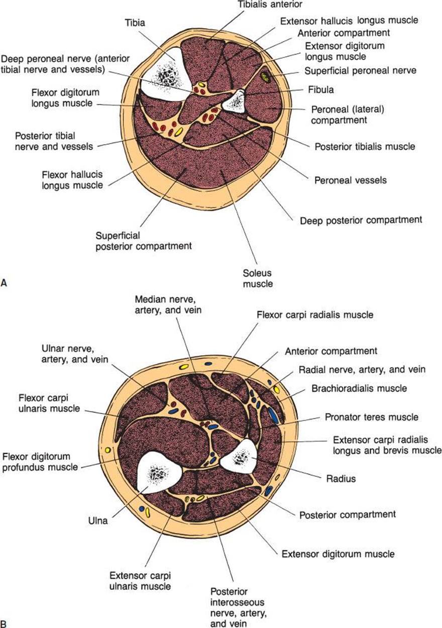
FIGURE 48.1 Compartments of the leg (A) and forearm (B).
DIFFERENTIAL DIAGNOSIS
Differential diagnostic considerations include deep vein thrombosis, necrotizing fasciitis, arterial embolus or insufficiency, gas gangrene, myositis, and rhabdomyolysis.
ED EVALUATION
Laboratory and radiologic studies cannot confirm or rule out the diagnosis of compartment syndrome but may help in assessing for other etiologies and secondary complications such as rhabdomyolysis. Significant elevation of serum creatinine phosphokinase may indicate severe muscle damage or ischemia but is not helpful for early diagnosis (2). The critical diagnostic test for compartment syndrome is the direct measurement of intracompartmental pressures. This is especially important in those patients who are unreliable because of distracting injury or altered sensorium. These direct tissue measurements can also be used to provide ongoing monitoring in patients who are at high risk for development of a compartment syndrome, as in the case of a crush injury. Various pressure measurement techniques have been described, such as the central venous pressure manometer, the wick catheter, the slit catheter, and the portable Stryker handheld monitor. Experimental noninvasive methods of compartment pressure measurement have been described but are not in widespread clinical use currently. These include near-infrared spectroscopy, which measures the levels of muscle hemoglobin and myoglobin; laser Doppler flowmetry; and 99Tcm-methoxyisobutyl scintigraphy (2). The direct measurement techniques all provide acceptable accuracy, although the self-assembled needle manometer has been shown to be the least accurate and least reproducible (2). Most compartments are superficial, but access to deeper ones such as the gluteal and deep posterior compartments may require a spinal needle.
Whatever the method employed, once the compartment pressure has been obtained, there is ongoing debate over which pressure levels warrant a fasciotomy. Absolute pressures of >30 mm Hg have been suggested as an indication for fasciotomy. Alternatively, a “delta P” (the difference between the diastolic pressure and the compartment pressure) of <30 mm Hg has been suggested as being more specific for the requirement of a fasciotomy (2,7). These pressure measurements must be assessed in the context of hypotension, anemia, hypoxia, or other factors that would increase the tissue sensitivity for ischemia at less elevated pressures. Nerve conduction studies and arteriography are used primarily to distinguish primary nerve and arterial injuries and not useful in the acute diagnosis of compartment syndrome.
It should be noted that pulse oximetry has been advocated as a simple, noninvasive indicator of vascular compromise. This is not true. Clinical studies using pulse oximetry have shown a sensitivity of 40% for vascular compromise at clinically significant elevated compartment pressures (11). With a >50% risk of a false-negative result, pulse oximetry is not recommended in the evaluation of this disease process.
KEY TESTING
Compartment pressure measurement:
• If elevated compartment pressure is suspected, pressure testing is strongly suggested (unless there is clear evidence of compromise, in which case operative fasciotomy may occur by the surgeon without taking the time to assess intracompartmental pressures).
• Many techniques are described (wick, slit catheter, Stryker). The Stryker pressure manometer is a self-contained unit that assesses intracompartmental pressure using a needle and a calibrated digital readout. Knowledge of both the anatomic layout of the fascial compartments as well as the mechanics of the manometer itself is required for accurate use.
• Absolute compartment pressures >30 mm Hg/delta-P <30 mm Hg are generally used cutoffs as indications for fasciotomy. Normal compartment pressure is <10 mm Hg.
ED MANAGEMENT
The key to management of ED patients with compartment syndrome is early consideration and diagnosis. Surgical fasciotomy is the definitive treatment and surgical consultation should be initiated early. Surgical decompression should ideally be performed emergently, as soon as possible after the diagnosis is made (2,7). Supportive care is aimed at resuscitation and stabilization of the patient and preparation for appropriate operative intervention.
In patients who are suspected of having compartment syndrome owing to casts, bivalving and spreading of the cast may be necessary and can produce an almost 65% reduction in applied external pressures while maintaining alignment (12). Complete removal of the cast can further reduce pressure by up to 85% (4). Elevation of an extremity at risk to the level of the heart is recommended, as this promotes venous drainage without compromising arterial inflow (4). Ice should be avoided, as it can further compromise microvascular blood flow. The emergency physician should remove aggravating factors specific to the involved extremity, such as MAST suits or pneumatic splints, and treat systemic factors such as hypotension, anemia, and hypoxia.
CRITICAL INTERVENTIONS
• Measure intracompartmental pressures.
• Initiate surgical consultation early.
• Elevate the involved extremity to the level of the heart in the least restrictive dressing possible.
DISPOSITION
Patients with suspected compartment syndrome require emergent surgical or orthopedic consultation. Patients are typically admitted for observation and repeated clinical examinations and pressure measurements if they do not undergo immediate surgical intervention. Patients with multiple trauma may require intensive care unit admission for management of other associated injuries.
Common Pitfalls
• Failure to consider the diagnosis of compartment syndrome.
• Failure to recognize early signs and symptoms of compartment syndrome.
• Attributing the signs and symptoms of compartment syndrome to other types of injuries.
• Failure to do a thorough extremity and neurologic examination, especially in patients who are intoxicated or unreliable.
ACKNOWLEDGMENTS
The authors gratefully acknowledge the contributions of John A. Siefert to the content of this chapter.
REFERENCES
1. Leversedge FJ, Moore TJ, Peterson BC, et al. Compartment syndrome of the upper extremity. J Hand Surg Am. 2011;36(3):544–559.
2. Gourgiotis S, Villias C, Germanos S, et al. Acute limb compartment syndrome: A review. J Surg Educ. 2007;64(3):178–186.
3. Jepson PN. Ischaemic contracture: Experimental study. Ann Surg. 1926;84:785–795.
4. Perron AD, Brady WJ, Keats TE. Orthopedic pitfalls in the ED: Acute compartment syndrome. Am J Emerg Med. 2001;19(5):413–416.
5. Matsen FA III, Krugmire RB Jr, King RV. Nicholas Andry Award. Increased tissue pressure and its effect on muscle oxygenation in level and elevated human limbs. Clin Orthop Relat Res. 1979;(144):311–320.
6. Frink M, Hildebrand F, Krettek C, et al. Compartment syndrome of the lower leg and foot. Clin Orthop Relat Res. 2010;468(4):940–950.
7. Kostler W, Strohm PC, Sudkamp NP. Acute compartment syndrome of the limb. Injury. 2004;35(12):1221–1227.
8. Rush RM Jr, Arrington ED, Hsu JR. Management of complex extremity injuries: Tourniquets, compartment syndrome detection, fasciotomy, and amputation care. Surg Clin North Am. 2012;92(4):987–1007.
9. Shadgan B, Menon M, Sanders D, et al. Current thinking about acute compartment syndrome of the lower extremity. Can J Surg. 2010;53(5):329–334.
10. Brahmamdam P, Plummer M, Modrall JG, et al. Hand ischemia associated with elbow trauma in children. J Vasc Surg. 2011;54(3):773–778.
11. Mars M, Hadley GP. Failure of pulse oximetry in the assessment of raised limb intracompartmental pressure. Injury. 1994;25(6):379–381.
12. Garfin SR, Mubarak SJ, Evans K, et al. Quantification of intracompartmental pressures and volume under plaster casts. J Bone Joint Surg. 1981;63:449–453.