
Stacy Sawtelle
Glaucoma is a group of disorders characterized by increased intraocular pressure (IOP) sufficient to cause loss of vision. This definition has recently evolved, however, and currently isolated increased IOP is not considered to constitute glaucoma; rather it is the resulting damage to the optic nerve that defines the disease (1,2). Visual change, pain, or elevated IOP without signs of injury to the optic nerve is now termed acute angle-closure crisis (AACC) rather than acute angle-closure glaucoma (AACG) (2,3).
The underlying pathologic mechanisms of AACG, increased aqueous humor production and decreased aqueous humor outflow, cause varying degrees of increased IOP. Patients with only mildly elevated IOP can have findings consistent with glaucoma (low-tension glaucoma) whereas patients with significantly elevated IOP may lack criteria for glaucoma (ocular hypertension) (4). Glaucoma is classified in terms of both anatomy of the anterior chamber (angle) and presence or absence of comorbid disease. The anatomic classification describes whether the angle of the anterior chamber is open or closed. Each anatomic classification may be further subdivided into primary or secondary depending on the absence or presence, respectively, of pre-existing disease (4). Primary AACG is particularly important to the emergency physician, as it carries a high rate of visual morbidity if not quickly diagnosed and treated. Glaucoma is the second most common cause of blindness worldwide (5).
ANATOMY AND PATHOPHYSIOLOGY
The eye is a complex organ controlled by opposing effects from the sympathetic nervous system (SNS) and the parasympathetic nervous system (PNS). Stimulation of the ciliary body receptors (β1) results in production of aqueous humor. The aqueous humor flows from the posterior chamber to the anterior chamber via the space between the posterior surface of the iris and the lens (Fig. 61.1). The area of this opening varies with miosis, mydriasis, and accommodation. The muscles controlling these functions also affect the trabecular meshwork in the anterior chamber angle and, thus alter the opening of the canal of Schlemm.

FIGURE 61.1 Normal anterior chamber angle. Aqueous passes from the posterior chamber through the pupil into the anterior chamber and out through the trabecular meshwork.
The iris contains two muscle groups, the radial and circular, whereas the ciliary body contains one group, the ciliary muscle. The radial muscle, which contains α1-receptors, contracts when stimulated by the SNS, resulting in mydriasis. The circular muscle of the iris and the ciliary muscle both contain muscarinic receptors under the control of the PNS. Stimulation of the circular muscle causes contraction and thus miosis. Ciliary muscle contraction reduces the tension on the zonules of Zinn, thereby reducing tension on the lens. Zonule laxity allows the lens to “round up” for accommodation of near vision; however, this also increases the contact between the lens and posterior iris, potentially inhibiting flow of aqueous humor from the posterior to the anterior chamber. Conversely, relaxed tension on the meshwork reduces resistance to outflow at the angle by enhancing the opening from the anterior chamber into the canal of Schlemm.
Elevated IOP in glaucoma most often results from an obstruction to aqueous outflow rather than excessive production (6). Obstruction to outflow from the anterior chamber occurs in both open-angle glaucoma and AACG. The location and mechanism of the resistance to outflow distinguish these two conditions. In open-angle glaucoma, the trabecular meshwork of the anterior chamber angle remains open, but the aqueous humor is unable to reach the canal. In AACG, the peripheral iris contacts the trabecular meshwork and blocks the flow of aqueous into the canal.
Several anatomic features predispose an eye to AACG, and patients may have any combination of risk factors. A shallow anterior chamber results in increased baseline narrowing of the entrance to the angle, with a greater area of contact between the lens and iris. This lens-to-iris apposition impedes the flow of aqueous from the posterior chamber to the anterior chamber through the pupil, produces a forward bowing of the peripheral iris, and effectively closes the angle (Fig. 61.2). Those with a shallow anterior chamber angle, at increased risk for the development of AACG, include the elderly, women, patients of Asian or Inuit descent, and farsighted patients (2,4,5,7). A pharmacologic or physiologic event in a susceptible person typically triggers an attack of acute angle closure by upsetting the equilibrium of aqueous humor dynamics.

FIGURE 61.2 Angle closure with pupillary block. The angle is closed as the iris is pushed up against the trabecular meshwork.
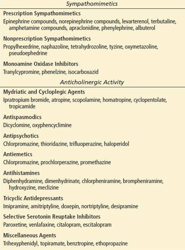
Pupillary dilatation frequently precipitates an attack of acute angle closure. As the pupil dilates, the peripheral iris becomes more flaccid and may be pushed against the trabecular meshwork, blocking aqueous outflow. This may occur when a person is in a dimly lit environment or under emotional stress perhaps due to increased sympathetic tone. Medications that stimulate α1- and/or β1-receptors (sympathomimetics) and those that oppose M3 receptors (anticholinergics) cause pupillary dilatation and may precipitate AACC (Table 61.1) (3,4,8).
TABLE 61.1
Common Pharmacologic Preparations That May Precipitate AACG
CLINICAL PRESENTATION
Most patients present with a complaint of sudden onset of severe pain localized to the orbit, or brow or more generally with a severe headache, often associated with nausea and vomiting. Occasionally, the presenting clinical picture is more systemic than ocular, leading to an erroneous or missed diagnosis (9). The physician should consider AACG in patients with headache and high-risk patients with undifferentiated gastrointestinal illness. Blurred vision or the appearance of rainbow-colored halos around lights may occur shortly after the onset of pain (Table 61.2).
TABLE 61.2
Diagnosis of AACG

The patient with AACC has a unilateral red eye with congested episcleral and conjunctival blood vessels, a nonreactive middilated pupil, corneal edema, a shallow anterior chamber, and a high IOP (Fig. 61.3). The IOP is elevated, usually ranging between 60 and 90 mm Hg (IOP <21 mm Hg is considered normal). If the attack has been prolonged, however, the IOP may be lower, due to ciliary body ischemia and decreased aqueous production. Corneal involvement varies with the duration of the attack but is often hazy or steamy appearing from epithelial edema. The anterior chamber appears shallow. Although mild anterior chamber inflammation is common, keratic precipitates (aggregates of white blood cells) on the corneal endothelium are not a typical finding.
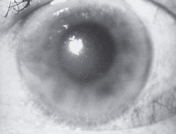
FIGURE 61.3 This eye demonstrates the hallmarks of acute angle-closure glaucoma: perilimbal injection, corneal edema, and a middilated nonreactive pupil.
The course of untreated AACG varies. An attack may damage the optic nerve as well as the corneal endothelium, lens, and retinal ganglion cell layer. In some undiagnosed patients, the attack will continue and, if untreated, can result in permanent blindness within 2 to 3 days. Visual prognosis improves with the initiation of prompt, effective treatment, but any damage to the optic nerve is irreversible.
DIFFERENTIAL DIAGNOSIS
The differential diagnosis of the acutely painful red eye is extensive and is described in detail in Chapter 56. Because headache, nausea, and vomiting may be dominant symptoms in glaucoma, the differential should also include central nervous system and gastrointestinal disorders. Most conditions are readily distinguished by a careful history and physical examination. Other ophthalmic disorders that more closely mimic AACG are the secondary glaucomas: glaucomatocyclitic crisis, neovascular glaucoma, glaucoma secondary to iritis or uveitis, and lens-induced glaucoma.
Glaucomatocyclitic crisis produces recurrent attacks of visual blurring and halos but, in contrast to AACG, pain is not a prominent feature. In addition, the anterior chamber is deep and keratic precipitates develop on the cornea after 2 to 3 days (10).
Neovascular glaucoma presents with a chief complaint of a painful red eye months after vision impairment in the affected eye. It is usually associated with disorders such as, diabetic proliferative retinopathy and retinal vascular occlusion. Retinal hypoxia serves as the underlying stimulus for neovascularization. If the condition progresses, a fibrovascular membrane may obstruct the drainage angle, leading to secondary AACG (4).
Acute iritis with secondary glaucoma presents with ocular pain, photophobia, and blurred vision. The onset is gradual in contrast to AACG. Clinical findings may mimic angle closure, with perilimbal conjunctival injection and corneal edema. However, a miotic pupil, keratic precipitates, and a deep anterior chamber differentiate acute iritis with secondary glaucoma from AACG.
Phacolytic glaucoma presents similarly to AACG, but the anterior chamber depth (ACD) is normal and there are cells in the anterior chamber. Keratic precipitates are also present, and a mature or hypermature cataract can be seen. The cause of phacolytic glaucoma is the release of soluble lens protein into the aqueous, which leads to elevated IOP when it occludes the trabecular meshwork and canal (4,11).
Lens particle glaucoma is associated with spontaneous or traumatic disruption of the lens capsule. As with acute iritis and phacolytic glaucoma, the anterior chamber is of normal depth. In addition, the pupil is often miotic, and lens cortical material is present in the anterior chamber. The particulate lens material reduces outflow in the trabecular meshwork, with resultant increased IOP (4,11).
ED EVALUATION
A careful history is an essential part of the ED evaluation and should include onset of symptoms (sudden, gradual, subsequent to accidental or surgical trauma); history of similar symptoms previously (brief episodes of pain, blurred vision, and/or halos around lights); visual impairment in the affected eye and the unaffected eye; pain in or around the eye; discharge or secretions from the eye; and a detailed medication history.
The ocular examination is described in detail in Chapter 54. The following aspects of the ocular examination deserve special emphasis in relation to AACC and glaucoma. Visual acuity is probably one of the most important evaluations done during any ocular examination. If a distance or near visual acuity cannot be recorded owing to inability to read the chart, the following should be recorded (including the distance at which each is tested): finger counting, and the presence or absence of perception of hand motion and light. Conjunctival injection is often present but is nonspecific. The pattern of injection, however, may be helpful. Perilimbal injection suggests acute iritis or angle closure. Segmental injection characterizes episcleritis; diffuse injection is typical of keratoconjunctivitis. Finally, the presence of purulent discharge suggests an infectious cause. Corneal edema is apparent in many cases of AACG and is due to the rapid rise of IOP. Assessment of the ACD is essential in the evaluation of the painful red eye and in the diagnosis of AACG. In the ED setting, an adequate estimation of the ACD can be accomplished with the oblique flashlight test and slit-lamp examination.
Oblique Flashlight Test
In this test (Fig. 61.4), the examiner shines the light across the eye from the temporal side toward the pupil. In an eye with a normal depth anterior chamber, the entire iris is illuminated. In an eye with a narrow angle or shallow ACD, a shadow is cast on the nasal side of the iris (12).

FIGURE 61.4 Flashlight test for wide and narrow angles. A: Wide angle: Entire iris is illuminated. B: Narrow angle: Only temporal half of iris is illuminated. (Photo courtesy of D. R. Anderson, MD.)
Slit-Lamp Examination
The ACD can also be estimated during the slit-lamp examination. A narrow beam of light is directed obliquely at the surface of the iris. The corneal thickness is estimated, as is the distance between the corneal beam and the iris beam, and the ACD is calculated. If the peripheral ACD is less than one-fourth the thickness of the adjacent cornea, the angle is narrow and at risk of occlusion (4,12,13).
The size and shape of the pupil and pupillary reaction to light are recorded for each eye. Unequal pupil size (anisocoria) should be noted. A middilated, nonreactive pupil is characteristic of AACC and glaucoma. In contrast, many other conditions have a miotic pupil that is minimally reactive.
A mature (opaque) or hypermature (liquid cortex) lens may be associated with lens-induced glaucoma. Phacodonesis, movement of the lens, indicates a dislocated or subluxed lens. This may occur secondary to trauma, or it may be part of a systemic syndrome such as Marfan syndrome. A dislocated lens can increase pupillary block and result in a secondary ACG (11).
Intraocular Pressure
The range of IOP considered normal is 10 to 21 mm Hg, although the actual range found in population studies is dependent on a number of factors and varies significantly among different ethnicities (14). Three commonly used tonometers available in the ED are the air-puff noncontact tonometer, the Schiötz tonometer, and the Tono-Pen. The Goldmann applanation tonometer is frequently used by ophthalmologists and is considered the gold standard for measurement of IOP.
Air-Puff Noncontact Tonometer
The air-puff tonometer has the advantage of measuring the IOP without the application of topical anesthesia or contact between the instrument and the eye. A puff of air is blown against the cornea and the instrument records the force needed to flatten the cornea and displays the correlated IOP (4,15).
Schiötz Tonometry
Schiötz, or indentation, tonometry determines IOP by measuring the indentation of the cornea produced by a known weight. The patient is placed in a supine position and a drop of a local anesthetic is instilled. The patient is asked to look upward and fix on some object in the distance. While separating the eyelids, the instrument is gently placed in a vertical position directly over the cornea. The plunger is allowed to rest completely on the eye. With the instrument held steady, the pointer will stay fixed at a value on the scale. Each instrument is accompanied by a conversion table that expresses the scale readings in mm Hg. Inadvertent pressure on the globe can result in a falsely elevated reading. Scleral rigidity and corneal thickness can affect the measurement (4).
Tono-Pen
A handheld electronic applanation tonometer, the Tono-Pen is frequently used in EDs. After initial calibration, the sheathed tip of the Tono-Pen is repeatedly tapped on the anesthetized cornea. The instrument calculates and displays the average of 4 to 10 readings, as well as variability percentages (4). The Tono-Pen generally correlates well with Goldmann applanation tonometry, but the Tono-Pen underestimates IOP when it is high and overestimates IOP when the pressure is low (4,16).
Goldmann Applanation Tonometry
In applanation tonometry, the cornea is flattened and the instrument measures the area flattened and the force required to do so. A local anesthetic and fluorescein are instilled. Correctly positioned at the slit lamp, the patient looks straight ahead. Illuminated by the cobalt blue light, the examiner gently contacts the anterior corneal surface with the tip of the Goldmann tonometer (Fig. 61.5). On contact, two fluorescein semicircles are seen. A calibrated dial is then adjusted until the semicircles are aligned such that the inner borders of each semicircle match up. The IOP is estimated from the value displayed on the dial (4).

FIGURE 61.5 Applanation tonometry. A: The tonometer tip should just contact the patient’s cornea. B: Viewed through the slit lamp are two fluorescent semicircles aligned edge to edge.
KEY TESTING
• Visual acuity
• ACD (use slit lamp or oblique flashlight test)
• IOP
ED MANAGEMENT
AACC, as the name suggests, is an ophthalmic emergency. Once the diagnosis has been established in the ED, the immediate goal is twofold: to obtain appropriate consultation with an ophthalmologist and to begin treatment to decrease the IOP. The latter is directed at decreasing aqueous production, increasing aqueous outflow, and reducing vitreous volume with dehydrating agents. A combination of topical agents and systemic medications directed toward these three goals is often effective in reducing IOP and is first-line therapy (17). A number of agents can be used to acutely lower the IOP, but these medications are not without side effects and treatment should be tailored to each patient’s medical status.
Topical Therapy
Timolol Maleate
Timolol maleate is a nonselective β-blocker that lowers IOP by decreasing the production of aqueous humor. It should be used with caution in patients with contraindications to β-blockers, such as asthma, heart block, and heart failure, as systemic absorption may occur. Side effects include bronchospasm, decreased pulse rate, and altered mental status. The recommended dosage is timolol 0.5% solution, one drop at 5-minute intervals initially for three doses, and then one drop every 12 hours.
Pilocarpine Hydrochloride
Pilocarpine is a direct-acting parasympathomimetic agent that causes miosis by stimulation of M3 receptors in the iris. Miosis improves the mechanical obstruction in AACG because the iris is pulled taut and away from the trabecular meshwork. Systemic side effects such as sweating, tremors, bradycardia, and hypotension, though uncommon, have been observed as a result of excessive administration of pilocarpine. Pilocarpine may be ineffective if IOP is more than 50, in which case its application can be delayed until the IOP is lowered with other agents (4). The recommended dosage is pilocarpine 2%, two drops every 5 minutes until the pupil constricts, and then one drop every 6 hours.
Apraclonidine
Apraclonidine is a topical α-2 agonist used in chronic glaucoma; it lowers IOP by reduction of aqueous humor production by an indirect sympatholytic mechanism. Its effects are additive to those of topical β-blockers. Success in AACG treatment has also been reported, and its use should be considered (18). The recommended dosage is apraclonidine 1% solution, one drop every 5 minutes for three doses.
Prednisolone Acetate
Topical corticosteroids reduce inflammation and may be recommended by an ophthalmologist. Steroids may be used as a bridge to surgery. The recommended dosage is prednisolone acetate 1%, one drop every 30 to 60 minutes until surgical treatment is completed.
Topical Prostaglandins
Topical prostaglandin drops are thought to decrease pressure by increasing outflow of aqueous humor. The recommended dose is latanoprost 0.005% solution, one drop daily (19).
Systemic Therapy
Acetazolamide
Acetazolamide is a carbonic anhydrase inhibitor that inhibits aqueous humor formation. Because respiratory acidosis can occur with the use of carbonic anhydrase inhibitors, the drug should be avoided in patients with significant respiratory disease. Hypokalemia can also be seen with acute therapy and should be appropriately monitored. The recommended dosage for treating AACG is acetazolamide 500 mg IV every 12 hours or 500 mg PO every 6 hours.
Mannitol
Mannitol is a hyperosmotic agent that lowers IOP by increasing the serum osmolality, thus creating a gradient between the blood and the vitreous, and drawing water from the vitreous cavity. The recommended dosage is mannitol 20%, 1 to 2 g/kg IV over 30 to 60 minutes.
Glycerol and Isosorbide
Glycerol and isosorbide are hyperosmotic agents that can be administered by mouth in patients who are able to tolerate oral medications. Glycerol should be avoided in diabetic patients because it can produce hyperglycemia and ketosis. The recommended dosage is glycerol 50%, 1 to 1.5 g/kg PO (on ice with juice) or isosorbide 45%, 1.5 to 2 g/kg PO (4).
Corneal Indentation
When the IOP is higher than 50 mm Hg, topical miotics such as pilocarpine may be ineffective owing to ischemia of the iris constrictors (4). In this situation one might consider a maneuver to lower IOP known as corneal indentation. As the cornea is indented, the aqueous is displaced peripherally, temporarily opening the angle. This results in a prompt decrease in IOP. Typically, a cotton-tipped applicator, a glass rod, or an applanation prism is used, although any smooth instrument is appropriate (20). After application of a topical anesthetic, the tool is used to apply firm pressure to the inferior part of the cornea in 30-second intervals (30 seconds on 30 seconds off). The IOP is typically decreased after 3 to 4 cycles. Corneal indentation applied to the superior aspect of the cornea may result in epithelium disruption, complicating laser peripheral iridotomy (21).
Surgical Therapy
The definitive treatment of ACG is surgical release of pupillary block. Peripheral laser iridotomy has largely replaced incisional iridectomy due to its low-risk profile and ease of performance (20). In cases where the laser equipment is not readily available and the patient is refractory to medical management, the ophthalmologist may choose to perform an anterior chamber paracentesis to avoid further delays in lowering the IOP. At times, it is necessary to perform a surgical iridectomy emergently when the IOP is medically unresponsive and in which corneal clouding prevents laser iridotomy. Argon laser peripheral iridoplasty and lens extraction are other measures used in specific cases of ACG (20). The unaffected eye should be examined for an anatomically narrow chamber and if present, prophylactic iridotomy is generally performed, since half of patients with AACG will have an attack in the other eye within 5 years (2).
CRITICAL INTERVENTIONS
• Initiate therapy to reduce the IOP.
• Consult an ophthalmologist in all cases of AACC and AACG.
DISPOSITION
AACG is an ophthalmic emergency, and an ophthalmologist should be consulted immediately.
Owing to vagal stimulation as a consequence of the rapid and severe elevation of IOP, patients with AACG are often systemically ill and require admission. Analgesic and antiemetic agents should be given if there is severe pain, nausea, or vomiting.
Parenteral administration of medications should be carried out in the hospital so that the patient can be closely observed for possible adverse reactions. Initially, IOP should be monitored every 30 minutes. If the eye is responding and IOP is decreasing, IOP can be measured every 1 to 2 hours while treatment is continued. However, if the eye is not responding well to medical maneuvers and the IOP remains elevated, laser iridotomy or iridoplasty should be performed by the ophthalmologist as soon as possible.
If emergent consultation with an ophthalmologist is unavailable at the initial hospital, therapy should be initiated and the patient should be transferred expeditiously, since delays in treatment are detrimental to the visual prognosis.
Common Pitfalls
• Failure to consider the diagnosis of AACG and to check IOP.
• Failure to obtain specialty consultation. Any patient with a painful red eye and decreased vision has an ophthalmic emergency and requires prompt consultation with an ophthalmologist.
ACKNOWLEDGMENTS
Special thanks to Dr. William Bozeman and Dr. R. Scott Everett for past contributions to this chapter.
REFERENCES
1. Foster PJ, Buhrmann R, Quigley HA, et al. The definition and classification of glaucoma in prevalence surveys. Br J Ophthalmol. 2002;86:238–242.
2. American Academy of Ophthalmology Glaucoma Panel. Preferred Practice Pattern® Guidelines. Primary Angle Closure. San Francisco, CA: American Academy of Ophthalmology; 2010. Available at www.aao.org/ppp.
3. Lachkar Y, Bouassida W. Drug-induced acute angle closure glaucoma. Curr Opin Ophthalmol. 2007;18:129–133.
4. Yanoff M, Duker JS, eds. Ophthalmology. 3rd ed. St. Louis, Mo: Mosby Elsevier; 2008.
5. Quigley HA, Broman AT. The number of people with glaucoma worldwide in 2010 and 2020. Br J Ophthalmol. 2006;90:262–267
6. Marquis RE, Whitson JT. Management of glaucoma: Focus on pharmacological therapy. Drugs Aging. 2005;22:1–21.
7. Yip JL, Foster PJ. Ethnic differences in primary angle-closure glaucoma. Curr Opin Ophthalmol. 2006;17:175–180.
8. Lai JS, Gangwani RA. Medication-induced acute angle closure attack. Hong Kong Med J. 2012;18:139–145.
9. Gordon-Bennett P, Ung T, Stephenson C, et al. Misdiagnosis of angle closure glaucoma. BMJ. 2006;333(7579):1157–1158.
10. Hahn IH, Stillman MC. A case of glaucomatocyclitic crisis in the emergency department. Ann Emerg Med. 2006;47:167–169.
11. Epstein DL. Diagnosis and management of lens-induced glaucoma. Ophthalmology. 1982;89:227–230.
12. Vargas E, Drance SM. Anterior chamber depth in angle-closure glaucoma. Clinical methods of depth determination in people with and without the disease. Arch Ophthalmol. 1973;90:438–439.
13. Van Herick W, Shaffer RN, Schwartz A. Estimation of width of angle of anterior chamber. Incidence and significance of the narrow angle. Am J Ophthalmol. 1969;68:626–629.
14. Colton T, Ederer F. The distribution of intraocular pressures in the general population. Surv Ophthalmol. 1980;25:123–129.
15. Farhood QK. Comparative evaluation of intraocular pressure with an air-puff tonometer versus a Goldmann applanation tonometer. Clin Ophthalmol. 2013;7:23–27.
16. Frenkel RE, Hong YJ, Shin DH. Comparison of the Tono-Pen to the Goldmann applanation tonometer. Arch Ophthalmol. 1988;106:750–753.
17. Ramli N, Chai SM, Tan GS, et al. Efficacy of medical therapy in the initial management of acute primary angle closure in Asians. Eye(Lond). 2010;24:1599–1602.
18. Krawitz PL, Podis SM. Use of apraclonidine in the treatment of acute angle closure glaucoma. Arch Ophthalmol. 1990;108:1208–1209.
19. Perry CM, McGavin JK, Culy CR, et al. Latanoprost: An update of its use in glaucoma and ocular hypertension. Drugs Aging. 2003;20:597–630.
20. Boey PY, Singhal S, Perera SA, et al. Conventional and emerging treatments in the management of acute primary angle closure. Clin Ophthalmol. 2012;6:417–424.
21. Masselos K, Bank A, Francis IC, et al. Corneal indentation in the early management of acute angle closure. Ophthalmology. 2009;116:25–29.