
Valerie Dobiesz, Samreen Vora, and Carissa Tyo
Tracheostomy placement is a mainstay in airway management of critically ill patients. A tracheostomy tube is placed because of mechanical obstruction, inability to clear secretions, or need for prolonged ventilation. A tracheostomy provides access for mechanical ventilation, clearance of secretions, and protection of the airway. Placement may be done in the operating room using an open technique, or at the bedside in the intensive care unit (ICU) using a percutaneous dilational tracheostomy (PDT) technique. This surgical procedure has been known for centuries, with references to this practice in Eber’s Papyrus (1550 BC) and the ancient Rig Veda texts (two of the three oldest known medical works) (1,2).
Complications of these devices range from minor inconveniences for the patient to life-threatening emergencies and may occur immediately postoperatively or may be delayed in nature. Complications include local bleeding, obstruction, infection, stenosis, tracheoesophageal fistula, pneumothorax, pneumomediastinum, air leaks, dislodgement, false passage, tracheostomy tube fractures with aspiration, and decannulation. Rarely, an extra-long tube can lead to right mainstem bronchus intubation recognized in the immediate postoperative period. The overall mortality rate is <1% with a total complication rate of up to 65%. A greater mortality rate and complication rate are seen in emergency situations, ICU patients, and pediatric patients (3,4). Because the tracheostomy tube is frequently encountered in the emergency department (ED), emergency physicians must be aware of the purpose, structure, and proper maintenance of these devices, so they will be prepared for complications.
HEMORRHAGE
Major hemorrhage during and soon after tracheostomy is rare but can be life threatening. Bleeding complications account for 20% of ED tracheostomy visits (5). There are numerous vessels in the vicinity that can bleed and cause problems, including the superficial blood vessels, thyroid vessels, carotid arteries, anterior jugular veins, and the innominate (brachiocephalic) artery. The lacerated thyroid gland can bleed, as can granulation tissue. Incisional or stomal bleeding may be controlled with cauterization or packing the wound with petroleum jelly gauze.
Of particular concern is the potential for a tracheoarterial fistula. This fistula is typically caused by erosion of the tip of the tracheostomy tube into the innominate artery, producing massive bleeding that is universally fatal if not recognized and treated immediately. Tracheo-innominate artery fistula (TIF) is reported in approximately 0.1% to 1% of surgical tracheostomy patients and 0.35% of PDT patients; moreover, delayed hemorrhage is underreported (6). Although rare, TIF has a mortality rate of 50% to 75% despite emergent management (5). The clinical presentation may include a sentinel bleed in the form of hemoptysis or mild bleeding at the tracheostomy site hours or days before massive bleeding occurs.
The ED evaluation should include a rapid assessment for airway patency and for signs of shock or coagulopathy. Basic laboratory studies and a type and screen are helpful in this regard.
The ED management should initially focus on resuscitation, if necessary. Vigorous resuscitation with isotonic fluids and possibly blood should be started for any brisk bleeding while emergent surgical consultation is obtained for definitive management. Bronchoscopy is the diagnostic method of choice. Temporizing measures should be attempted to control the bleeding, such as hyperinflation of the tracheostomy cuff to compress bleeding vessels against the sternal wall. If cuff inflation is unsuccessful, digital pressure should be applied to the anterior tracheal wall through the tracheostomy, compressing the anterior tracheal wall against the sternum until definitive care can be provided (7,8).
OBSTRUCTION
A patient with a tracheostomy who presents with respiratory distress must first be assessed for either a partial or complete obstruction of the tracheostomy tube. Patients with tracheostomies lack the humidification of air inspired by nasal passages, and this often results in thick, dry secretions. Improper care and maintenance of tracheostomy tubes in the outpatient setting are common. Mucus, inspissated secretions, blood, or dislodgement of the tracheostomy tube may cause life-threatening obstruction. Secretions may act as a ball valve, allowing air in but restricting outward ventilation (9).
The clinical presentation may range from slow, progressive shortness of breath to sudden obstruction and respiratory arrest.
The initial ED management should include verification of proper placement of the tracheostomy tube, with repositioning as needed. Treatment with high-flow oxygen should be initiated. Saline, hypertonic saline, or N-acetylcysteine may be used to help loosen secretions, followed by suction with an endotracheal suction catheter. Nebulized sodium bicarbonate raises the pH of secretions and weakens the saccharide structure of the mucus, and may offer an additive benefit when combined with nebulized N-acetylcysteine (10).
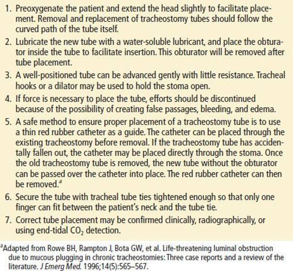
The inner cannula should be removed and cleaned, as this is a common source of obstruction. However, if the problem persists, the outer cannula should be removed and replaced with a new tracheostomy tube. Mechanical problems with the tube such as cuff leak or tube fracture may also necessitate tube replacement or consultation for bronchoscopy (9,11). Replacement of the tracheostomy tube in nonemergent circumstances is described in Table 72.1. However, the process may need to be abbreviated in the prehospital or emergency setting. A patient in respiratory distress not relieved by conservative methods such as suction, oxygen, and bronchodilators should be treated by immediate removal of the tracheostomy tube and tube replacement. Most obstructions occur in patients with long-standing tracheostomies, and those patients generally have uncuffed tracheostomy tubes that can be easily removed (12). If a new tracheostomy tube needs to be emergently replaced, the procedure may be simplified by passing a catheter or gum elastic bougie through the old tracheostomy tube and using it to guide the removal and replacement of the tube (12).
TABLE 72.1
Replacement of the Tracheostomy Cannula
INFECTION
Serious infection of the stomal site is rare, despite most fresh wounds being colonized by nosocomial organisms (13). The patient’s underlying medical condition, the presence of an indwelling device, and exposure to flora-colonizing ventilator tubing predisposes to infection, including lower respiratory infections. Prophylactic antibiotics, however, are not indicated.
The clinical presentation ranges from cellulitis or drainage around the tracheostomy tube to tracheitis, bronchitis, pneumonia, or sepsis.
The ED management of peristomal cellulitis consists of outpatient antibiotics for mild cases. Rarely, the wound infection may spread inferiorly, causing mediastinitis, mediastinal abscess, and paratracheal abscess. Patients with signs or symptoms of systemic infection or pain with breathing and swallowing should be evaluated for these complications, usually by imaging with computed tomography (CT) scan, and given intravenous antibiotics. Antibiotics should cover common respiratory flora, though methicillin-resistant Staphylococcus aureus must also be considered.
STENOSIS
Constant pressure from the cuff may cause pressure necrosis, ulceration, and formation of granulation tissue that subsequently causes tracheal stenosis (14). This is a late complication presenting weeks to months after decannulation.
The clinical presentation is insidious. Typically, patients do not become symptomatic until the endotracheal lumen has been reduced by 50% to 75%, at which point they may complain of cough, dyspnea, and inability to clear secretions.
The ED evaluation includes consultation or referral, because the diagnosis of stenosis is typically made with bronchoscopy. Once identified, significant stenosis is treated with operative dilation or resection of granulation tissue.
TRACHEOESOPHAGEAL FISTULA
Tracheoesophageal fistula is a rare but potentially life-threatening complication. It is caused either by perforation of the posterior wall of the trachea during the tracheotomy or erosion into the esophagus by the tracheostomy tube.
The clinical presentation may include signs and symptoms of cuff leak, aspiration of food contents, abdominal distention, or copious secretions (4).
The ED evaluation includes making arrangements for bronchoscopy or swallowing studies to confirm the diagnosis. This complication has also been associated with PDT (1,15).
PERCUTANEOUS DILATIONAL TRACHEOSTOMY
An alternative technique to surgical tracheostomy is a PDT, which was first described in 1985 and has become a popular alternative and has recently been proposed as the new gold standard (16–19). This technique has several advantages compared to surgical tracheostomies, including lower rates of pneumothorax, major hemorrhage, and laryngeal granulation (18). PDT can be done at the bedside, thereby reducing the operative risk and the cost. A major advantage is the ability to gain and maintain competence with the procedure in the elective setting (20). Long-term complications are the same as for standard surgical tracheostomy, although there is a slight reduction in tracheal stenosis with PDT. This procedure also has some unique risks not encountered with surgical tracheostomy, such as paratracheal insertion of a tracheostomy tube and posterolateral tracheal wall laceration (21). However, these complications are not common and are rarely lethal with a reported mortality rate of 0.17% (3,14).
REPLACEMENT OF TRACHEOSTOMY CANNULA
Tracheostomy tubes may be cuffed or uncuffed and may come with an inner cannula that can be removed for cleaning (Table 72.1). Indications for replacement of the tracheostomy tube in the ED include cuff rupture or leak and complete or partial obstruction. Changing the tracheostomy should be avoided for at least 1 week after the creation of the stoma, if possible. A replacement tube should be the same type and size as the original. In an emergency, replacement with a smaller size is preferred if maintaining a patent airway depends on it. If the appropriate-sized tracheostomy tube is unavailable in an emergency situation, a standard endotracheal tube may serve as a temporary replacement, typically a size 6 mm to 7.5 mm cuffed tube in an adult.
If the tracheostomy tube cannot be replaced and the airway is in jeopardy, the physician should consider oral endotracheal intubation. The original indication for the tracheostomy may hamper attempts at endotracheal intubation (e.g., laryngeal malignancy). If respiratory distress persists after adequate tube replacement, the physician must not overlook other causes such as pneumothorax, pulmonary embolus, pneumonia, or congestive heart failure.
CRITICAL INTERVENTIONS
• Obstruction of a tracheostomy tube can be life threatening. Remove the inner cannula and clear the obstruction to restore airway patency.
• Gain rapid control of hemorrhage, and consult a surgeon for definitive care.
DISPOSITION
Patients with minor bleeding or infection around a tracheostomy tube site may be treated and discharged with appropriate follow-up. Those patients with partial obstruction caused by deposits or secretion buildup on the tracheostomy tube may also be discharged after the tube is cleaned or changed and the symptoms are resolved.
Patients with persistent bleeding, more serious infection, or difficulty breathing mandate consultation and admission. Those patients with suspected tracheoesophageal fistula or early stenosis may be further evaluated on an inpatient or outpatient basis, depending upon the severity of symptoms.
Common Pitfalls
• Failure to rapidly clear or remove an obstructed tracheostomy tube.
• Inadequate control of hemorrhage around a tracheostomy tube. A tracheoarterial fistula should be suspected if a patient presents with brisk bleeding from the tracheostomy tube site.
• Overly aggressive application of force when replacing a tracheostomy tube must be avoided to prevent the creation of a false tract.
REFERENCES
1. De Leyn P, Bedert L, Delcroix M, et al. Tracheotomy: Clinical review and guidelines. Eur J Cardiothorac Surg. 2007;32(3):412–421.
2. Frost EA. Tracing the tracheostomy. Ann Otol Rhinol Laryngol. 1976;85(5 Pt 1):
618–624.
3. Hamid AA, Sattar F, Din SE, et al. Complications of tracheostomy. J Postgrad Med Inst. 2011;18(3):385–390.
4. Sue RD, Susanto I. Long-term complications of artificial airways. Clin Chest Med. 2003;24(3):457–471.
5. Hackeling T, Triana R, Ma OJ, et al. Emergency care of patients with tracheostomies: A 7-year review. Am J Emerg Med. 1998;16(7):681–685.
6. Dempsey GA, Grant CA, Jones TM. Percutaneous tracheostomy: A 6 yr prospective evaluation of the single tapered dilator technique. Br J Anaesth. 2010; 105(6):782–788.
7. Mirza S, Cameron DS. The tracheostomy tube change: A review of techniques. Hosp Med. 2001;62(3):158–163.
8. Wright CD. Management of tracheoinnominate artery fistula. Chest Surg Clin N Am. 1996;6(4):865–873.
9. Tayal VS. Tracheostomies. Emerg Med Clin North Am. 1994;12(3):707–727.
10. Rogers DF. Mucoactive agents for airway mucus hypersecretory diseases. Respir Care. 2007;52:1176–1193.
11. Feller-Kopman D. Acute complications of artificial airways. Clin Chest Med. 2003;24(3):445–455.
12. Rowe BH, Rampton J, Bota GW, et al. Life-threatening luminal obstruction due to mucous plugging in chronic tracheostomies: Three case reports and a review of the literature. J Emerg Med.1996;14(5):565–567.
13. Van Heurn LW, Brink PR. The history of percutaneous tracheotomy. J Laryngol Otol. 1996;110(8):723–726.
14. Stauffer JL, Olson DE, Petty TL, et al. Complications and consequences of endotracheal intubation and tracheotomy: A prospective study of 150 critically ill adult patients. Am J Med. 1981;70(1):65–76.
15. Dost P, Thurauf N, Armbruster W, et al. Perforation of the posterior tracheal wall during percutaneous dilatational tracheotomy. ORL J Otorhinolaryngol Relat Spec. 2000;62(3):167–169.
16. Al-Ansari MA, Hijazi MH. Clinical review: Percutaneous dilatational tracheostomy. Crit Care. 2006;10(1):202.
17. Ault MJ, Ault B, Ng PK. Percutaneous dilatational tracheostomy for emergent airway access. J Intensive Care Med. 2003;18:222–226.
18. Ciaglia P, Firsching R, Syniec C, et al. Elective percutaneous dilatational tracheostomy: A new simple bedside procedure; preliminary report. Chest. 1985; 87(6):715–719.
19. Kornblith LZ, Burlew CC, Moore EE, et al. One thousand bedside percutaneous tracheostomies in the surgical intensive care unit: Time to change the gold standard. J Am Coll Surg. 2011;212(2):163–170.
20. McCague A, Wong, D. Percutaneous dilational tracheostomy in the emergent setting. Int J Clin Med. 2013;4(2):96–98.
21. Simon M, Metschke M, Braune SA, et al. Death after percutaneous dilational tracheostomy: A systematic review and analysis of risk factors. Crit Care. 2013; 17(5):R258.