
David S. Howes and Leah W. Skjei
Chronic obstructive pulmonary disease (COPD) comprises a spectrum of chronic respiratory illnesses characterized by cough, sputum production, dyspnea, airflow limitation, impaired gas exchange, and inflammation (1–3). The mortality rate is greater than 50% within 10 years of diagnosis. COPD is the fourth leading cause of death in the United States and is expected to become the third leading cause of death worldwide by 2020 (4). The overall prevalence of symptomatic COPD in selected countries is 10.1% (5).
The term COPD includes conditions, other than asthma, that have the common feature of airflow obstruction and inflammation of the small airways and alveoli. The inflammatory response in COPD is different than that seen in asthma and involves the recruitment of neutrophils, macrophages, and cytotoxic T lymphocytes. Inflammation of the small airways leads to airway wall thickening and fibrosis, resulting in decreased airway diameter and increased resistance to flow. This is chronic obstructive bronchitis (1,6). Chronic bronchitis is characterized clinically by a productive cough caused by excessive production of bronchial mucus that must be present for 3 months in a year in at least 2 successive years. Pulmonary emphysema, in contrast, is defined from a pathologic standpoint by inflammatory infiltrates in the alveolar wall, leading to an irreversible enlargement of the alveolar air spaces with destruction of the alveolar wall and pulmonary capillary bed (1,6). The result is a reduction in the elastic pressure that generates expiratory flow (6). Most patients with COPD will have remodeling of both their small airways and their alveoli due to abnormal inflammation, and will therefore exhibit characteristics of both chronic bronchitis and emphysema.
The predominant risk factor for COPD is tobacco use; half of smokers develop airflow obstruction (4). However, 5% to 10% of patients with COPD have never smoked, implicating other causal factors (1). Other risk factors include inhalation of organic and inorganic dusts and fumes, that is environmental pollution, passive smoke inhalation, and occupational exposure (7). α1-Antitrypsin deficiency is a genetic risk factor and is present in 1% to 2% of patients with COPD (4). Regardless of etiology, the ultimate outcome is irreversible airflow limitation.
The natural course of COPD is one of progressively worsening dyspnea, hypoxemia, and diminished exercise tolerance, with recurrent exacerbations. Most patients with COPD have modest reversible airway obstruction. Although the forced expiratory volume in 1 second (FEV1) is classically considered the best single prognostic indicator of disability and death, disease severity is better assessed by considering FEV1, body mass index (BMI), and exercise performance (3). The onset of chronic hypoxemia leads to progressive clinical deterioration caused by cor pulmonale, which worsens already diminished lung function. Recurrent episodes of infection and respiratory failure ultimately result in death. Long-term ambulatory oxygen therapy prolongs life in patients with advanced COPD.
CLINICAL PRESENTATION
All patients with COPD exhibit airflow obstruction, but the pure forms of emphysema and chronic bronchitis have distinctive clinical features. Patients with emphysema tend to hyperventilate to compensate for the decreased ability of the lungs to oxygenate the blood due to their decreased alveolar surface area. These classic “pink puffers” work hard to establish a near-normal partial pressure of arterial oxygen (PaO2), and they appear barrel-chested, dyspneic, and tachypneic. They use pursed lips breathing to create positive end-expiratory pressure (PEEP) to prevent early airway closure. Breath sounds are markedly diminished, with a prolonged expiratory phase.
In contrast, patients with chronic bronchitis tolerate hypoxemia well and make no effort to hyperventilate. Chronic hypoxia induces a secondary polycythemia that, combined with the cyanosis of marked desaturation of hemoglobin, produces a plethoric appearance. These patients tolerate an elevated partial pressure of arterial carbon dioxide (PaCO2) and have a less labored respiratory pattern. With the decreased work of breathing, the patient with chronic bronchitis does not experience muscle wasting like the emphysematous patient. These features produce the picture of the “blue bloater.” Lung examination reveals rhonchi, rales, and variable wheezing. The severity of COPD as classified by Global Initiative for Chronic Lung Disease (GOLD) is listed in Table 77.1 (8).
TABLE 77.1
Global Initiative for Chronic Lung Disease Severity of Chronic Obstructive Pulmonary Disease

Most patients who present with an acute exacerbation of COPD do not fit either picture precisely. Patients with COPD exacerbation present with worsening of baseline dyspnea, cough, and/or sputum that is beyond normal day-to-day variations, is acute in onset, and may warrant a change in regular medication (8). Physical examination may reveal a variable degree of respiratory distress, tachypnea, accessory muscle use, retractions, and cyanosis. On auscultation, one may hear diminished breath sounds, a prolonged expiratory phase, wheezing, rales, or rhonchi. Signs of cor pulmonale should be sought: a centrally displaced point of maximal impulse, heart gallop, tricuspid murmur, peripheral edema, jugular venous distention, hepatojugular reflux, and hepatomegaly. This complication of long-standing hypoxemia is associated with substantial mortality.
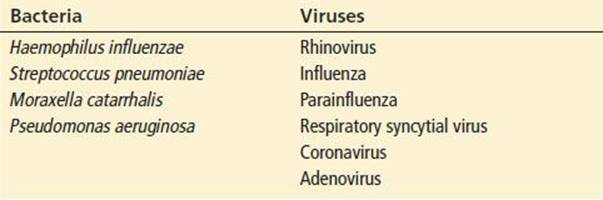
The causes of exacerbation include respiratory infection, noncompliance with (or under dosing of) medications, changes in weather, exposure to certain drugs (e.g., sedatives or β-blockers), cardiac dysrhythmias, left ventricular dysfunction, and environmental exposure to allergens or other irritants. Most exacerbations are caused by infections, either viral or bacterial, with bacterial infections accounting for 50% of COPD exacerbations. Air pollution and other environmental conditions account for approximately 15% to 20% (9). Table 77.2 lists common respiratory agents associated with COPD exacerbations (10–12). Patients typically present with a gradual, but progressive, deterioration, taking hours to days.
TABLE 77.2
Bacterial and Viral Agents Associated with Chronic Obstructive Pulmonary Disease Exacerbations
Acute decompensation may be the result of a number of other conditions. Spontaneous pneumothorax in COPD carries a significantly higher complication and mortality rate than in patients with a normal cardiorespiratory status. The patient with chronic bronchitis is predisposed to embolic and thrombotic phenomena because of the hyperviscosity associated with polycythemia. Other disorders, such as congestive heart failure (CHF), pneumonia, acidosis, or renal or hepatic failure, may also overwhelm the COPD patient’s limited reserves, resulting in respiratory failure.
DIFFERENTIAL DIAGNOSIS
Other conditions must also be considered in the patient who presents to the emergency department (ED) with wheezing, cough, dyspnea, or respiratory failure. Pneumonia or acute bronchitis may present with varying degrees of shortness of breath, cough, and bronchospasm, and previously undiagnosed COPD may be discovered after an acute pneumonic process has resolved. The onset of respiratory symptoms in the presence or absence of chest pain, when associated with risk factors such as recent surgery, malignancy, immobility, or extended travel, or leg findings consistent with deep venous thrombosis, strongly suggest pulmonary embolism.
Acute left ventricular dysfunction may mimic many findings of COPD including cough, dyspnea, and hypoxemia, and patients may exhibit wheezing, referred to as “cardiac asthma.” Orthopnea, paroxysmal nocturnal dyspnea, and cardiomegaly favor CHF, but COPD patients with cor pulmonale may demonstrate jugular venous distention and peripheral edema. Classically, chest radiography helped distinguish between these entities, with a lack of cardiomegaly and pulmonary venous redistribution and the presence of bullae and hyperinflation of the lungs suggesting COPD. However, patients with both CHF and COPD who present with dyspnea can present a diagnostic challenge. The rapid laboratory quantification of B-type natriuretic peptide (BNP) may allow more accurate distinction between an acute exacerbation of COPD versus CHF. BNP is secreted from ventricles in response to pressure and volume expansion. Low levels of BNP are consistent with exacerbations of COPD, asthma, bronchitis, or pneumonia, whereas a BNP level of 100 pg/mL or greater is a strong predictor of CHF. Pulmonary embolism or COPD triggering cor pulmonalemay also produce elevated BNP levels of 200 to 600 pg/mL (3). Therefore, it is best to use the patient’s physical examination, history, and ancillary studies, together with the BNP level to make the most informed clinical diagnosis and therapeutic decisions.
ED EVALUATION
If the patient’s respiratory status allows a brief history to be taken, attention should be directed to the rapidity of onset, duration of symptoms, precipitating factors, the dosage of medications, and when medications were last taken. Information should be sought regarding the character of the sputum, the course of previous exacerbations, and any history of concomitant disease processes.
The physical examination focuses on the patient’s mental status and the degree of respiratory distress. Hypercapnia and hypoxia can cause confusion, somnolence, and irritability. The cardiac and pulmonary examinations are vital, and repeated examinations are mandatory after therapeutic interventions to monitor the response to treatment.
Pulse oximetry is useful in monitoring oxygen saturation and the effectiveness of oxygen supplementation. Arterial blood gas (ABG) analysis provides information on the PaCO2 and pH, reflecting adequacy of ventilation. The pH is the best single laboratory marker for gauging the severity of acute respiratory insufficiency, because it reflects the speed and degree of change in PaCO2 (8). Baseline or prior ABG results may be useful for comparison; in general, hypercapnia with acidemia suggests acute respiratory failure.
The chest radiograph (CXR) is usually done at the bedside, unless the patient is stable and can tolerate being transported to the radiology suite for posteroanterior (PA) and lateral films. The CXR is useful when findings of COPD are present, but it is even more helpful in evaluating for other disease processes, such as pneumothorax, atelectasis, infiltrate, lung mass, or CHF.
Measurements of airflow do not always yield accurate assessments of the severity of illness but may be useful in judging the response to therapy. The peak expiratory flow rate (PEFR) is most commonly used in the ED setting, but, in contrast to the asthmatic patient’s response, it does not correlate well with COPD severity during exacerbations, and does not improve as rapidly as the asthmatic patient’s in response to therapy.
Laboratory studies are generally of limited value. β-Agonists may cause hypokalemia, although this is rarely clinically significant. A complete blood count may reveal anemia, which can compound the problem of hypoxemia, or a high white blood cell count, which may be a clue to pneumonia or other systemic infections. If there is suspicion for CHF or cardiac injury, BNP and troponin assays may be ordered. D-Dimer may be useful for evaluating low-risk patients when pulmonary embolism is being considered.
A 12-lead electrocardiogram (ECG) may suggest right atrial enlargement, low voltage, right ventricular hypertrophy, or strain (cor pulmonale). Acute ischemic patterns must be appreciated to identify an acute coronary event that may be triggering or complicating the acute COPD attack. Continuous ECG monitoring may be useful in revealing dysrhythmias that may aggravate an acute exacerbation.
Thoracic ultrasound is sensitive for ruling out pneumothorax when used by experienced operators. However, it should not be used to diagnose pneumothorax in patients with COPD because there is a high false-positive rate in this population (13).
Thoracic ultrasound is sensitive for ruling out pneumothorax when used by experienced operators. However, it should not be used to diagnose pneumothorax in patients with COPD because there is a high false-positive rate in this population (13).
KEY TESTING
• Order an ABG or VBG to obtain the pH, and thereby gauge the severity of the patient’s acute respiratory insufficiency.
• Obtain a CXR to evaluate for infection, pneumothorax, mass, and edema.
• Consider a CBC and basic metabolic panel to screen for anemia, a high WBC, and hypokalemia.
• A BNP should be ordered if CHF is suspected (14).
• A troponin assay and an EKG should be ordered if symptoms or signs of cardiac ischemia or dysrhythmia are present.
• A D-dimer is useful in evaluating low-risk patients for whom PE is being considered.
ED MANAGEMENT
The effective management of the COPD patient with an acute decompensation includes rapid estimation of severity, prompt therapy, frequent reevaluation, and recognition of treatable entities that may have precipitated the ED visit or that complicate the patient’s course. The patient should be placed on a cardiac monitor and intravenous (IV) access should be established. Dysrhythmias such as rapid atrial fibrillation usually reflect coexisting heart disease that is worsened by acute hypoxemia; current evidence suggests that most patients with a COPD exacerbation who exhibit ventricular dysrhythmias have underlying left ventricular diastolic dysfunction. Table 77.3 provides a summary to the approach to patients with moderate-to-severe COPD exacerbations.
TABLE 77.3
Overview of Emergency Department Management of Patients with Moderate-to-Severe Chronic Obstructive Pulmonary Diseasea

Treatment with oxygen is critical because hypoxemia is the major immediate threat to life. The goal is to maintain a PaO2 >60 mm Hg or a pulse oximetry reading of 90% to 92% to minimize further risk of hypercapnea and respiratory acidosis (15). A controlled oxygen-delivery system is preferable; a Venturi-mask system can deliver a precise oxygen concentration ranging from 24% to 50%. Adjustments in oxygen delivery should be guided by continuous pulse oximetry and ABG determinations as needed. In the acutely hypoxemic patient, the administration of oxygen is paramount and far outweighs the risk of hypercarbia caused by blunting of the hypoxemic drive in a chronic CO2 retainer.
Sympathomimetics, specifically β2-agonists, are the initial pharmacologic agents of choice for acute COPD exacerbations. Effectiveness is dependent in part on the reversible component of the patient’s obstructive lung disease; however, a significant benefit of bronchodilator therapy is to decrease dynamic hyperinflation by improving lung emptying during expiration (3). Side effects include tremor, agitation, insomnia, headache, cardiac tachyarrhythmias, and mild hypokalemia. Nebulizer aerosolization is the preferred delivery method in the ED. Although there are many β2-agonists available, albuterol, is the most widely used agent. Albuterol, 2.5 mg, is used either in repeated treatments every 20 to 60 minutes, or by continuous nebulization in severe cases. Levalbuterol, the active (R) isomer of racemic albuterol, is available for treatment of COPD by nebulizer; the “on-label” dosage is 1.25 mg every 6 to 8 hours, although dosages of up to 1.25 mg every 30 minutes have been reported in clinical studies. The inactive (S) isomer found in racemic albuterol is thought to promote hypersensitivity and bronchospasm in susceptible individuals, but though patients treated with levalbuterol may require less medication and experience a longer therapeutic duration of action as compared with those given racemic albuterol (16), the considerable added expense outweighs the questionable clinical significance of these minor differences (16).
The slower onset of action renders anticholinergic agents to second-line treatment in the acute setting. Ipratropium bromide, a congener of atropine, has little systemic absorption when inhaled, thus minimizing undesirable anticholinergic effects, and has been shown to have an additive effect when combined with a β2-agonist. This agent may be administered at a dose of 500 μg per nebulizer treatment.
Corticosteroids are considered useful in acute exacerbations of COPD. Even patients who do not respond to steroids when stable may still benefit from them during acute attacks. Because steroid use may suppress the adrenal–pituitary axis, patients with current or recent use may require steroids for the stress of any acute illness. IV methylprednisolone at a dose of 125 mg or oral prednisone 60 mg are commonly used in the treatment of exacerbations of COPD. Patients may be given oral prednisone 30 to 40 mg daily for 7 to 14 days if they are discharged from the ED (10).
Before the widespread availability of oral or inhaled β2-agonists, methylxanthines, most commonly theophylline, were used as monotherapy for COPD. However, methylxanthines have since been proven to be of no benefit in acute COPD exacerbations, and have potentially serious side effects (17).
Other medications may be utilized in the management of acute COPD. Magnesium sulfate has modest bronchodilatory effects and may be given a trial as adjunctive therapy to patients with severe exacerbations who have normal renal function. Some authors have shown benefit of magnesium sulfate only when administered before β-agonists (18).
Cessation of tobacco use may well be the most important intervention for COPD. Brief counseling can be effective, and an effort should be made to advise the patient to stop smoking and seek further counseling from a primary care physician (1,3). COPD patients in the ED are also candidates to receive polyvalent pneumococcal and influenza vaccines to potentially decrease the risk of future exacerbations (19).
COPD exacerbations are commonly (>50%) associated with acute respiratory tract infection. The most common agents are outlined in Table 77.2 (10–12). Antibiotic therapy is indicated if dyspnea is associated with a change in the volume or purulence of sputum or with a new infiltrate on CXR. Antibiotics should also be considered for any patient with a severe COPD exacerbation, or an exacerbation requiring admission. Antibiotic treatment has been associated with better inpatient outcomes (20). Former first-line agents such as trimethoprim/sulfamethoxazole, amoxicillin, and doxycycline have proven to be inferior to advanced-generation quinolones, second- and third-generation cephalosporins, and amoxicillin/clavulanate. Antibiotic therapy increases treatment success and decreases mortality when used to treat acute exacerbations of COPD associated with increased sputum purulence and volume production (12,21,22). Increasing resistance of Streptococcus pneumoniae to macrolides also limits use of this antimicrobial (12). The choice of antibiotic should be based on local resistance patterns, particularly among the bacterial species commonly isolated from sputum during exacerbations: Streptococcus pneumoniae, Haemophilus influenzae, and Moraxella catarrhalis (19). Broad-spectrum coverage should be instituted to reduce the incidence of antibiotic resistance.
For patients who do not respond to acute interventions and who begin to show signs of respiratory failure (such as deterioration of mental status, worsening hypoxemia [PaO2 of <50 mm Hg], or worsening respiratory acidosis [pH 7.25]), assisted ventilation is indicated. Noninvasive positive-pressure ventilation (NIPPV) should be considered for patients who present with moderate exacerbations of COPD and should be instituted for patients with severe exacerbations of COPD who have a relatively normal mental status and who do not require immediate endotracheal intubation. Contraindications to NIPPV include impending respiratory arrest, cardiovascular instability, and vomiting. NIPPV decreases the work of breathing and has been shown to decrease the rate of endotracheal intubation, to decrease hospital mortality, and to shorten hospital stay in patients with severe exacerbations of COPD (23). Recent studies have shown that NIPPV may safely and effectively be used in patients with altered mental status who are arousable and able to follow simple commands (24). However, for patients who are not candidates for these modalities or for whom noninvasive ventilation has failed, endotracheal intubation with mechanical ventilation is the treatment of choice.
After performing endotracheal intubation and implementing mechanical ventilation, the clinician must be aware of the potential complications associated with positive-pressure ventilation in the COPD patient. Complications include hypotension and cardiovascular collapse caused by decreased venous return secondary to increased intrathoracic pressure. The increased intrathoracic pressure occurs in patients who “stack” breaths because of the inability to completely exhale the mechanically provided inspired volume that results in greater intrathoracic volume and pressure. If hypotension does not resolve with administration of IV fluids, the patient’s endotracheal tube should be temporarily disconnected from the ventilator to relieve excessive positive intrathoracic pressure. Mechanical ventilation should be adjusted to allow for a short inspiratory and long expiratory time with a low tidal volume of 5 to 7 mL/kg. The frequency of ventilation may be gradually increased to allow for resolution of the acute respiratory acidosis and gradual normalization of pH. Extrinsic PEEP may be added to nearly equal intrinsic or auto-PEEP in an effort to keep distal small airways from collapsing with attendant gas trapping. Short-term administration of paralytics eliminates patient-ventilator dyssynchrony and reduces the risk of hyperinflation.
It is paramount that proper sedation is provided to the patient if paralytics are administered. However, hypotension can result from the administration of sedative agents that reduce sympathetic tone. Hypotension should be treated with IV fluids and pressors in more extreme cases.
CRITICAL INTERVENTIONS
• Obtain an arterial or venous pH to determine the severity of acute respiratory insufficiency, because it reflects the speed and degree of change in PaCO2. Hypercapnia with acidemia suggests acute respiratory failure.
• Administer oxygen in the acutely hypoxemic patient: the administration of oxygen far outweighs the risk of hypercarbia owing to blunting of the hypoxemic drive in a chronic CO2 retainer.
• Use a controlled oxygen delivery system (Venturi mask) to maintain a PaO2 >60 mm Hg or a pulse oximetry reading of 90% to 92% to minimize further risk of hypercapnia and respiratory acidosis in stable hypoxemic patients.
• Adjust oxygen delivery based on continuous pulse oximetry and serial ABGs.
• Start antibiotic therapy in patients with increased sputum volume and purulence, and patients being admitted with severe COPD exacerbations.
• Use NIPPV in patients with moderate-to-severe exacerbations to decrease the work of breathing when endotracheal intubation is not immediately indicated.
DISPOSITION
Many patients who present with an acute exacerbation of COPD require admission to the hospital. However, if the patient responds to therapy and the vital signs and pulmonary function tests approach baseline, discharge may be considered if good follow-up is assured. The bronchodilator regimen should be maximized: mono, dual, or triple therapy may be selected from long-acting β2-agonists, long-acting anticholinergics, and inhaled corticosteroids (25). A short-acting β-agonist should be prescribed as a rescue inhaler. Systemic steroids should be continued for a short course, as they reduce the relative risk of treatment failure by about 30% as compared to placebo (19). Antibiotics should be prescribed for exacerbations associated with increased sputum volume or purulence. It should be recognized that approximately 15% of patients with COPD who are discharged from the ED require readmission.
Accepted criteria for hospitalization of patients with COPD include poor response to outpatient therapy, severe limitation of function, inability to eat or sleep because of dyspnea, presence of significant comorbid conditions, worsening respiratory failure, new or progressive cor pulmonale, and the need for invasive procedures, especially those requiring analgesics or sedatives. Guidelines for admission to the intensive care unit are not as well defined. Treatment in an intensive care unit is indicated when the close observation, monitoring, and therapeutic interventions (including assisted ventilation) required by these patients cannot be provided elsewhere.
If transfer to another institution is required, it must be done carefully, because transfer is often associated with an interruption in treatment and increased patient fatigue. The transport team must be capable of managing the airway if decompensation occurs.
Common Pitfalls
• The COPD patient should not be expected to have disease that reverses as rapidly as the asthmatic patient. Look for a slower course of improvement, and have a lower threshold for admission.
• If patients have not slept well in the last 2 days, they are tired and the risk of respiratory failure is high. Such patients must be admitted to the hospital.
• Precipitating or complicating conditions (e.g., CHF, PE) may be overlooked when treating only the exacerbation of COPD.
• Oxygen should never be withheld because of concern about respiratory depression. If inadequate ventilation occurs, ventilatory support should be provided.
• It is easy to underestimate the severity of illness in patients with chronic dysfunction. Utilize radiography, pulse oximetry, and ABG analyses. Respect the patient’s estimation of the severity of illness.
• If hypotension occurs during mechanical ventilation, the most common cause is “stacked” breaths caused by incomplete exhalation, with attendant rise in intrathoracic pressures and impaired venous return; the patient must be disconnected from the ventilator and allowed to “exhale.”
ACKNOWLEDGMENTS
Thanks to previous edition author Marc A. Bellazini.
REFERENCES
1. Barnes PJ. Chronic obstructive pulmonary disease: New treatments for COPD. Thorax. 2003;58(9):803–808.
2. Fabbri LM, Rabe KF. From COPD to chronic systemic inflammatory syndrome? Lancet. 2007;370(9589):797–799.
3. MacNee W, Calverley PMA. Chronic obstructive pulmonary disease: Management of COPD. Thorax. 2003;58(3):261–265.
4. Devereux G. ABC of chronic obstructive pulmonary disease: Definition, epidemiology, and risk factors. BMJ. 2006;332(7550):1142–1144.
5. Buist AS, McBurnie MA, Vollmer WM, et al. International variation in the prevalence of COPD (the BOLD Study): A population-based prevalence study. Lancet. 2007;370(9589):741–750.
6. Cosio MG, Saetta M, Agusti A. Immunologic aspects of chronic obstructive pulmonary disease. N Engl J Med. 2009;360:2445–2454.
7. Lamprecht B. McBurnie MA, Vollmer WM, et al. COPD in never smokers: Results from the population based burden of obstructive lung disease study. Chest. 2011;139:752–763.
8. Rabe KF, Hurd S, Anzueto A, et al. Global strategy for the diagnosis, management, and prevention of chronic obstructive pulmonary disease: GOLD executive summary. Am J Respir Crit Care Med.2007;176(6):532–555.
9. Sethi S, Murphy T. Infection in the pathogenesis and course of chronic obstructive pulmonary disease. N Engl J Med. 2008;359:2355–2365.
10. Currie GP, Wedzicha JA. ABC of chronic obstructive pulmonary disease: Acute exacerbations. BMJ. 2006;333(7558):87–89.
11. Ko FWS, Ip M, Chan PKS, et al. A 1-year prospective study of the infectious etiology in patients hospitalized with acute exacerbations of COPD. Chest. 2007;131(1):44–52.
12. Martinez FJ, Anzueto A. Appropriate outpatient treatment of acute bacterial exacerbations of chronic bronchitis. Am J Med. 2005;118(suppl7A):39S–44S.
13. Slater A, Goodwin M, Anderson KE, et al. COPD can mimic the appearance of pneumothorax on thoracic ultrasound. Chest. 2006;129(3):545–550.
14. Collins SP, Ronan-Bentle S, Storrow AB. Diagnostic and prognostic usefulness of natriuretic peptides in emergency department patients with dyspnea. Ann Emerg Med. 2003;41(4):532–545.
15. Plant PK, Elliott MW. Chronic obstructive pulmonary disease: Management of ventilatory failure in COPD. Thorax. 2003;58(6):537–542.
16. Truitt T, Witko J, Halpern M. Levalbuterol compared to racemic albuterol: Efficacy and outcomes in patients hospitalized with COPD or asthma. Chest. 2003;123(1):128–135.
17. Barr RG, Rowe BH, Camargo CA. Methylxanthines for exacerbations of chronic obstructive pulmonary disease: Meta-analysis of randomized trials. BMJ. 2003; 327:643–646.
18. Abreu González J, Hernández García C, Abreu González P, et al. Effect of intravenous magnesium sulfate on chronic obstructive pulmonary disease exacerbations requiring hospitalization: A randomized placebo-controlled trial [Spanish]. Arch Bronconeumol. 2006;42(8):384–387.
19. Niewoehner D. Outpatient management of severe COPD. N Engl J Med. 2012;362:
1407–1416.
20. Stefan MS, Rothberg MB, Shieh MS, et al. Association between antibiotic treatment and outcomes in patients hospitalized with acute exacerbation of COPD treated with systemic steroids. Chest.2013;143:82–90.
21. Fabbri LM, Boschetto P, Mapp CE. COPD guidelines: The important thing is not to stop questioning. Am J Respir Crit Care Med. 2007;176(6):527–528.
22. Wise RA, Tashkin DP. Optimizing treatment of chronic obstructive pulmonary disease: An assessment of current therapies. Am J Med. 2007;120(8 suppl 1):S4–S13.
23. Keenan SP, Sinuff T, Cook DJ, et al. Which patients with acute exacerbation of chronic obstructive pulmonary disease benefit from noninvasive positive-pressure ventilation: A systematic review of the literature. Ann Int Med.2003;138(11):861–870.
24. Scala R, Naldi M, Archinucci I, et al. Noninvasive positive pressure ventilation in patients with acute exacerbations of COPD and varying levels of consciousness. Chest. 2005;128(3):1657–1666.
25. Sutherland ER, Cherniack RM. Management of chronic obstructive pulmonary disease. N Engl J Med. 2004;350:2689–2697.