
Sanjay Bhatt
Coughing up blood is a significant cause of concern to both the provider and patient. While a common symptom in adults, it is an uncommon presentation in children. The differential diagnosis can be diverse and includes pulmonary pathology as well as pseudohempotysis caused by ear, nose, and throat (ENT) or gastrointestinal pathology. Although typically indicative of primary pulmonary pathology, hemoptysis may result from numerous systemic illnesses. It is important to note that the severity of bleeding does not correlate with the gravity of the underlying disease.
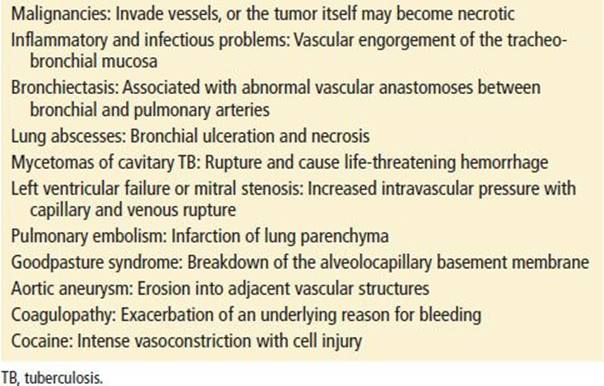
Hemoptysis is attributed to multiple underlying pathophysiologic mechanisms. Bleeding sources include bronchial and pulmonary arteries. In general, bronchial arteries are under high-pressure systems whereas pulmonary are under low-pressure systems. Massive bleeding, which accounts for 1.5% of cases, usually arises from injury to the high-pressure bronchial arteries (1–3). The cause of bleeding depends on the underlying etiology. Exsanguination is rarely the cause of death in hemoptysis; rather, blood can obstruct distal alveoli thereby impairing gas exchange. In these cases, death can result from hypoxemia. There are several possible causes for bleeding; Table 80.1 lists the multitude causes of bleeding in a patient presentation to the emergency department (ED).
TABLE 80.1
Mechanisms of Bleeding in Hemoptysis
CLINICAL PRESENTATION AND DIFFERENTIAL DIAGNOSIS
Patients may present to the ED with a chief complaint of “coughing up blood.” The extent of this bleeding can range from minor to copious. While the vast majority of patients have stable vital signs, those with pulmonary compromise may reflect some degree of tachycardia. Those with severe bleeding may show signs of hypoxemia, hemodynamic instability, and/or airway compromise. Patients with systemic diseases, such as Goodpasture syndrome, may present with hematuria and hemoptysis.
Pseudohemoptysis should be excluded before the pulmonary causes of hemoptysis are entertained. An appropriate history and physical examination can eliminate gastrointestinal and otolaryngologic sources. The vomitus of gastrointestinal bleeding tends to be darker in color and may contain food particles. In comparison, true hemoptysis is typically bright red and frothy. Mouth, throat, and nasopharyngeal sources of bleeding may be visualized with direct, indirect, or fiberoptic laryngoscopy.
The etiology of hemoptysis worldwide can vary by country (4). In the United States, the most common causes include infections, malignancy, bronchiectasis, bronchitis, and cardiovascular disorders (5–7). Chronic obstructive pulmonary disease is an independent risk factor for hemoptysis (1). Worldwide, while tuberculosis (TB) is still the most common cause of hemoptysis, it is much less common in the United States (8). In areas such as New York City or Los Angeles that house a large TB population, this diagnosis must be considered.
While uncommon, patient with systemic lupus erythematous (SLE) may present with alveolar hemorrhage and resultant hemoptysis (9). Travelers or immigrants, especially those from Asia, the Middle East, and South America, may have parasitic infections (3). Catamenial hemoptysis, which is hemoptysis during menstruation, may occur in women with thoracic endometriosis (1,10). In those who are immunocompromised, bacterial pneumonia is a common diagnosis while in the HIV population, Kaposi’s sarcoma must be considered (1).
Independent of coagulation parameters, patients on immunosuppressive medications, or those with a history of transbronchial biopsy or lung transplant, are at an increased risk for hemoptysis after bronchoscopy (11). It is important to note that the mortality rate among lung transplant patients who present with hemoptysis is high (12). Occult malignancy should always be considered, especially in those with a long-standing smoking history.
Three potential biologic weapons that may present with hemoptysis include plague, tularemia, and T2 mycotoxin (2). Table 80.2 lists the common causes of hemoptysis by category.
TABLE 80.2
Common Causes of Hemoptysis

In pediatric patients, lower respiratory tract infections account for almost 40% of all presentations of hemoptysis. Other causes include cystic fibrosis (CF), foreign-body aspiration, bronchial adenomas, and trauma. All providers must be aware of accidental as well as deliberate trauma; bleeding caused by deliberate suffocation has been described (1). CF may be associated with recurrent, severe bleeding in both children and young adults. Hemoptysis in children with CF occurs with increasing age and advancing disease and almost 60% of children will have at least one episode. Most of these episodes are not life-threatening (13). Although congenital heart disease and mitral stenosis after rheumatic fever are still reported causes of hemoptysis, corrective surgery for the former and decreasing incidence of the latter make them infrequent causes of bleeding (13). Other uncommon causes include bronchiolitis obliterans organizing pneumonia (7), pulmonary embolism, Wegener granuloma, primary pulmonary hemosiderosis, and arteriovenous malformations (AVMs) (5,13).
ED EVALUATION
Patients with massive hemoptysis require initial focus on the airway, breathing, and circulation (ABCs) of support; as noted above, especially given concern for asphyxiation and lesser so exsanguination. The ABCs take precedence to making a definitive diagnosis. While providers agree that massive hemoptysis describes the expectoration of a large amount of blood or a rapid rate of bleeding, the thresholds that define massive hemoptysis remain controversial. Thresholds range from 100 to 1000 mL (14) over 24 hours though none has been generally accepted. Some clinicians argue that abnormal gas exchange and hemodynamic instability, generally tachycardia or tachypnea, should be part of the definition.
Nevertheless, patients who present with what is termed “massive” hemoptysis have a mortality rate close to 50% (6). Similar to the adult definition, the pediatric definition of massive hemoptysis can vary; some sources define the rate of bleeding as 8 mL/kg/24 hrs (13). While massive versus submassive bleeding carries both diagnostic and prognostic implications, this differentiation is difficult to perform in the ED. Similar to adult presentation, mortality is primarily due to asphyxiation and lesser so exsanguination.
Most series list TB and bronchiectasis as the most common causes of massive hemoptysis; other diseases include lung abscesses, malignancy, Goodpasture syndrome, Behçet disease, AV malformations, trauma, coagulopathies, and aspergillomas (3).
As almost any amount of bleeding may prove detrimental to patients with limited cardiopulmonary reserve or significant underlying disease, baseline cardiopulmonary status, and overall physical condition must weigh into a physician’s decision to pursue aggressive measures. History, physical examination, and laboratory studies can help evaluate the patient’s ability to respond to the physiologic stress of hemoptysis and the therapy it necessitates.
Once a patient has been stabilized, efforts to determine the cause of hemoptysis should include a thorough history and physical examination. Laboratory studies should include: a complete blood count to determine the chronicity and magnitude of bleeding and the need for a platelet transfusion; a urinalysis and creatinine values in those whom pulmonary renal syndromes are being considered; arterial blood gas analysis and a pulse oximetry to determine oxygenation; coagulation studies to determine whether fresh-frozen plasma may be of benefit; and sputum evaluation in those whom TB is considered, radiograph studies should include a chest x-ray to evaluate for a multitude of diagnosis including neoplasm and focal infections. A chest computed tomography may better identify malignancies, infections, and pulmonary emboli. The combination of high-resolution CT scan and bronchoscopy is the standard by which a diagnosis is established (2,4,15). Definitive diagnosis may require sputum cytology, pulmonary function testing, angiography, or specialized sputum studies (2,15).
Aggressive management must occur at the same time as evaluation. Some diagnostic modalities, most notably bronchial artery embolization, are both diagnostic and therapeutic (4,16–18). Some centers are now using a new technique of bronchoscopic endobronchial sealing with cyanoacrylate (19).
Even after exhaustive study, 10% of cases will have no identifiable etiology (3). These patients are classified as having idiopathic or cryptogenic hemoptysis; in these cases, it is presumed that subtle airway or parenchymal disease is the culprit. Close follow-up to exclude repeat presentation and malignancy is especially important (20).
KEY TESTING
• Pulse oximetry
• CXR
• CBC
• Coagulation studies
ED MANAGEMENT
The primary goals of therapy are threefold: airway control to prevent asphyxia and less likely exsanguination, termination of bleeding, and diagnosis and treatment of the underlying cause. As noted above, patients with massive hemoptysis require initial focus on the ABCs of support. Airway control and adequate oxygenation are the immediate priorities. Endotracheal intubation, which may be technically challenging especially in this setting, is the intervention of choice, especially in those with poor gas exchange, hemodynamic instability, rapid ongoing hemoptysis, and at physician discretion. Placing a large 8.0 lumen tube can facilitate suctioning and the passage of a fiberoptic laryngoscope. Bag-valve-mask ventilation may be difficult in the patient with massive bleeding as blood may further block the tracheobronchial tree. The provider should consider selective intubation and, if readily available may consider a double-lumen endotracheal tube. Unilateral intubation, often performed in conjunction with a localizing bronchoscopic examination, can ventilate the nonbleeding lung. A localizing bronchoscopic examination is essential because of the importance of ventilating the proper lung as well as ensuring that the right upper lobe bronchus is not blocked as a result of the procedure. Another option is selective intubation using a fiberoptic laryngoscope. If the suspected bleeding lung or pathologic lesion is on the left side, the right mainstem bronchus may be selectively intubated by advancing an endotracheal tube past the carina. Normal lung anatomy frequently allows passage of the tube into the right mainstem bronchus. As noted above, given the potential problem that the right upper lobe bronchus may become blocked, it is imperative that direct visualization is performed during or soon after the intervention.
In addition, patients with massive hemoptysis should be placed in a dependent, lateral decubitus position. Thus, in a patient with massive bleeding from the left lung, the patient should be intubated into the right mainstem and placed in a left lateral decubitus position. This position may protect the uninvolved lung from filling with blood and, in so doing, maximize gas exchange.
Special care is given to IV fluids and blood and resuscitation measures. Isotonic fluids and blood products should be rapidly infused. In addition to intravenous isotonic fluids, packed red blood cells, platelets, and fresh-frozen plasma are imperative in the proper clinical situations. Coagulation and platelet abnormalities should be corrected with fresh-frozen plasma and platelet infusions, as indicated. Significant coughing may exacerbate the bleeding and can be reduced with small doses of intravenous morphine. Antibiotics should be administered for suspected infectious etiologies. Pulmonary, ENT, interventional radiology, and surgical consultants will assist in localizing the bleeding site. Bronchoscopy and angiography may be both diagnostic and therapeutic. Bleeding control may be achieved with selective bronchial artery embolization, topical vasoconstrictors, and catheter tamponade. Selective bronchial artery embolization is an effective nonsurgical treatment for massive hemoptysis but carries a risk of systemic and spinal artery embolization (3,16). Although high success rates are frequently cited, spontaneous recanalization may occur and result in a recurrence of bleeding. Flexible endoscopy may be done at the bedside and rigid endoscopy may be performed in the operating room. Bronchoscopically administered topical hemostatic tamponade with a cellulose mesh has been reported as effective (21). Finally, recombinant factor VII may be effective in select cases (22). Hemoptysis may be best controlled by addressing the underlying source. As an example, mitral valve surgery may be considered in patients with severe stenosis. Bleeding associated with CF may respond to intravenous pitressin therapy and bronchial artery embolization (23).
Medical therapy is successful in most patients-–even those with massive hemoptysis. However, when massive bleeding is caused by AV malformations, aortic aneurysms, chest trauma, pulmonary rupture, bronchial adenomas, or fungal lesions that do not respond to medical therapy, surgical options should be considered (2,3).
CRITICAL INTERVENTIONS
• For massive hemoptysis, place the patient in a lateral decubitus position. This will protect the uninvolved lung from filling with blood and allow adequate gas exchange.
• Use large endotracheal tubes when intubating (8.0 in the average adult) to facilitate suctioning and a fiberoptic laryngoscope.
• Selectively intubate the left or the right mainstem bronchus in the proper setting.
• Adequately resuscitate patient with IV isotonic fluids and blood products as appropriate.
DISPOSITION
Most patients respond to medical treatment and require admission to an intensive care setting; a smaller number of patients will require emergent operative intervention. As in all situations of potentially exsanguination, the operating room must be readily available. When definitive care is not available at the presenting institution, a patient may need to be transferred. The transferring physician should optimize the hemodynamic and respiratory status before transfer. Personnel involved in transferring a patient must be competent in emergency airway management. Admission to the hospital is indicated for all patients who are at high risk for deterioration; that includes those with limited pulmonary reserve, respiratory compromise, mycetomas or cavitary TB, and immunocompromised patients with likely infectious etiologies.
Patients with minimal hemoptysis may be discharged after the initial ED evaluation and institution of therapy. Follow-up bronchoscopy is indicated, especially in men, smokers, patients over the age of 40, those with a localized abnormality on chest radiography or CT, and those in whom bleeding has lasted more than 7 days.
Common Pitfalls
• Hemoptysis must be recognized and differentiated from nasopharyngeal and gastrointestinal bleeding.
• The bleeding rate must be accurately estimated. Special concern is appropriate even with only a few ounces of hemoptysis,
• Cardiopulmonary reserve and clinical status must be evaluated. A small amount of hemoptysis may have potentially devastating consequences for a patient with significant underlying disease.
• Close monitoring is essential; clinical deterioration may respond to measures including repositioning the patient and endotracheal intubation.
• In situations requiring transfer to another facility for higher level of care, measures must be taken to assure airway and hemodynamic stability, including prophylactic intubation when indicated.
• Radiographic lesions may be subtle; the radiologist’s final report must be followed-up closely in all patients with hemoptysis.
REFERENCES
1. Bidwell JL, Pachner RW. Hemoptysis: Diagnosis and management. Am Fam Physician. 2005;72(7):1253–1260.
2. Corder R. Hemoptysis. Emerg Med Clin North Am. 2003;21:421–435.
3. Jean-Baptiste E. Clinical assessment and management of massive hemoptysis. Crit Care Med. 2000;28:1642–1447.
4. Anderson PE. Imaging and interventional radiological treatment of hemoptysis. Acta Radiol. 2006;47(8):780–792.
5. Coss-Bu JA, Sachdeva RC, Bricker JT, et al. Hemoptysis: A 10 year retrospective study. Pediatrics. 1997;100:E7.
6. Hirshberg B, Biran I, Glazer M, et al. Hemoptysis: Etiology, evaluation and outcome in a tertiary referral hospital. Chest. 1997;112:440–444.
7. Mroz B, Sexauer W, Meade W, et al. Hemoptysis as the presenting symptom in bronchiolitis obliterans organizing pneumonia. Chest. 1997;111(6):1775–1778.
8. Ashraf O. Hemoptysis, a developing world perspective. BMC Pulm Med. 2006; 6:1.
9. Santos-Ocampo AS, Mandell BF, Fessler BJ. Alveolar hemorrhage in systemic lupus erythematosus—presentation and management. Chest. 2000;118:1083–1090.
10. Joseph J, Sahn SA. Thoracic endometrial syndrome: New observations from an analysis of 110 cases. Am J Med. 1996;100:164–170.
11. Diette GB. The higher risk of bleeding in lung transplant recipients from bronchoscopy is independent of traditional bleeding risks: Results of a prospective cohort study. Chest. 1999;115:397–402.
12. Planitier L. Hemoptysis in lung transplant recipients: A series of 15 cases. Chest. 2006;129(6):1715–1718.
13. Pianosi P, Al-sadoon H. Hemoptysis in children. Pediatr Rev. 1996;17:344–348.
14. Amirana M, Frater R, Tirschwell P, et al. An aggressive surgical approach to significant hemoptysis in patients with pulmonary tuberculosis. . Am Rev Respir Dis. 1968;97(2):187–192.
15. Tsoumakidou M. A prospective analysis of 184 hemoptysis cases: Diagnostic impact of chest X-ray, computed tomography, bronchoscopy. Respiration. 2006;73(6):
808–814.
16. Antonelli M. Bronchial artery embolization for the management of nonmassive hemoptysis in cystic fibrosis. Chest. 2002;121:796–801.
17. Brinson G, Noone P, Mauro M, et al. Bronchial artery embolization for the treatment of hemoptysis in patients with cystic fibrosis. Am J Respir Crit Care Med. 1998;157:1951–1958.
18. Swanson KL. Bronchial artery embolization: Experience with 54 patients. Chest. 2002;121:789–795.
19. Bhattacharyya P. New procedure: Bronchoscopic endobronchial sealing; a new mode of managing hemoptysis. Chest. 2002;121:2066–2069.
20. Bruzzi JF. Multi-detector row CT of hemoptysis. Radiographics. 2006;26(1):3–22.
21. Valipour A. Bronchoscopy-guided topical hemostatic therapy for the management of life-threatening hemoptysis. Chest. 2005;127(6):2113–2118.
22. Macdonald JA. Successful use of recombinant factor VII in massive hemoptysis due to community-acquired pneumonia. Chest. 2006;130(2):577–579.
23. Bilton D, Webb A, Foster H, et al. Life-threatening hemoptysis in cystic fibrosis: An alternative therapeutic approach. Thorax. 1990;45:975–976.