
David Hartman and David T. Overton
The cardiac conduction system consists of pacemaker cells, conducting cells, and contractile cells. Pacemaker cell possess the capacity to spontaneously depolarize (termed automaticity). The sinoatrial (SA or sinus node) is normally the predominant pacemaker of the heart, is located at the junction of the right atrium and the superior vena cava, and has an intrinsic basal rate of 60 to 100 bpm. The SA node is innervated by both sympathetic and parasympathetic nerves that alter its discharge rate. The arterial blood supply is either from the right coronary artery (55%) or the left circumflex artery (45%).
The atrioventricular (AV) node is located beneath the right atrial endocardium superior to the insertion of the septal leaflet of the tricuspid valve and can also function as a pacemaker, with an intrinsic rate of 45 to 60 bpm. The arterial blood supply of the AV node is from the posterior descending artery of the right coronary artery (90%) or the left circumflex artery (10%). This anatomy becomes clinically relevant in the AV nodal dysrhythmias associated with acute inferior myocardial infarction. The AV node fires in the absence of sinus node impulses, or it may usurp control from the sinus node. Other cells in the bundle branches and Purkinje network can function as pacemakers as well, but their intrinsic rate is quite low, approximately 30 to 40 bpm.
Electrical impulses in the heart travel over a network of conducting cells. Impulses normally originate in the sinus node. Passage through the AV node is relatively slow, accounting for a normal physiologic delay in ventricular depolarization. The AV node blends into the bundle of His, which divides into bundle branches. The bundle branches arborize into a network of Purkinje fibers that transmit impulses to the ventricular myocardial contractile cells. Contractile cells in the atria and the ventricles do not depolarize spontaneously.
The electrocardiographic P wave represents atrial depolarization. The P–R interval (normally 0.12 to 0.20 seconds in the adult) reflects intra-atrial, AV nodal, and His–Purkinje conduction. The QRS complex, representing ventricular depolarization, has a normal duration of 0.04 to 0.10 seconds, and the following T wave represents ventricular repolarization. The U wave is seen in a variety of circumstances such as hypokalemia, although its electrophysiologic basis is uncertain. Two basic pathophysiologic mechanisms underlie the production of both tachydysrhythmias and bradydysrhythmias: (i) disorders of impulse formation and (ii) disorders of impulse conduction. Depressed automaticity may result in bradydysrhythmias, such as sinus bradycardia or sinus arrest. If SA node automaticity is sufficiently depressed, escape rhythms originating elsewhere in the heart may assume control. Depressed impulse conduction may lead to AV or fascicular blocks.
ED EVALUATION AND MANAGEMENT
When approaching patients with bradydysrhythmias, as with any patient, the priorities of airway, breathing, and circulation must be addressed first. The patient should be placed on a cardiac monitor, and an electrocardiogram (ECG) should be obtained expeditiously. The urgency and means of treating bradydysrhythmias depend on how symptomatic the dysrhythmia is, the clinical setting in which the dysrhythmia occurs, the propensity for the dysrhythmia to progress, and concurrent drug therapy. Bradydysrhythmias may produce hypotension, lightheadedness, dizziness, fatigue, mental status changes, angina, congestive heart failure, syncope, or even seizures. The elderly may be more susceptible to symptomatic bradycardia. Prompt therapy using electronic pacing or intravenous medications is required for these symptoms, which are caused by end-organ hypoperfusion. Many dysrhythmias are asymptomatic and require no treatment. A dysrhythmia that occurs during an acute myocardial infarction often mandates a different treatment approach than a dysrhythmia incidentally discovered during a routine physical examination. Electrolyte imbalances and a wide variety of drugs (e.g., digitalis, β-blockers, calcium channel blockers, cocaine, methylprednisolone, fentanyl, and amiodarone) may cause bradydysrhythmias, and each case has its own implications for treatment (1–7).
Sleep apnea may also be a cause of bradycardia (8), as may seizure disorders (9). Propofol has also been implicated in causing acute refractory bradycardia especially at doses >4 mg/kg/h for a prolonged time (10). Many infectious diseases have been reported to cause a relative bradycardia, including typhoid, Legionnaires, malaria, and dengue fever (11). Symptomatic bradycardias are increasingly common with advancing age. General supportive measures such as oxygenation, ventilation, and correction of electrolyte abnormalities are usually the first step. Specific drug therapy or artificial cardiac pacing may also be required.
Among drug therapies, the cornerstone is atropine sulfate, a vagolytic agent that enhances sinus node automaticity and AV nodal conduction. The usual adult dose is 0.5 to 1.0 mg intravenous push every 3 to 5 minutes to a total maximum dose of 0.04 mg/kg (3 mg) in the adult. Atropine is also effective when administered via the endotracheal route. The suggested endotracheal dose is 2 to 2.5 times the recommended intravenous dose diluted in 10 mL of normal saline. Because atropine can increase myocardial oxygen consumption, it should be used with caution in the presence of myocardial ischemia. Adverse effects include sinus tachycardia, ventricular tachycardia, or ventricular fibrillation, as well as manifestations of anticholinergic toxicity. In patients with high-degree AV block, one may also see, in rare instances, a marked reduction in the heart rate after atropine use.
Glucagon has been found to be beneficial in treating bradydysrhythmias secondary to β-blockers and calcium channel blockers. The drug appears to be effective in both toxic and nontoxic patients. Glucagon stimulates the SA node, resulting in a modest increase in the heart rate. Glucagon increases automaticity at the AV node, which may be helpful in slow junctional rhythms. Glucagon has also been shown to increase cardiac contractility, increasing coronary perfusion. The peak action of glucagon occurs approximately 5 to 10 minutes after intravenous administration. Suggested initial dosing ranges are 0.05 to 0.10 mg/kg in children and 0.05 to 0.15 mg/kg in adults. This dose may be repeated based on the clinical situation and the characteristics of the offending medication.
Isoproterenol, a β-adrenergic sympathomimetic drug with potent inotropic and chronotropic effects, is only used rarely currently, having been largely supplanted by the widespread availability of transcutaneous pacing. Still, isoproterenol may be useful as a temporary measure when other options fail. It is administered as an infusion at a rate of 2 to 10 mg/min. Isoproterenol markedly increases myocardial oxygen demand and may cause significant adverse effects, including ventricular tachycardia or fibrillation.
If drug therapy is ineffective, artificial pacing may be instituted. Transcutaneous pacing is more easily performed than internal pacing and should be attempted first. Emergent temporary pacing may improve organ perfusion and cognitive function (12).
Disorders of Impulse Formation
Atrial Bradydysrhythmias
Sinus dysrhythmia is a physiologic finding commonly seen in healthy young people. In it, the sinus discharge rate decreases with expiration and increases with inspiration. Sinus dysrhythmia is thought to be caused by changes in vagal tone during respiration. P wave morphology and P–R intervals are usually constant. No treatment is indicated.
Sinus bradycardia, arbitrarily defined as a sinus rhythm of <60 bpm, may be the result of organic heart disease and may cause symptoms, but it is also a common finding in healthy patients and particularly in conditioned athletes (13). The significance of sinus bradycardia depends on the clinical setting. It is common during therapy with digitalis and β-blockers and may not warrant intervention. In contrast, the following findings should suggest nonphysiologic sinus bradycardia: (i) profound bradycardia in an individual not engaged in strenuous endurance training, (ii) sinus pauses longer than 3 seconds on a Holter monitor, or (iii) syncope or near-syncope at rest or after strenuous exercise. When sinus bradycardia is symptomatic, treatment with atropine is usually successful, although cardiac pacing is sometimes necessary.
The sick sinus syndrome encompasses a spectrum of conditions, including severe sinus bradycardia, SA block, sinus arrest, and the bradycardia–tachycardia syndrome. The latter refers to the intermittent occurrence of bradydysrhythmias and tachydysrhythmias (e.g., atrial fibrillation, flutter, or paroxysmal supraventricular tachycardia) in the same patient. The bradydysrhythmia typically occurs immediately after resolution of an episode of tachycardia. Symptoms such as syncope or chest pain may result from either the tachycardia or the bradycardia. Pharmacologic therapy that is effective for the tachycardia may exacerbate the bradycardia, so cardiac pacing may need to be initiated before proceeding with pharmacologic therapy.
Atrioventricular Nodal Bradydysrhythmias
An AV junctional rhythm may be a physiologic rhythm initiated by the AV node in the absence of an adequate sinus stimulus or may result from an abnormally rapid AV junctional focus that usurps control from the sinus node (e.g., accelerated junctional rhythm). Junctional rhythms usually exhibit a QRS morphology similar to the patient’s sinus rhythm. The P waves are usually inverted if they are conducted in a retrograde manner. The P waves can fall before, during, or after the QRS complex, depending on the location of the focus within the AV junction and the degree of retrograde AV block. When the P wave occurs before the QRS complex, the P–R interval is usually <0.12 seconds. The absence of P waves may be caused by obliteration by the QRS complex or by retrograde block.
AV junctional escape beats occur singly or multiply in the absence of stimuli arriving at the AV node (Fig. 84.1). Their hallmark is occurrence after an interval longer than the dominant cycle. The underlying disorder may be sinus bradycardia, sinus arrest, sinus exit block, or AV block. Digitalis or β-blocker therapy may also be responsible. Junctional escape rhythms may also be incidental findings in otherwise healthy people with increased vagal tone. Treatment is not indicated for asymptomatic patients with infrequent escape beats. If symptoms occur, the clinician should treat the underlying rhythm rather than attempt to obliterate the escape beats, as pharmacotherapy to obliterate the AV nodal escape beats may lead to asystole. Treatment involves withholding offending drugs or using atropine or artificial pacing.
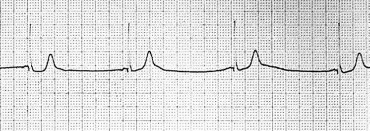
FIGURE 84.1 Junctional escape rhythm.
Ventricular Bradydysrhythmias
A ventricular rhythm with a rate of <50 bpm is termed a ventricular escape rhythm or idioventricular rhythm. Ventricular escape rhythm represents a physiologic safety mechanism that arises in the absence of stimuli from above, as in sinus bradycardia, sinus arrest, or, more commonly, AV block. Most patients with ventricular escape are symptomatic because the heart rate is low. Treatment is directed at the underlying dysrhythmia (i.e., atropine, or most often, cardiac pacing). Lidocaine, amiodarone, and other drugs that may abolish the ventricular rhythm are contraindicated because they have the potential for causing cardiac standstill.
Also known as idioventricular tachycardia or slow ventricular tachycardia, accelerated idioventricular rhythm has a rate of 50 to 100 bpm. Accelerated idioventricular rhythm is one cause of isorhythmic dissociation with occasional fusion beats demonstrating the continued presence of an intact AV node (Fig. 84.2). This rhythm may be associated with acute myocardial infarction (particularly after reperfusion), but it may also be seen in otherwise healthy patients. In the absence of symptoms, specific therapy is not warranted.
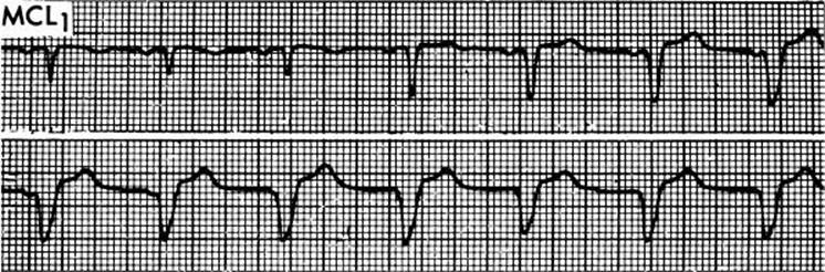
FIGURE 84.2 Idioventricular rhythm with isorhythmic dissociation and fusion beats.
Disturbances of Conduction
Disorders of Sinoatrial Conduction
The tissue surrounding the sinus node may delay or prevent conduction of sinus node impulses to the atria and AV node. Such conduction disorders are termed SA block and are classified in a manner analogous to AV block. Because of the limitations of the surface ECG, their precise diagnosis is more difficult than the varieties of AV block.
In first-degree SA block, there is a delay in the propagation of the sinus node impulse out to the atrial myocardium. Because every beat is transmitted, no abnormality is detected on the surface ECG.
Similar to AV block, second-degree SA block may be divided into types I and II. In type I (Wenckebach), there is, with each succeeding beat, a progressive delay in conduction from the sinus node to the atria, until conduction in totally blocked and a P wave is dropped (Fig. 84.3). The P–P interval shortens progressively before the dropped P wave (analogous to the situation in second-degree AV block, in which the R–R interval shortens before the dropped QRS complex).

FIGURE 84.3 Type I (Wenckebach) SA block.
In type II second-degree SA block, there is intermittent failure of the sinus impulse to reach the atria, resulting in a missing P wave and a sudden lengthening of the P–P interval. The resulting P–P interval is usually a multiple of the previous one that is doubled in the case of 2:1 block (Fig. 84.4). This disorder can be mimicked by blocked premature atrial contractions, where the P wave is obscured by the preceding T wave (Fig. 84.5).

FIGURE 84.4 Type II SA block 2:1.

FIGURE 84.5 Blocked premature atrial contractions mimicking AV or SA block.
Third-degree SA block is where there is a failure of any sinus node impulses to be conducted to the atria (Fig. 84.6). Third-degree SA block is usually indistinguishable from sinus arrest, where there is a failure of impulses to arise from the sinus node.
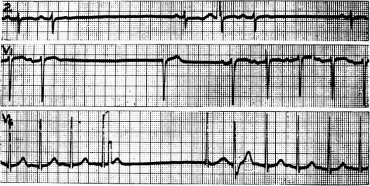
FIGURE 84.6 Third-degree SA block, with escape rhythm.
Disorders of Atrioventricular Conduction
Traditionally, AV blocks have been divided into first, second, and third degrees. However, AV block is a relative phenomenon, and the physiologic delay normally present in the AV node is one end of a continuum. For example, in the patient with atrial flutter, “normal” AV conduction usually results in 2:1 block. In addition, the degree of block is rate dependent. One patient with mild AV disease may exhibit second-degree block at a particularly fast supraventricular rate, and another patient with more advanced AV nodal disease may exhibit 1:1 conduction if the supraventricular rate is slower.
A P–R interval of >0.20 seconds defines first-degree AV block. All P waves are conducted, 1:1 AV conduction is maintained, and the P–P and R–R intervals are consistent. Most commonly, the delay in AV conduction is within the AV node, and the QRS complex is of normal duration. Delays within the His bundle or His–Purkinje system are less common and are usually associated with widened bundle-branch block QRS patterns.
First-degree AV block may be associated with electrolyte disturbances or the use of digitalis, β-blockers, or calcium channel blockers. It becomes more prevalent as patients age. First-degree AV block may be seen during acute myocardial infarction, particularly inferior myocardial infarction. Treatment includes correction of electrolyte abnormalities or removal of the responsible drug. During acute myocardial infarction, close observation is warranted to detect progression to higher degrees of block. Otherwise, specific treatment is not indicated.
Second-degree AV block can be divided into Mobitz types I and II. Wenckebach distinguished between these two in the pre-ECG era, using only physical observation (14).
Mobitz type I second-degree AV block (Wenckebach) usually results from conduction delay in the AV node, although delays may occur in the bundle of His, bundle branches, or Purkinje system (Fig. 84.7). For this reason, the QRS complex in type I block is most often of normal duration and configuration. Mobitz type I AV block may be associated with inferior myocardial infarction or the use of digitalis, β-blockers, and calcium channel blockers. Mobitz type I AV block is usually transient, asymptomatic, and it has a good prognosis. If the patient is symptomatic, atropine can be given to enhance AV nodule conduction. Pacing is usually not necessary. Offending drugs may need to be withheld.

FIGURE 84.7 Mobitz type I (Wenckebach) second-degree AV block.
Mobitz type II second-degree AV block is less common. The site of block is usually within the His–Purkinje system, and a bundle-branch block QRS pattern is usually seen. In type II block, the P–R intervals are constant until single or multiple beats are suddenly dropped (Fig. 84.8). Type II block is usually symptomatic and has a higher likelihood of progressing to complete AV block. If associated with acute anterior myocardial infarction, type II block carries an ominous prognosis. Most authorities agree that Mobitz type II second-degree AV block requires permanent cardiac pacing. Because emergency drug treatment is often ineffective, temporary pacing may be necessary as a stabilizing measure.
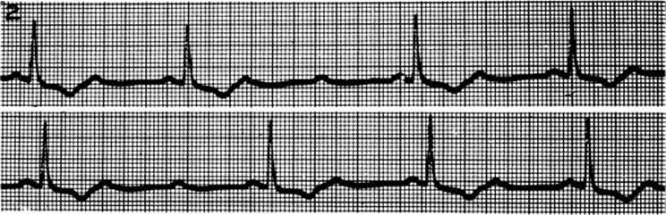
FIGURE 84.8 Mobitz type II second-degree AV block.
A particular clinical problem is presented by the patient with 2:1 AV block. Although it is often assumed that such patients have type II block, type I block often presents as 2:1 block. The distinction is important given the difference in prognosis and treatment between the two entities. Although definitive proof may require intracardiac recordings, the following principles may aid in the clinical distinction:
• Type I block is more common than type II.
• A narrow QRS complex usually reflects type I block (although not all type I blocks have a narrow QRS complex). Type II block usually has a wide QRS complex.
• Coexisting acute inferior myocardial infarction suggests type I block, whereas anterior myocardial infarction suggests type II block.
• Digitalis, β-blocker, or calcium channel blocker therapy tends to be associated with type I block.
• In type I block, other areas of the rhythm strip may reflect other patterns of type I conduction such as 3:2 or 4:3.
In third-degree (complete) AV block, no atrial impulses reach the ventricles, and a subsidiary, escape pacemaker usually emerges. AV dissociation results with constant but independent P–P intervals and R–R intervals. The P–R intervals are variable, and P waves have no discernible relation to QRS complexes (Fig. 84.9). The site of block may be within the AV node, His bundle, bundle branches, or Purkinje system. In general, the lower the site of the block the more severe the symptoms.

FIGURE 84.9 Third-degree atrioventricular block.
Third-degree AV block may be intermittent or self-limited. If the AV block is a complication of drug therapy, it usually resolves on withdrawal of the offending agent. Third-degree AV block may be associated with acute myocardial infarction but occasionally is an asymptomatic finding in an otherwise healthy person.
Like other bradydysrhythmias, the emergent treatment of third-degree AV block depends on symptomatology and clinical setting. Symptomatic patients may improve with atropine, particularly if the block is within the AV node. Symptomatic patients who are unresponsive to pharmacologic therapy require emergent pacing. Stable patients with acute inferior infarction may require only a prophylactic pacemaker. However, acute anterior infarction complicated by third-degree AV block is more ominous and requires emergent pacing.
The term AV dissociation, although commonly used interchangeably with the term third-degree AV block, encompasses a much broader range of rhythms. AV dissociation implies that the atria and ventricles are beating independently, a situation that includes not only complete AV block but also accelerated junctional and idioventricular rhythms (including ventricular tachycardia). In isorhythmic dissociation (Fig. 84.10), the sinus rate and the escape or ectopic rate are nearly identical, producing an electrocardiographic pattern characterized by varying P–R intervals as the sinus and ectopic foci slowly change their relative firing rates. Occasional fusion beats are an indication that the cause is not complete AV block.

FIGURE 84.10 Atrioventricular dissociation with isorhythmic dissociation owing to a junctional focus.
Artificial Pacemakers
Temporary cardiac pacemakers are normally used in emergent or urgent circumstances, while most permanent pacemakers are placed electively. The emergency physician should be familiar with the indications and use of temporary pacemakers and with the principles of permanent pacemakers and their complications (see Chapter 86).
Temporary Pacemakers
Therapeutic emergency cardiac pacing is indicated in any hemodynamically unstable bradycardia that fails to respond to pharmacologic therapy. In addition, prophylactic emergent cardiac pacing may be indicated, even in the absence of symptoms, for patients with acute myocardial infarction in the following circumstances:
• First-degree AV block with new-onset bundle-branch block.
• Second-degree AV block type II.
• Third-degree AV block.
• Right bundle-branch block with left anterior fascicular block or left posterior fascicular block (either old or new).
• Left bundle-branch block (old or new) and placement of a Swan–Ganz catheter (because of the risk of inducing iatrogenic right bundle-branch block and then complete block).
Transcutaneous cardiac pacing involves the application of cutaneous electrodes to the chest and delivery of electrical impulses through the chest wall to the myocardium. Transcutaneous pacing is the technique of choice for emergent pacing in the emergency department and is the only option available for prehospital use.
The advantages of transcutaneous pacing are its ease and speed of use and the absence of serious side effects. The disadvantages include an inability to capture in some patients and the discomfort experienced by conscious patients because of chest wall contractions. The latter can usually be treated satisfactorily with analgesics or sedation. Transcutaneous pacing should be considered a stabilizing treatment, to be replaced with other pacing techniques, when possible.
Transvenous cardiac pacing involves the placement of the pacing electrode into the right ventricle by way of a central vein, often the subclavian or internal jugular. Therefore, the procedure carries the risk of central venous catheterization but is the temporary modality that most closely approximates the function of a permanent pacemaker. Transvenous cardiac pacing does not involve painful muscle contractions or the hazard of direct cardiac puncture and can be used for relatively prolonged periods. Unfortunately, emergent transvenous pacemaker placement in the emergency department setting is often a lengthy and unsuccessful procedure associated with a high incidence of noncapture. Ideally, placement is performed under fluoroscopic guidance. Ultrasonographic guidance may make this procedure more effective, both in the initial placement of the central line and also in the correct placement of the pacing wire in the right ventricle. With the advent of transcutaneous pacing, transvenous pacing has been relegated to those situations in which fluoroscopy is available, stabilization has not been achieved by another modality, or attempts at transcutaneous pacing have been unsuccessful.
Transthoracic cardiac pacing is now rarely performed. It involves the placement of a pacing wire through the skin directly into the right ventricular cavity. Because of significant complications such as pericardial tamponade, pneumothorax, and coronary vessel injury, transthoracic pacing should usually be considered only in unusual circumstances.
Once capture has been achieved, regardless of the device used, the pacemaker settings will be dependent on the clinical situation. An initial rate of 80 to 100 bpm is appropriate for the majority of the patients. The initial output setting should be at maximum and decreased once capture is achieved. The mode should initially be asynchronous (sensitivity off) in those patients requiring emergency pacing for cardiac arrest. The mode should be set to synchronous in patients with bradydysrhythmias, as ventricular fibrillation may occur if the pacemaker fires during the vulnerable period (T wave).
CRITICAL INTERVENTIONS
• Initiate artificial pacing in unstable patients if drug therapy is ineffective.
DISPOSITION
Virtually all patients with symptomatic bradydysrhythmia require admission to the hospital. Patients with type II second-degree AV block and complete AV block usually warrant admission even if asymptomatic. Admission should be to a monitored bed in a unit with the capability of initiating emergent pacing.
If transfer to another facility is necessary, pacing should first be initiated if indicated and available. En route, the patient should have continuous cardiac monitoring and access to advanced cardiac life support. Transcutaneous pacing capability en route is also desirable.
Common Pitfalls
• Assuming that a 2:1 second-degree AV block is of the Mobitz type II variety, potentially leading to unnecessary treatment and intervention.
• Failing to take rate into account when evaluating AV blocks and fascicular blocks.
• Misdiagnosing blocked premature atrial contractions as significant AV block, potentially leading to unnecessary treatment and intervention.
• Blocking an escape rhythm, with the potential for causing cardiac standstill.
REFERENCES
1. Arima H, Sobue K, Tanaka S, et al. Profound sinus bradycardia after intravenous nicardipine. Anesth Analg. 2002;95:53–55.
2. Calvo-Romero JM, Lima-Rodriguez EM. Bradycardia associated with ophthalmic beta-blockers. J Postgrad Med. 2003;49:186.
3. Castro VJ, Nacht R. Cocaine-induced bradyarrhythmia: An unsuspected cause of syncope. Chest. 2000;117:275–277.
4. Essebag V, Hadjis T, Platt RW, et al. Amiodarone and the risk of bradyarrhythmia requiring permanent pacemaker in elderly patients with atrial fibrillation and prior myocardial infarction. J Am Coll Cardiol. 2003;41:249–254.
5. Kaur A, Yu SS, Lee AJ, et al. Thalidomide-included sinus bradycardia. Ann Pharmacother. 2003;37:1040–1043.
6. Akikusa JD, Feldman BM, Gross GJ, et al. Sinus bradycardia after intravenous pulse methylprednisolone. Pediatrics. 2007;119(3):e778–e782.
7. Hawley P. Case report of severe bradycardia due to transderaml fentanyl. Palliat Med. 2013;27(8):793–795. Published online January 22, 2013, doi: 10.1177/0269216312472383.
8. Ji KH, Kim DH, Yun CH. Severe obstructive sleep apnea syndrome with symptomatic daytime bradyarrhythmia. J Clin Sleep Med. 2009;5(3):246–247.
9. Monte C, De Krom M, Weber W, et al. The ictal bradycardia syndrome. Acta Neurol Belg. 2007;107:22–25.
10. Kam PC, Cardone D. Propofol infusion syndrome. Anaesthetia. 2007;62(7):690–701.
11. Lateef A, Fizler DA, Tambrajah PA. Dengue fever and relative bradycardia. Emerg Infect Dis. 2007;13(4):650–651.
12. Inoue T, Takayanagi K, Sakai Y, et al. Cardiac pacing as emergency care for serious bradyarrhythmias with circulatory shock. J Med. 2001;32:321–331.
13. Walker J, Calkins H, Nazarian S. Evaluation of cardiac arrhythmia among athletes. Am J Med. 2010;123(12):1075–1081.
14. Wenckebach KF. Zur analyse des unregelmassigen Pulses. Z Klin Med. 1899;37: 475–488.