
E. Kevin Heist, MD, PhD, Dan Blendea, MD, Jeremy N. Ruskin, MD, Moussa Mansour, MD
CASE PRESENTATION
A 20-year-old man without previous medical history suffered a witnessed cardiac arrest in a locker room shortly after refereeing a wrestling match. He received bystander CPR; when paramedics arrived, he was found to be in preexcited atrial fibrillation with a rapid rate (290 bpm) and was externally shocked to sinus rhythm. His ECG in sinus rhythm demonstrated manifest preexcitation, with deeply negative delta waves in the inferior leads, suggestive of a posteroseptal accessory pathway (AP) (Figure 11-1). He was taken to EP study, which demonstrated the presence of a posteroseptal AP, with anterograde effective refractory period 400/210 ms, and 1:1 A:V conduction via the AP with rapid atrial pacing down to 220 ms, as well as inducible, nonsustained orthodromic AV reciprocating tachycardia (AVRT). Mapping of ventricular activation during sinus rhythm (preexcited) utilizing endocardial access localized the earliest ventricular potentials to the proximal coronary sinus (CS) os, where a small CS diverticulum was identified by contrast venography. Extensive radiofrequency ablation at this region resulted in only transient loss of preexcitation, and cryoablation at this site was also ineffective. The patient then returned for a second procedure with preprocedural CT imaging and percutaneous subxyphoid epicardial access. The earliest preexcited ventricular electrogram using both endocardial and epicardial mapping was identified in the epicardial posteroseptal region (Figures 11-2 and 11-3). After coronary angiography was performed to demonstrate that this site was not adjacent to a coronary artery, radiofrequency ablation at this site successfully and durably ablated AP conduction (Figure 11-4), and the patient has had no further arrhythmic symptoms.1
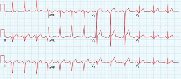
FIGURE 11-1 Twelve-lead ECG after cardioversion, demonstrating manifest preexcitation with deeply negative delta waves in leads II, III, and aVF.
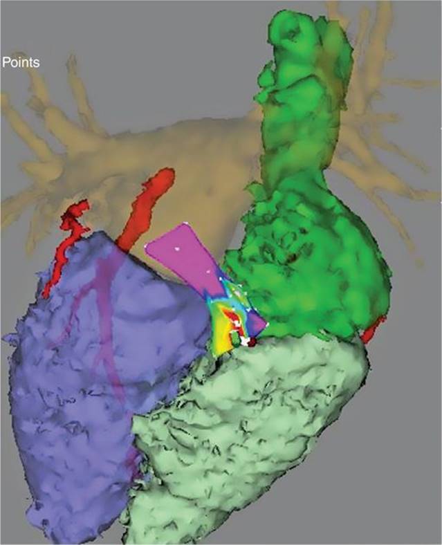
FIGURE 11-2 Site of successful epicardial mapping and ablation. This shows a fusion of electroanatomic mapping from the epicardial space (with colors indicating timing of earliest preexcited ventricular electrogram) with a previously acquired CT scan of the heart. The left and right atria and ventricles can be seen in the CT image, as well as the relevant portions of the right and left circumflex coronary arteries, which are adjacent to the ablation site.
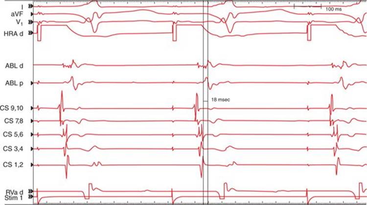
FIGURE 11-3 Surface and intracardiac electrograms from the successful ablation site. Mapping was performed during high right atrial (HRA) pacing. Tracings are shown from surface leads I, V1, and aVF, as well as HRA, ablation (ABL) proximal and distal electrograms, coronary sinus (CS) proximal (9,10) to distal (1,2), and right ventricular apex (RVa). The earliest accessory pathway (AP) electrograms are noted 18 ms before the onset of the delta wave visualized on the surface leads.
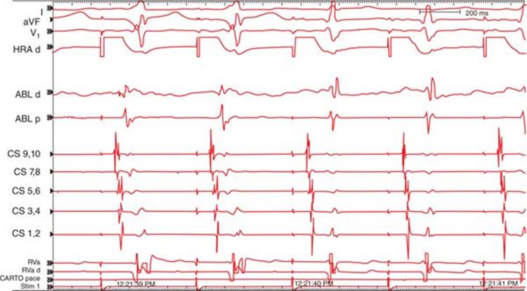
FIGURE 11-4 Loss of preexcitation during ablation. These electrograms (same annotation as Figure 11-3) were obtained during ablation at the same site as Figure 11-3. The first 2 beats show preexcitation on the surface leads, which is no longer evident on the 3rd and subsequent beats. In addition, the short delay between the local atrial and ventricular electrograms in the preexcited beats noted on the ablation catheter lengthens with loss of preexcitation. Loss of preexcitation was noted after 6.5 seconds of ablation at this site and was durable after completion of ablation and administration of IV adenosine.
EPIDEMIOLOGY AND PATHOPHYSIOLOGY
Manifest preexcitation is evident in approximately 0.1% to 0.3% of ECGs,2 although this underestimates the true prevalence of AP conduction, as many APs are concealed, either due to AV nodal conduction preceding AP conduction or due to lack of anterograde AP conduction. Classic APs result from electrically excitable fibers (bundles of Kent) which pass through the fibrous AV ring, although other types of APs (ie, atriofascicular pathways) have also been extensively described in the literature.3
The initial interventional approaches to patients with symptomatic Wolff-Parkinson-White (WPW) syndrome involved cardiac surgical incisions in the region of the AP.4 Because these incisions were transmural, the exact intramural location of the AP (endocardial, midmyocardial, or epicardial) was generally not relevant for clinical success in most cases. This changed with the advent of catheter ablation,5however, as current ablation catheters may not always create transmural lesions, particularly in regions of thick myocardium, such as the AV ring (particularly relevant to AP ablation) and the ventricular septum and left ventricle (relevant to ventricular tachycardia ablation).
If microscopic studies could be performed on all APs, it would be possible to anatomically define the course of each AP, and endocardial/epicardial location could be precisely determined. Because this is obviously not possible in patients, the location of a given AP is typically defined based on activation mapping of the region of earliest atrial and/or ventricular preexcitation and AP potentials, and particularly by success (or lack of success) of ablation at a given site. Epicardial APs are typically defined as those which require epicardial access for successful ablation, as well as APs which map to and/or are successfully ablated from the coronary sinus (CS) and its tributaries, which is an epicardial structure. This is thus a functional rather than a true anatomic classification of endocardial/epicardial location of APs, and it should be noted that pericardial mapping (other than mapping within the CS) is rarely performed for a given AP unless ablation from an endocardial approach is unsuccessful. APs requiring pericardial access for successful ablation have generally been reported as single cases or small case series, and so it is currently difficult to estimate their true incidence.1,6-9 In comparison, APs which have been successfully ablated from within the CS or its tributaries have been described with much greater frequency.10
It should be noted that in many cases it is possible to create electrically transmural lesions at the AV ring utilizing only endocardial catheter ablation. Examples of this include cavo-tricuspid isthmus ablation for typical right atrial flutter (which is virtually always successful using endocardial ablation with current techniques and requires transmural electrical block) and mitral isthmus ablation at the AV ring adjacent to the left inferior pulmonary vein (which is successful in some cases using only endocardial ablation, while other cases require combined endocardial and intra-CS ablation11). Based on this evidence, it would be reasonable to hypothesize that an AP in the vicinity of the cavo-tricuspid isthmus could be successfully ablated using an endocardial approach in almost all cases regardless of endocardial/epicardial course, while an epicardial or intra-CS AP in the vicinity of the mitral isthmus could be ablated using an endocardial approach in some but not all cases.
The large majority of APs which are thus functionally classified as epicardial are posteroseptal, as is true for the case presented here. It should be noted that the thickness of the myocardium at the AV ring is typically greatest in the posteroseptal region. It is therefore not clear whether anatomically epicardial APs are truly more common in the posteroseptal region compared to other regions, or whether the greater myocardial thickness in the posteroseptal region results in greater difficulty in creating transmural ablation lesions, more often necessitating an epicardial approach for posteroseptal APs. It should be noted that the proximal CS (and its tributaries such as the takeoff of the middle cardiac vein) contains a muscular coat capable of electrical conduction and contraction,10 and this may also contribute to the presence of epicardial APs in the posteroseptal location. Other unusual epicardial AP connections such as from the left or right atrial appendage to the ventricle (which in some cases may require a surgical approach for successful interruption of the AP), have also been reported.12
APPROACH TO EP STUDY AND ABLATION
Although ECG criteria have been devised to estimate the likelihood that a particular VT will require pericardial access for successful ablation,13 APs requiring pericardial access are much less common, and no definitive criteria for such APs has been devised. Given that the majority of these APs are posteroseptal, however, preexcitation suggestive of a posteroseptal AP14 (typically negative delta waves in the inferior leads) suggests a greater likelihood of the requirement for epicardial ablation. It should be noted, however, that only a small minority of posteroseptal APs require pericardial access for successful ablation; most can be ablated from endocardial sites, or from the proximal CS or its tributaries.10 Given the greater risk of complications from pericardial access15 compared to traditional endocardial access, it is our opinion that mapping should be attempted from an endocardial/intra-CS approach for virtually all patients requiring AP ablation prior to obtaining pericardial access. For operators skilled in pericardial access and with the proper equipment available, it is certainly reasonable to obtain pericardial access during the same procedure if endocardial and intra-CS mapping/ablation is not successful and there is reason to believe that this may be due to an epicardial AP (this will generally require reversal of any IV anticoagulation used for the endocardial procedure). Suggestive findings of the need for epicardial access may include a low frequency “far field” electrogram with endocardial mapping at the earliest preexcited site with lack of a sharp AP potential and unsuccessful ablation at that site, particularly for posteroseptal APs (and after extensive CS mapping has been performed). It should also be kept in mind that midseptal sites do not have a corresponding epicardial site, and unsuccessful ablation at a midseptal site should prompt mapping of the septum of the alternate atrium, rather than pericardial access.
Percutaneous pericardial access for AP ablation is essentially identical to the approach described by Sosa and colleagues for ablation of ventricular tachycardia.16,17 This has been described in great detail in the published literature, but generally involves careful subxyphoid introduction of a round tipped spinal needle using small amounts of contrast to identify the pericardial space, and then advancement of a guidewire and sheath into this space. Fortunately, most AP ablation procedures are performed on patients who have not previously undergone cardiac surgical procedures, and so the pericardial adhesions which may challenge attempts at percutaneous epicardial VT ablation procedures18 are much less commonly encountered with AP ablation. Once pericardial access has been obtained, epicardial mapping of the AP can be performed in the same manner as endocardial mapping. In almost all cases, endocardial mapping should also be performed, and epicardial ablation only performed if an epicardial site is more favorable (based on typical criteria for AP ablation such as earliest site of preexcitation, presence of AP potentials, etc.) than the best endocardial site.
As is true for other types of epicardial ablation, there is the risk of damage to nearby structures.15 Proximity to coronary arteries should always be assessed by coronary angiography prior to ablation in most AP locations, and high output pacing prior to ablation to determine proximity to the phrenic nerve may also be important, although typical posteroseptal AP locations are usually distant from either phrenic nerve. The esophagus is also in proximity to the posterior AV groove in many patients, and the strategies currently in use for esophageal protection during AF ablation19 may also be relevant during ablation of posterior APs from an epicardial approach.
REFERENCES
1. Ho I, d’Avila A, Ruskin J, Mansour M. Images in cardiovascular medicine. Percutaneous epicardial mapping and ablation of a posteroseptal accessory pathway. Circulation. 2007;115(16):e418-421.
2. Wellens HJ, Rodriguez LM, Timmermans C, Smeets JP. The asymptomatic patient with the Wolff-Parkinson-White electrocardiogram. Pacing Clin Electrophysiol. 1997;20(8):2082-2086.
3. Gandhavadi M, Sternick EB, Jackman WM, Wellens HJ, Josephson ME. Characterization of the distal insertion of atriofascicular accessory pathways and mechanisms of QRS patterns in atriofascicular antidromic tachycardia. Heart Rhythm. 2013;10(9):1385-1392.
4. Misaki T, Watanabe G, Iwa T, et al. Surgical treatment of patients with Wolff-Parkinson-White syndrome and associated Ebstein’s anomaly. J Thorac Cardiovasc Surg. 1995;110(6):1702-1707.
5. Jackman WM, Wang XZ, Friday KJ, et al. Catheter ablation of accessory atrioventricular pathways (Wolff-Parkinson-White syndrome) by radiofrequency current. N Engl J Med. 1991;324(23):1605-1611.
6. Schweikert RA, Saliba WI, Tomassoni G, et al. Percutaneous pericardial instrumentation for endo-epicardial mapping of previously failed ablations. Circulation. 2003;108(11):1329-1335.
7. Valderrabano M, Cesario DA, Ji S, et al. Percutaneous epicardial mapping during ablation of difficult accessory pathways as an alternative to cardiac surgery. Heart Rhythm. 2004;1(3):311-316.
8. de Paola AA, Leite LR, Mesas CE. Nonsurgical transthoracic epicardial ablation for the treatment of a resistant posteroseptal accessory pathway. Pacing Clin Electrophysiol. 2004;27(2):259-261.
9. Sapp J, Soejima K, Couper GS, Stevenson WG. Electrophysiology and anatomic characterization of an epicardial accessory pathway. J Cardiovasc Electrophysiol. 2001;12(12):1411-1414.
10. Sun Y, Arruda M, Otomo K, et al. Coronary sinus-ventricular accessory connections producing posteroseptal and left posterior accessory pathways: Incidence and electrophysiological identification. Circulation. 2002;106(11):1362-1367.
11. Shah AJ, Pascale P, Miyazaki S, et al. Prevalence and types of pitfall in the assessment of mitral isthmus linear conduction block. Circ Arrhythm Electrophysiol. 2012;5(5):957-967.
12. Mah D, Miyake C, Clegg R, et al. Epicardial left atrial appendage and biatrial appendage accessory pathways. Heart Rhythm. 2010;7(12):1740-1745.
13. Valles E, Bazan V, Marchlinski FE. ECG criteria to identify epicardial ventricular tachycardia in nonischemic cardiomyopathy. Circ Arrhythm Electrophysiol. 2010;3(1):63-71.
14. d’Avila A, Brugada J, Skeberis V, Andries E, Sosa E, Brugada P. A fast and reliable algorithm to localize accessory pathways based on the polarity of the QRS complex on the surface ECG during sinus rhythm. Pacing Clin Electrophysiol. 1995;18(9):1615-1627.
15. Koruth JS, Aryana A, Dukkipati SR, et al. Unusual complications of percutaneous epicardial access and epicardial mapping and ablation of cardiac arrhythmias. Circ Arrhythm Electrophysiol. 2011;4(6):882-888.
16. d’Avila A, Koruth JS, Dukkipati S, Reddy VY. Epicardial access for the treatment of cardiac arrhythmias. Europace. 2012;14: Suppl 2:ii13-ii18.
17. Sosa E, Scanavacca M, D’Avila A, et al. Endocardial and epicardial ablation guided by nonsurgical transthoracic epicardial mapping to treat recurrent ventricular tachycardia. J Cardiovasc Electrophysiol. 1998;9(3):229-239.
18. Tschabrunn CM, Haqqani HM, Cooper JM, et al. Percutaneous epicardial ventricular tachycardia ablation after noncoronary cardiac surgery or pericarditis. Heart Rhythm. 2013;10(2):165-169.
19. Contreras-Valdes FM, Heist EK, Danik SB, et al. Severity of esophageal injury predicts time to healing after radiofrequency catheter ablation for atrial fibrillation. Heart Rhythm. 2011;8(12):1862-1868.