
David G. Benditt, MD, Barry L.S. Detloff, BS, Oana Dickinson, MD
CASE PRESENTATION
A 39-year-old otherwise healthy active woman was seen in consultation at the Syncope/Arrhythmia Clinic at the University of Minnesota Medical Center complaining of multiple “faints” and numerous near-faints. The faints that she did experience were always similar in presentation; they tended to occur more often early in the day and developed within a few minutes after she stood up from a seated or lying position. Typically, within seconds of arising she experienced dull ache in her shoulder and arms, lightheadedness, and “blurry vision.” These symptoms usually resolved promptly. However, from time-to-time 1 or 2 minutes later she would sense palpitations, sweatiness, cold skin, and occasional nausea. Observers noted marked pallor. In response, she had learned to lie down on the floor. However, even though lying down prevented complete loss of consciousness, she volunteered that afterward she felt “drained” and wanted to rest.
The patient does not recall fainting as a child, and there is no family history of transient loss of consciousness or premature death. She does not recollect ever having a faint triggered by prolonged standing, blood draws, emotional upset, or pain. Her only medications, apart from vitamin supplements, are occasional pain relievers and a vasoconstrictor for migraine control. Physical examination in the clinic was normal with a seated blood pressure of 104/69 mm Hg and a heart rate of 80 beats/min. Her ECG was normal, as was an echocardiogram.
ETIOLOGY AND PATHOPHYSIOLOGY
Faints occurring as the result of transient neural reflex-mediated systemic hypotension (so-called “neural-reflex syncope”) are the most common form of syncope encountered in clinical practice (40%-50% of cases), with vasovagal syncope being the most frequent of all forms of neural-reflex syncope. Others include carotid sinus syndrome and situational faints (eg, cough syncope, postmicturition syncope) (Figure 46-1).

FIGURE 46-1 Summary of the most common forms of neurally mediated reflex syncope. In each case the basis of the faint may be “vasodepressor” or “cardioinhibitory” or both (“mixed”).
Neural reflex faints (particularly the vasovagal faint) not only comprise the vast majority of syncope events in younger, otherwise healthy patients but also remain a major contributing cause of syncope in older individuals (Figure 46-2).3,4 However, due to a higher prevalence of heart and vascular disease in older individuals, establishing the diagnosis may be difficult. In any case, no matter the patient’s age, it is the “reflex” nature of the faint, most often detected by the patient’s symptoms documented during a carefully obtained medical history, that most effectively distinguishes neural-reflex faints from other causes of collapse.

FIGURE 46-2 Bar graph depicting approximate frequency of various causes of syncope by age. Note that neurally mediated syncope (NMS, white) remains a common cause in all age groups. Orthostatic syncope increases with advancing age (light blue). (Modified with permission from Parry SW, Tan MP: An approach to the evaluation and management of syncope in adults, BMJ 2010 Feb 27;340(7744): 468-473.)
NOMENCLATURE
In neurally mediated reflex syncope, systemic hypotension with consequent cerebral hypoperfusion may be caused by either vasodilation or severe bradycardia acting alone, or as is more often the case, both working together.1 When vasodilatation is the primary cause of symptomatic hypotension, the faint is termed “vasodepressor” and is the result of some degree of diminished arterial resistance, but an even greater component of increased venous capacitance (particularly in the splanchnic bed) with consequent diminished venous return to the heart. If bradycardia is the principal cause of hypotension, then the faint is termed “cardioinhibitory,” and is due to the occurrence of a predominantly vagally-mediated slow heart rate (often prolonged asystolic pauses [Figure 46-3]) of sufficient severity to compromise cardiac output and cerebral blood flow. Most often both mechanisms are present, and the pathophysiology of the syncope is said to be “mixed.”1,2
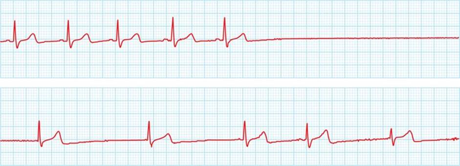
FIGURE 46-3 Sinus arrest suggestive of a cardioinhibitory syncope in a young patient with typical vasovagal symptoms. However, it should be recognized that a vasodepressor mechanism may also be present but cannot be confirmed in the absence of documenting the presence of hypotension during cardiac pacing to overcome the bradycardia.
Given the pathophysiology described above, “vasodepressor” syncope is best considered a subset of the vasovagal reaction in which systemic hypotension is primarily due to vaso- and venodilatation; in the vasodepressor faint, cardioinhibition is either nonexistent or modest. The physiology of the “vasodepressor” faint was investigated in the classic study by Weissler et al in 1957.5 These investigators pointed out that muscarinic blockade and correction of bradycardia (by atropine) had little impact on susceptibility to syncope. Thus a “vasodepressor” faint should be considered a physiologic unique entity (albeit often found in a “mixed” form with a cardioinhibitory feature) and should not be simply employed as another term for vasovagal syncope or “neural reflex faint.” However, and rather unfortunately, the term “vasodepressor” often continues to be misused; this erroneous usage is confusing and should be abandoned.
CLINICAL EVALUATION
Initially, the medical history of the patient described here was interpreted to suggest that this patient was experiencing both “immediate” and subsequently the more classic “delayed” form of orthostatic hypotension. However, her age and absence of evident triggers (eg, drugs, neurologic disease) made orthostatic syncope unlikely. Furthermore, her symptom complex (ie, palpitations, sweating, feeling cold, pallor, and later, fatigue) were more compatible with a neurally mediated reflex vasovagal reaction triggered by abrupt movement to upright posture. Consequently, an autonomic study using noninvasive ECG and hemodynamic monitoring (Finometer, Finapres Medical Systems, Arnhem, The Netherlands) was undertaken. In brief, after a 10-minute period of being quietly seated in a chair, “active” standing resulted in evidence for immediate orthostatic hypotension (Figure 46-4A). The test was repeated in both the drug-free state and after sublingual nitroglycerin (0.4 mg) pretreatment; both resulted in her experiencing typical symptoms of “dizziness” and brief visual disturbance associated with an immediate but transient fall of systemic blood pressure but without substantial bradycardia. The observation strongly favored a predominantly “vasodepressor” response. The patient indicated that the sequence of events was typical of her spontaneous attacks.
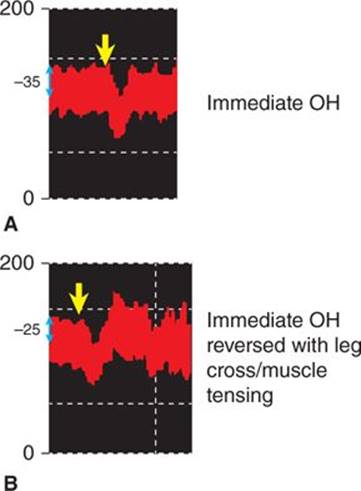
FIGURE 46-4 Impact of muscle tensing on immediate orthostatic hypotension (OH) response. Vertical blue arrows indicate time of movement from sitting to standing. The almost “immediate” fall of systemic pressure is apparent. PCM = physical counter maneuver. (A) Control test. Approximate drop in blood pressure is 35 mm Hg. (B) PCM test. Approximate drop of blood pressure is 25 mm Hg. Leg and buttock muscle tensing results in prompt normalization of pressure with slight overshoot evident.
DIAGNOSIS
The medical history is crucial to recognition of reflex syncope. Factors initiating the vasovagal form of neural reflex syncope, whether it be cardioinhibitory or vasodepressor in nature, are often elusive. Nevertheless, certain triggers are well recognized, including extreme emotion, relative dehydration, excessively warm confining environments, and acute pain. In addition, onset may be associated with prolonged periods of upright posture. However, the finding in the patient reported here, that abrupt postural change was to the best of our knowledge the only trigger, is uncommon. Nevertheless, it was the history that essentially excluded the primary contending diagnosis (ie, “delayed” form of orthostatic hypotension) and led to undertaking confirmatory laboratory testing.
Identifying vasodepressor hypotension is difficult in free-living individuals, as current ambulatory blood pressure recording technology is unsuitable to the task, and ambulatory ECG monitoring is not helpful. With regard to the latter, however, until newer ambulatory sensor systems able to detect blood pressure in a continuous manner become available,6 indirect evidence may be used to implicate a vasodepressor response. Specifically, the recording of sinus tachycardia prior to a reported syncope in which the heart rate is now essentially “normal,” suggests the possibility of a vasodepressor event. However, psychogenic pseudosyncope may exhibit a similar footprint. Consequently, findings during “active standing test” and/or head-up tilt table test may need to be relied upon even though they are at best surrogates for the spontaneous event.
Neurally mediated reflex faints, such as postural faints, are more likely to occur during periods of prolonged upright posture, especially if the affected individual is dehydrated or somewhat “volume down” (eg, in the morning after night-long fasting, or in the presence of excess diuretics or hot environments), or deconditioned.
The role of rapid postural change for triggering vasovagal reactions is difficult to assess using conventional tilt-table technique and is more amenable to evaluation by the “active standing test” or even more aggressively by the “squat-stand” test. In this regard, Rickards et al7 compared the immediate cardiovascular effects of tilt-table testing to those of the squat-stand test in healthy subjects. In essence, squat-stand triggered a greater and more prolonged fall of systemic blood pressure (systolic and diastolic), with the principal driver being the dramatic impact on reduction of diastolic pressure (an indirect marker of a more severe diminution of arterial peripheral resistance). Others8have hypothesized that squatting impairs blood flow to the legs and increases local accumulation of vasodilator metabolites. Upon movement to upright posture, and despite otherwise normal neural (baroreceptor) reflex adaptation, there is more persistent reduction of peripheral resistance than would be observed with tilt. The impact may be more prolonged hypotension in certain susceptible patients (such as the individual described here) and the consequent triggering of a vasovagal response, and particularly a vasodepressor response.
TREATMENT RECOMMENDATIONS
In most cases of vasovagal syncope, whether predominantly cardioinhibitory or vasodepressor, patient education and advice regarding maintenance of hydration (with low calorie electrolyte solutions), along with warning regarding the potential for falls and injury are the foundations of treatment. Thereafter it may be useful to introduce physical countermaneuvers. Only infrequently are drugs such as midodrine and fludrocortisone required. Pacing therapy is used only on very rare occasions and generally in older patients in whom a marked cardioinhibitory form of vasovagal syncope has been confirmed by ambulatory monitoring (not by tilt table testing alone).
In the case of our patient, physical counter-maneuvers9,10 were explained as part of the autonomic testing procedure; buttock and thigh muscle tensing clearly ameliorated the postural drop of blood pressure and eliminated near-syncope symptoms associated with abruptly moving to an upright posture (Figure 46-4B). Thereafter, a conventional head-up tilt table test was undertaken. A drug-free tilt-test was well tolerated for 20 minutes at which time sublingual nitroglycerin (0.4 mg) was administered (Figure 46-5). The patient experienced her usual near-syncope symptoms with an associated drop of blood pressure but only a modest reduction of heart rate (see Figure 46-5, top); however, the near-syncope symptoms were rapidly reversed by having her perform thigh and buttock muscle tensing as had been taught earlier in the study (see Figure 46-5).

FIGURE 46-5 Recording depicting head-up tilt table findings. (Top) The white vertical bar at left indicates the time when the patient is tilted to 70 degrees. (Bottom) After approximately 15 minutes, the blood pressure was stable, and nitroglycerine (NTG) was administered sublingually. Within 2 to 3 minutes, marked hypotension and modest bradycardia (not shown) began to evolve. The patient developed typical vasovagal symptoms primarily on a vasodepressor basis. Thereafter a series of muscle tensing physical countermaneuvers (PCM) were initiated (noted by horizontal blue double arrows). Note the prompt increase in blood pressure with each intervention and subsequent blood pressure fall after each PCM termination, except for the last one. The patient’s symptoms were ameliorated by the maneuvers, and these maneuvers could permit an affected individual to seek a safe gravitationally neutral location (ie, sitting, lying down) until the episode self-terminates. The tilt-test was concluded after the last PCM and accounts for normalization of pressure at that time.
Based on her history and the autonomic testing results, and in addition to general education regarding her condition, the patient was initiated on a treatment regimen comprised of increased dietary salt and volume (focusing on low-calorie electrolyte-containing “sport drinks”) and physical counter-maneuvers (principally thigh and buttock muscle tensing) to minimize the “immediate orthostatic hypotensive” trigger. Follow-up at 8 months has been associated with absence of syncope and only infrequent near-syncope symptoms.
CONCLUSION
In conclusion, this case study highlights vasodepressor syncope as a unique entity and points out that this term is best considered a subset of the vasovagal reaction. Furthermore, the case illustrates the potential (albeit seemingly uncommon) for recurrent vasovagal reactions of the vasodepressor form to be triggered by immediate (“initial”) orthostatic hypotension.
ACKNOWLEDGEMENT
The authors wish to thank Professor Wouter Wieling, University of Amsterdam, Amsterdam, The Netherlands, for his assistance with this manuscript and his invaluable teaching over many years.
REFERENCES
1. Moya, A, Sutton R, Ammirati F, et al. The Task Force for the Diagnosis and Management of Syncope of the European Society of Cardiology (ESC). Guidelines for the diagnosis and management of syncope (version 2009). European Heart J. 2009;30: 2631-2671.
2. Wieling W, Thijs RD, van Dijk N, Wilde AA, Benditt DG, van Dijk JG. Symptoms and signs of syncope: a review of the link between physiology and clinical clues. Brain. 2009;132:2630-2642.
3. Samniah N, Sakaguchi S, Ermis C, Lurie KG, Benditt DG. Transient modification of baroreceptor response during tilt-induced vasovagal syncope. Europace. 2004;6:48-54.
4. Parry SW, Tan MP. An approach to the evaluation and management of syncope in adults. BMJ. 2010;340:468-473.
5. Weissler AM, Warren JV, Estes EH Jr, McIntosh HD, Leonard JJ. Vaodepressor syncope: factors affecting cardiac output. Circulation. 1957;15:875-882.
6. Benditt DG, Sakaguchi S, van Dijk JG. Autonomic nervous system and cardiac arrhythmias. In: Saksena S, Camm AJ, Boyden PA, Dorian P, Goldschlager N, Vetter VL, Zareba W, eds. Electrophysiology Disorders of the Heart,2nd ed. Philadelphia, PA: Elsevier Saunders; 2012:61-71.
7. Venugopal D, Patterson R, Jhanjee R, et al. Subcutaneous bioimpedance recording: assessment of a method for hemodynamic monitoring by implanted devices. J Cardiovasc Electrophysiol. 2009;20:76-81.
8. Rickards CA, Newman DG. A comparative assessment of two techniques for investigating initial cardiovascular reflexes under acute orthostatic stress. Eur J Appl Physiol. 2003;90:449-457.
9. Convertino VA, Tripp LD, Ludwig DA, Duff J, Chelette TL. Female exposure to high G: chronic adaptations of cardiovascular functions. Aviat Space Environ Med. 1998;69:875-882.
9. Krediet CT, Go-Schon IK, Kim YS, Linzer M, Van Lieshout JJ, Wieling W. Management of initial orthostatic hypotension: lower body muscle tensing attenuates the transient arterial blood pressure decrease upon standing from squatting. Clin Sci (Lond). 2007;113:401-407.
10. van Dijk N, Quartieri F, Blanc JJ, et al; PC-Trial Investigators. Effectiveness of physical counterpressure maneuvers in preventing vasovagal syncope: the Physical Counterpressure Manoeuvres Trial (PC-Trial). J Am Coll Cardiol. 2006;48:1652-1657.