
Kelly M.W. McDonnell, DO, Shawn Campbell, MEM, Kenneth A. Ellenbogen, MD, FHRS
CASE PRESENTATION
A 60-year-old man is seen in evaluation for an ICD shock. He has a history of sick sinus syndrome, nonischemic cardiomyopathy, ejection fraction of 30%, and New York Heart Association class II heart failure symptoms with a dual chamber, single coil ICD for primary prevention of sudden cardiac death. The patient has no history of device therapy but has reported intermittent palpitations prior to this event that he describes as “fast and regular” with sudden onset. Baseline laboratory studies and ECG were normal. The patient denied changes in his medications or baseline health status. The stored event is depicted in Figure 73-1 and demonstrates a tachycardia with 1:1 atrial to ventricular activity.
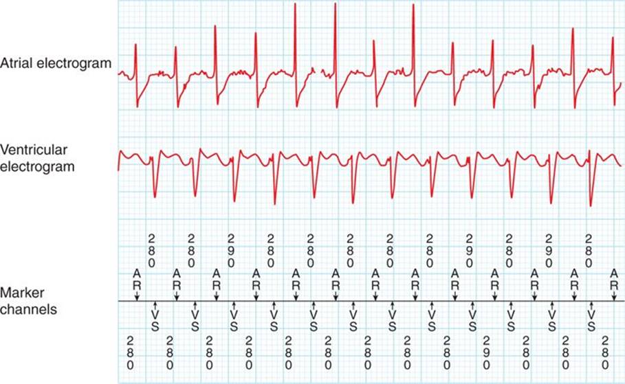
FIGURE 73-1 Documented tachycardia with atrial and ventricular electrograms and marker channel annotations with measured intervals in milliseconds.
CASE EXPLANATION
• This is a case of 1:1 tachycardia resulting in ICD therapy. Evaluation of an arrhythmia resulting in a shock requires careful evaluation of the stored electrograms, interval plots, tachycardia initiation, response to antitachycardia pacing (ATP), and arrhythmia termination to determine if the therapy was appropriate or inappropriate.
• Inappropriate shocks are shocks that occur for rhythms other than ventricular tachycardia or ventricular fibrillation and occur in up to 30% of patients with ICD shocks.1 They are most common due to supraventricular tachycardia (SVT), which gets classified by the device as ventricular tachycardia (VT). It is essential to determine the appropriateness of the shock to guide programming and medical therapy.
• The differential diagnosis of this event with 1:1 AV relationship includes ventricular tachycardia with retrograde atrial activation versus supraventricular tachycardia.
PATHOPHYSIOLOGY
The quintessential reason for implanting an ICD is to provide therapy for a life-threatening rhythm that if left untreated would result in sudden death. Differentiating VT from SVT is a key component of device algorithms to appropriately classify tachycardia events. Enhancements to tachycardia detection include rhythm stability, tachycardia onset, electrogram morphology, atrial to ventricular ratios, and atrial to ventricular timing. No algorithm is perfect in differentiating ventricular tachycardia from supraventricular tachycardia, but each detection enhancement aids in correctly identifying the rhythm.
• Rhythm stability helps differentiate atrial fibrillation with rapid ventricular response from ventricular tachycardia due to the variable R-R intervals seen in atrial fibrillation.
• Onset: Sinus tachycardia tends to gradually increase in rate; whereas other types of supraventricular tachycardia and ventricular tachycardia occur abruptly with a premature complex.
• Morphology templates: Newer algorithms store a baseline morphology of the intrinsic QRS. This is used during rhythm discrimination; SVT usually results in preserved QRS morphology, and VT results in a change in morphology due to the activation sequence. This algorithm is less reliable in patients with a baseline bundle branch block or when aberrancy develops during supraventricular tachycardia. Limitations to morphology algorithm, even when the template is regularly updated, include changes in the baseline QRS morphology, problems with alignment in the baseline and tachycardia morphology, and clipping of the signal.
• Atrial to ventricular ratios also assist. In general, A > V suggests SVT or atrial tachycardia, A = V suggest SVT or VT, and V > A suggests ventricular tachycardia.
DIAGNOSIS
The approach to evaluating an episode should be performed in a stepwise fashion.
• The episode text will outline the event, including the classification, duration, and therapy received as seen in Figure 73-2. The device has classified this rhythm as VT and provided therapy.
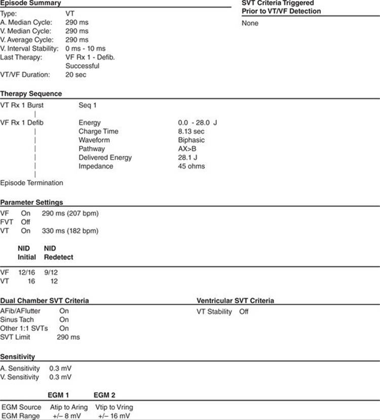
FIGURE 73-2 Event text summary with documentation of the programmed VT and VF zones and detection intervals.
• The interval plot shows the A-V relationship throughout the event and illustrates the rate in relationship to the programmed VT and VF zones (Figure 73-3). Initially there are slight variations between the atrial and ventricular rhythm followed by an abrupt onset of tachycardia. The interval plot shows that the tachycardia initiated with a V marker and then has a consistent ventricular to atrial relationship. The rate of the rhythm falls into the programmed ventricular tachycardia zone, which triggers the device to detect the event. After detection of the tachycardia, ATP is delivered, which results in AV dissociation and then acceleration of the ventricular rate into the ventricular fibrillation zone. Then a shock is delivered.

FIGURE 73-3 Interval plot showing baseline rhythm and then 1) onset of tachycardia, 2) burst pacing from ATP, and 3) termination of tachycardia with a 26.1-J shock.
• Initiation of tachycardia is seen in Figure 73-4. This event initiates with a PVC, which increases the likelihood of VT but does not exclude SVT (Figure 73-4).

FIGURE 73-4 Initiation of tachycardia with PVC (arrow).
 The PVC is labeled as VS and occurs at a coupling interval shorter than the previously paced rhythm. The presence of VA conduction is demonstrated with the PVC. Evaluating the marker channels, there is an AR (atrial refractory) immediately after the PVC, consistent with retrograde conduction to the atrium due to the PVC.
At onset does the V-V interval predict the cycle length of the next A-A interval (the ventricle is driving the rhythm) or does the A-A predict the V-V (the atrium is driving the rhythm)? In this case it appears that the atrial cycle length predicts the next ventricular rate but then becomes constant at 290 ms. This demonstrates that the ventricle and atrium are linked.
• Evaluate the response to ATP. The change in atrial activity during ventricular pacing makes atrial tachycardia less likely and increases the likelihood of ventricular tachycardia (Figure 73-5).
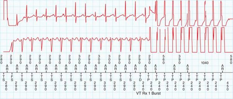
FIGURE 73-5 Initiation of ATP as indicated by TP (tachy pacing) on the marker channel. There is AV dissociation during ATP.
Atrial tachycardia occurs independent of the ventricle and therefore will not be affected by ventricular pacing, although it can terminate if retrograde conduction exists and the atrial rate is increased to the pacing rate during ATP.
ATP is also seen to accelerate the ventricular rate and slow the atrial rate (Figure 73-6), more consistent with ventricular tachycardia. This also suggests that there is a rate sensitive relationship between the VA conduction during VT. If this were SVT, the ventricular rate would have slowed in conjunction with the atrial rate.
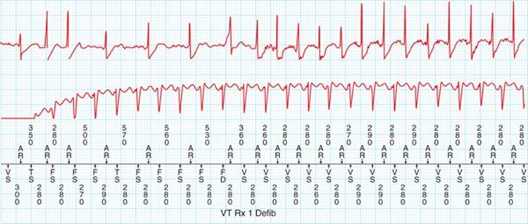
FIGURE 73-6 Termination of ATP with acceleration of the ventricular rate and initial slowing of the atrial rate.
• Termination: Rhythm stability and duration has been met, and the rhythm is terminated with a 26-J shock (Figure 73-7).

FIGURE 73-7 Termination of ventricular tachycardia with ICD shock.
This patient has ventricular tachycardia with 1:1 retrograde conduction.
MANAGEMENT AND PATIENT EDUCATION
This patient had appropriate detection of his ventricular tachycardia and received appropriate therapy. His programming should reflect changes for secondary prevention of ventricular tachycardia with a detection zone at least 40 ms slower than his slowest VT. The current programming provides this, so he does not require programming changes. The patient had no evidence of reversible causes of his ventricular tachycardia and no change in his baseline status. At this time he does not need further medical intervention. If he has recurrent events then he may be considered for antiarrhythmic therapy or ablation.
The patient must be advised that he is not to drive for a minimum of 6 months due to rapid, hemodynamically significant ventricular tachycardia.
FOLLOW-UP
The patient should continue to have routine scheduled office or remote follow-up with device checks every 3 months. This will allow for early identification in changes in the patient’s rhythm burden.
REFERENCES
1. Ellenbogen KA, Kay GN, Lau CP, Wilkoff BL. Clinical Cardiac Pacing, Defibrillation and Resynchronization Therapy. 4th ed. Philadelphia, PA: Elsevier; 2011.
2. Al-Ahmad A, Ellenbogen KA, Natale A, Wang PJ. Pacemakers and Implantable Cardioverter Defibrillators. Minneapolis, MN: Cardiotext; 2010.
3. Ellenbogen KE, Wood MA. Cardiac Pacing and ICDs. 5th ed. Hoboken, NJ: Blackwell; 2009.
4. Koneru JN, Swerdlow CD, Wood MA, Ellenbogen KA. Minimizing inappropriate or “unnecessary” implantable cardioverter-defibrillator shocks: appropriate programming. Circ Arrhythm Electrophysiol. 2011;4(5):778-790.