True heartburn (see also sections on indigestion, page 271 and anorexia, page 48) may be defined as a burning pain in the substernal area or midepigastrium, which is usually increased by swallowing and which is almost invariably due to esophagitis from gastric reflux. There are other causes, however, and the problem for the diagnostician is how best to recall these in the clinical situation.
From an etiologic standpoint inflammation is almost invariably the culprit, although myocardial infarction or angina pectoris are two frequent causes that are not inflammatory.
|
|
|
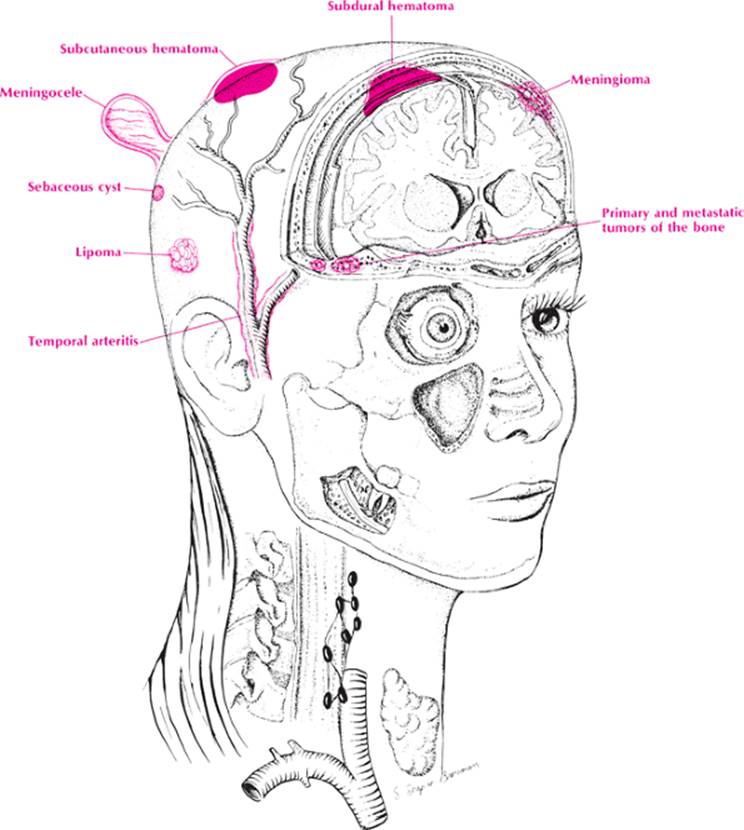
Head Deformities |
Anatomically, the best approach is to move in a targetlike fashion from the intrinsic portion of the esophagus and stomach peripherally. Thus, in the first zone, one encounters esophagitis, gastritis, and gastric ulcers. In the second zone, one encounters hiatal hernia (which, of course, predisposes to esophagitis), pericarditis, mediastinitis, and gastrojejunostomy complications. In the third zone, one visualizes cholecystitis (which probably induces a bile esophagitis), pancreatitis, myocardial infarction or coronary insufficiency, pleurisy, and intestinal obstruction. In the fourth zone, one recalls systemic diseases such as uremia, severe emphysema, cirrhosis, and congestive heart failure (CHF) (which probably causes gastritis or gastric ulcers).
|
|
|
Head mass |
Approach to the Diagnosis
The approach to the diagnosis of heartburn is similar to that of any GI complaint, but a few clinical tricks will help decide whether it is intrinsic or extrinsic, especially if the upper GI series is negative. Always order an esophagram. If the patient has the pain when in your office, administer a tablespoon or two of lidocaine (xylocaine viscous). If the patient gets relief in 5 to 10 minutes, the heartburn is probably caused by esophagitis. Further confirmation can be obtained by a Bernstein test. In this test, solutions of normal saline and 0.10 normal HCl are administered by intravenous tubing into the lower esophagus, alternating one with the other. If the patient invariably experiences pain when the 0.10 normal HCl is administered, esophagitis is confirmed. Esophagoscopy and gastroscopy will reveal most intrinsic lesions with certainty, but occasionally they are normal in esophagitis. Manometric studies of the esophagus are the best way to diagnose esophageal reflux. If the episodes are frequent but relatively brief, a trial of nitroglycerin may diagnose angina pectoris. Coronary insufficiency may also be confirmed by an exercise tolerance test. Cholecystogram and liver and pancreatic function studies may also be indicated.
|
|
|
Heartburn |
Other Useful Tests
1. Ambulatory pH monitoring (esophageal reflux)
2. Gallbladder sonogram (cholecystitis)
3. Thallium scan (coronary insufficiency)
4. Acid barium swallow (esophagitis)
5. Therapeutic trial of nitroglycerin (coronary insufficiency)
6. Holter monitoring (coronary insufficiency)
7. Coronary angiogram (coronary insufficiency)
Case Presentation #40
A 48-year-old black man complained of recurrent substernal burning pain radiating to his jaw for several months. He has a history of smoking 2 packs of cigarettes a day for 30 years and consumes alcohol in moderation.
Question #1. Utilizing your knowledge of anatomy and histology, what is your differential diagnosis?
The pain is only occasionally relieved by antacids but can be precipitated by exercise. Physical examination is unremarkable except for a blood pressure of 155/110 and grade two arteriosclerotic retinopathy.
Question #2. What is your diagnosis now?