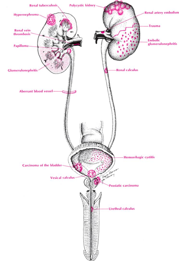
Using the anatomic approach, the physician can arrive at most of the causes of hematuria (Table 35). One need only visualize the urinary tract and proceed from the kidney on down to get a differential list. Let us apply the mnemonic VINDICATE to the kidney.
· V—Vascular diseases make one think of embolic glomerulonephritis, renal vein thrombosis, and (subacute bacterial endocarditis [SBE]).
· I—Infectious causes of hematuria are pyelonephritis (infrequently) and renal TB.
· N—Neoplasms that may present with hematuria are hypernephromas and papillomas and carcinomas of the renal pelvis. Wilms tumors present with hematuria less frequently.
· D—Degenerative diseases rarely present with hematuria as in other organ systems.
· I—Intoxicants such as sulfa drugs (that lead to nephrocalcinosis), mercury poisoning, and blood transfusion reactions are common causes of gross or microscopic hematuria.
· C—Congenital lesions such as polycystic kidneys and medullary sponge kidneys cause hematuria and predispose to stones and infections that may present with hematuria.
· A—Autoimmune conditions such as acute and chronic glomerulonephritis, Goodpasture disease, Wegener midline granulomatosis, and lupus erythematosus commonly present with hematuria.
|
|
|
Hematemesis and melena |
· T—Trauma to any organ causes hemorrhages, and the kidney is no exception. Hematuria after automobile or other accidents should signal the need for hospitalization, intravenous pyelogram (IVP), and close observation of vital signs. Hematuria may present with a crush injury to any muscle or a burn.
· E—Endocrine-metabolic diseases caused by stones. Most calcium stones are not caused by hyperparathyroidism, but it should always be considered a possibility. Urate stones are usually caused by gout, and cystine stones are always associated with congenital cystinuria.
|
|
|||||||||||||||||||||||||||||||||||||||||||||||||||||||||||||||||||||
|
Hematuria |
|||||||||||||||||||||||||||||||||||||||||||||||||||||||||||||||||||||
|
TABLE 35. Hematuria |
|||||||||||||||||||||||||||||||||||||||||||||||||||||||||||||||||||||
|
|||||||||||||||||||||||||||||||||||||||||||||||||||||||||||||||||||||
· Ureter. Stones, papillomas, and congenital defects (contributing to stones) are the most likely causes here.
· Bladder. Vascular disease is infrequently a cause, but cystitis (especially acute or “honeymoon” type) is a common cause. Stones, neoplasms (papillomas and transitional cell cardinomas), and foreign bodies are the next most likely causes. Trauma should not be forgotten, especially because of the numerous instances of various instruments being introduced into the bladder.
· Prostate. Neoplasms of the prostate occasionally cause hematuria, but most other etiologic conditions (prostatitis) are rarely associated with gross or microscopic hematuria.
· Urethra. Stones, neoplasms, and infections of the urethra may all cause hematuria, but very infrequently.
Using biochemistry as the basic chemistry, do not forget the coagulation disorders that may cause hematuria. Thus hematuria is often found in idiopathic thrombocytopenia purpura and in almost any disorder in which the platelet count drops below 40,000 cells/mm2. Hemophiliacs may present with hematuria. Patients given too much warfarin (Coumadin) will often get hematuria. Fibrinolysins and afibrinogenemia will also cause hematuria.
From this exercise, it should be evident that arriving at the causes of hematuria is not difficult if one visualizes the anatomy of the urinary tree and then considers each etiologic category in this light.
Approach to the Diagnosis
The clinical picture will point to the diagnosis in many cases. If there is a history of abdominal trauma, a contusion or laceration of the kidney or bladder should be suspected. Massive trauma anywhere prompts a tentative diagnosis of crush syndrome. Purpura or bleeding from other sites suggests a coagulation disorder. Severe colicky pain in the abdomen suggests kidney stone. A long history of hypertension suggests polycystic kidneys, renal artery stenosis, or glomerulonephritis. A history of fever and rheumatic valvular disease suggests SBE with renal embolism. Painless hematuria in an otherwise healthy looking adult suggests neoplasm, whereas painful hematuria with frequency and dysuria suggests cystitis. Hematuria and a flank mass would make a neoplasm or polycystic kidney likely.
The initial workup should include a CBC, urinalysis, urine culture, chemistry panel, flat plate of the abdomen to assess the presence of stones and kidney size, and personal examination of the urinary sediment. If a renal calculus is suspected, an IVP is ordered immediately and a urologist consulted. A three-glass test will help to localize the site of the bleeding. If there is blood in the initial specimen only, the urethra is probably the site of bleeding. If the blood is primarily in the final specimen, the bladder is most likely the site of bleeding. Equal blood discoloration in all specimens points to a renal lesion.
If renal TB is suspected, an acid-fast bacillus (AFB) smear and culture is done. If collagen disease is suspected, an ANA analysis and anti–double-strand DNA antibody titer is ordered.
If a renal carcinoma is suspected, a CT scan of the abdomen is probably the best study to order, but the advice of a urologist ought to be sought. Ultrasonography is useful in differentiating cysts from tumors. If a bladder neoplasm is suspected, cystoscopy will be done. If renal artery embolism or thrombosis is suspected, renal angiography may need to be done to clearly make the diagnosis.
Other Useful Tests
1. Chest x-ray (TB, Goodpasture disease)
2. Tuberculin test (TB)
3. Strain urine for stones
4. Serum complement (acute glomerulonephritis, lupus)
5. Anti-streptolysin O (ASO) titer (acute glomerulonephritis)
6. Addis count (glomerulonephritis)
7. Blood cultures (SBE)
8. Coagulation studies (hemophilia, collagen disease, allergic purpura)
9. Plasma haptoglobins (hemolytic anemias)
10. Coomb test (hemolytic anemias)
11. Platelet count (thrombocytopenic purpura)
12. Renal biopsy (chronic nephritis, neoplasm)
13. Surgical exploration
Case Presentation #42
A 31-year-old white man presents to the emergency room with severe right flank pain and a specimen of bloody urine. He is begging for a shot to relieve the pain.
Question #1. Utilizing your knowledge of anatomy of the urinary tract, what is your differential diagnosis?
The patient is given a shot of Demerol and admitted to the hospital. Physical examination aside from exquisite tenderness in the right flank is unremarkable. Complete workup including an IVP is negative. Repeated urinalysis reveals grossly bloody urine. However, on catheterization a normal specimen of urine is obtained.
Question #2. What is your diagnosis now?