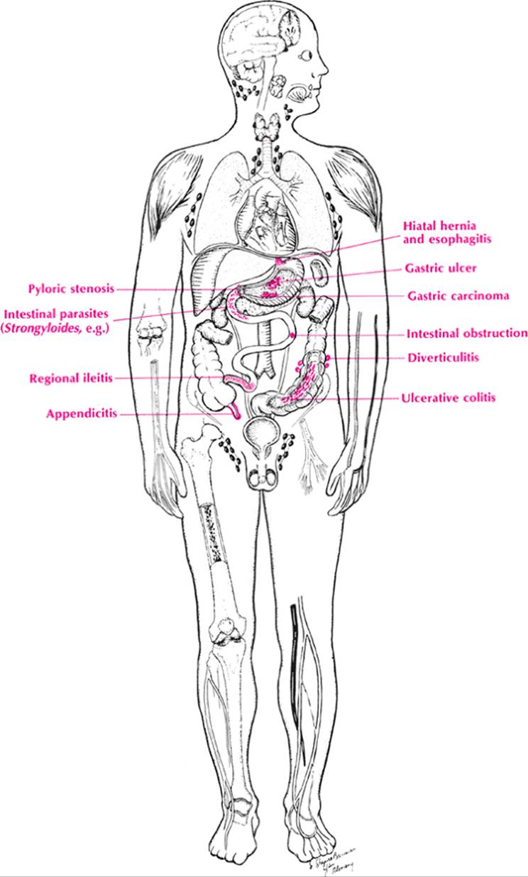
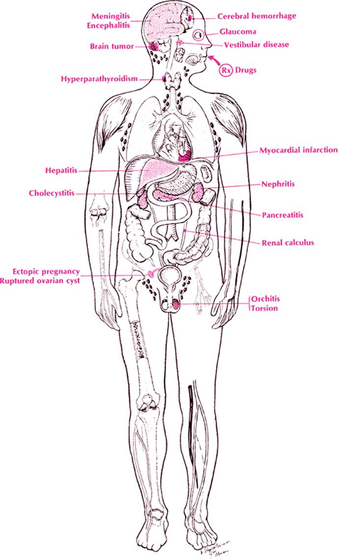
These two should be considered together, because nausea is just a forme fruste of vomiting. This symptom lends itself well to anatomicanalysis, particularly by the target method illustrated on page 324. The focus should be on the gastrointestinal (GI) tract. Starting from the top and working to the bottom, and at the same time cross-indexing this with etiologies (Table 46), one can review the most important causes of vomiting.
|
|
|
Nausea and vomiting |
In the nasopharynx, one encounters tonsillitis and foreign bodies. In the esophagus, achalasia, reflux esophagitis, and carcinoma are important, although they are more likely to produce dysphagia (see page 125). In the stomach, gastritis, gastric ulcers, and gastric carcinoma are important causes of vomiting. A polyp, carcinoma, or ulcer at the pylorus is most likely to produce vomiting because of gastric outlet obstruction. In children, one must not forget pyloric stenosis. In the duodenum, one must consider not only ulcers and duodenitis but also the afferent loop obstructions that occur after Billroth II surgery and the “dumping syndrome” in Billroth I and II surgery. Bile gastritis is also a cause. Intestinal obstruction from a variety of causes (e.g., volvulus, intussusception, malrotation, bezoar, carcinoma, and regional ileitis) must be considered in the jejunum and ileum. Parasites such as Strongyloides, Ascaris, and Taenia solium must also be considered in this part of the GI tract.
|
TABLE 46. Nausea and Vomiting |
||||||||||||||||||||||||||||||||||||||||||||||||||||||||||||||||||||||||||||||||||||||||||||||||||
|
||||||||||||||||||||||||||||||||||||||||||||||||||||||||||||||||||||||||||||||||||||||||||||||||||
An obstructed Meckel diverticulum or appendix may present with vomiting. In the large bowel, ulcerative colitis, amebiasis, and neoplasms should be considered. Mesenteric thrombosis can cause vomiting regardless of which portion of the intestine it involves. Acute viral or bacterial enteritis is associated with nausea and vomiting, but almost invariably there is diarrhea in botulism, salmonellosis, and shigellosis.
In the next circle in the target one encounters cholecystitis and cholelithiasis, pancreatitis, gastrinomas, pancreatic cysts, peritonitis, and myocardial infarction. In the next circle are the kidneys (e.g., renal stones), the thyroid, the pelvic organs (e.g., ectopic pregnancy), and the lungs (pneumonia with gastric dilatation). The next circle contains the vestibular apparatus (Ménière disease), the brain (e.g., tumor), and the testicles (e.g., torsion and orchitis).
The target method has served us well, but a biochemical evaluation of vomiting should also be done because many foreign substances or natural body substances occurring in high or low concentrations in the blood may affect the vomiting centers or cause a paralytic ileus. Thus uremia, increased ammonia and nitrogen breakdown products in hepatic disease, and hypokalemia and hyperkalemia may cause vomiting. Alterations in sodium, chloride, and CO2 may also cause vomiting. More important is hypercalcemia due to hyperparathyroidism or other causes.
In summary, vomiting is best analyzed anatomically. Physiologically, the symptoms of vomiting should suggest obstruction, either functional or mechanical. When all studies (see page 325) are normal, consider a neuropsychiatric disorder.
|
|
|
Nausea and vomiting, systemic causes |
Approach to the Diagnosis
The association of other symptoms and signs is essential in pinpointing the diagnosis of vomiting. For example, vomiting with tinnitus and vertigo suggests Ménière disease, whereas vomiting with hematemesis suggests gastritis, esophageal varices, and gastric ulcers. The laboratory workup should include a flat plate of the abdomen, upper GI series, esophagram, cholecystogram, gastric analysis, serum electrolytes, and amylase and lipase levels. Stools for occult blood, ova, and parasites are usually indicated. Gastroscopy and esophagoscopy are often indicated in the acute case, but an exploratory laparotomy should not be delayed if the patient's condition is deteriorating and pancreatitis has been excluded.
Other Useful Tests
1. CBC (anemia, infection)
2. Chemistry panel (liver disease, uremia)
3. Serial echocardiograms (ECGs) and cardiac enzymes (myocardial infarction)
4. Pregnancy test (ectopic pregnancy)
5. Arterial blood gases (pulmonary embolism)
6. Lung scan (pulmonary embolism)
7. Gallbladder sonogram (gallstones)
8. Small-bowel series (neoplasm, diverticulum, regional enteritis)
9. CT scan of the abdomen (neoplasm, abscess)
10. Laparoscopy (neoplasm of pancreas or liver)
11. Angiogram (mesenteric thrombosis)