|
TABLE 49. Paresthesias, Dysesthesias, and Numbness |
|||||||||||||||||||||||||||||||||||||||||||||||||||||||||||||||||||
|
|||||||||||||||||||||||||||||||||||||||||||||||||||||||||||||||||||
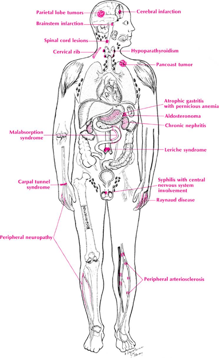
Anatomically, tingling and numbness or other abnormal sensations in the extremities result from involvement of the peripheral nerve, the nerve plexus (brachial or sciatic), the nerve root, the spinal cord, or the brain. When each of these is cross-indexed with the etiologies suggested by the mnemonic VINDICATE, most of the causes can be developed (Table 49). Only the most important conditions are mentioned in this discussion.
· Peripheral nerve. Peripheral neuropathies from alcohol, diabetes, and other causes are important in this category, but one should not forget vascular diseases that may cause paresthesias, such as peripheral arteriosclerosis, Raynaud syndrome, and Buerger disease. In addition, metabolic disorders such as tetany and uremia should be considered. Chronic acute inflammatory demyelinating polyneuropathy (Guillain–Barré syndrome) is brought to mind here. Finally, nerve entrapments such as carpal tunnel syndrome need to be checked.
· Nerve plexus. The brachial plexus may be involved by the scalenus anticus syndrome, a cervical rib, or Pancoast tumor. The sciatic plexus may be compressed by pelvic tumors.
· Nerve root. Herniated disks, spondylosis, tabes dorsalis, and infiltration of the spine by tuberculosis, metastatic tumor, and multiple myeloma need to be remembered here.
· Spinal cord. Spinal cord tumors, pernicious anemia, and tabes dorsalis are the most important conditions to recall here.
· Brain. Transient ischemic attacks (TIAs), emboli, and migraines are vascular diseases to remember in addition to the diseases that affect the spinal cord. The aura of epilepsy is also important. One would not want to miss brain tumors, abscesses, and toxic encephalopathy because these are potentially treatable.
Approach to the Diagnosis
This would be the same as the workup of weakness in one or more extremities. If the condition is in the hand, one would check for Tinel and Adson signs and x-ray the cervical spine for a cervical rib or disk degeneration. The next steps are nerve conduction studies and Electromyogram (EMG). Objective signs of radiculopathy are a clear indication for an MRI or cervical myelography, preferably combined with a CT scan. MRI may reveal tiny disk herniations. With associated pain in certain roots, diagnostic nerve blocks may be indicated. If there is coldness in the hand, a stellate ganglion block may be helpful.
If the condition is in the lower extremity, a careful examination of the arterial pulses, particularly the femoral, is performed. If these are abnormal, a flow study or femoral angiography may be indicated. X-rays of the spine to rule out a herniated disk or tumor of the spine are done routinely. One must not forget a pelvic examination in a female. If other neurologic signs are present, an MRI or CT scan may be necessary. When a disk herniation is still likely, myelography should be ordered. EMG has the same usefulness here as in the upper extremity. When a cerebral lesion is suspected, a CT scan, MRI, and four-vessel angiography should be considered.
Other Useful Tests
1. CBC (anemia)
2. Chemistry panel (hypoparathyroidism, electrolyte disturbance, uremia)
3. Fluorescent treponemal antibody absorption (FTA-ABS) test (neurosyphilis)
4. Serum B12 and folic acid levels (pernicious anemia)
5. Schilling test (pernicious anemia)
6. Blood lead level (lead neuropathy)
7. ANA analysis (collagen disease)
8. Glucose tolerance test (diabetic neuropathy)
9. Urine porphobilinogen (porphyria)
10. Hair analysis for arsenic
|
|
|
Paresthesias, dysethesias, and numbness |
|
|
|
Paresthesias, dysethesias, and numbness |
|
|
|
Paresthesias, dysethesias, and numbness |
11. Somatosensory evoked potentials (multiple sclerosis)
12. Spinal tap (neurosyphilis, multiple sclerosis)
13. Anticentromere antibody (scleroderma)
Case Presentation #72
A 25-year-old white male intern complained of intermittent numbness and tingling for several months of the lower extremities and, to a lesser extent, the upper extremities. He had occasional weakness in his left arm and hand but was told on an insurance examination that that was due to a scalenus anticus syndrome. He denies alcohol or substance abuse.
Question #1. Utilizing your knowledge of neuroanatomy, what is your differential diagnosis?
Further history reveals that he had an episode of optic neuritis at age 17. His neurologic examination reveals hyperactive reflexes of the left upper and lower extremities but is otherwise unremarkable.
Question #2. What is your diagnosis now?