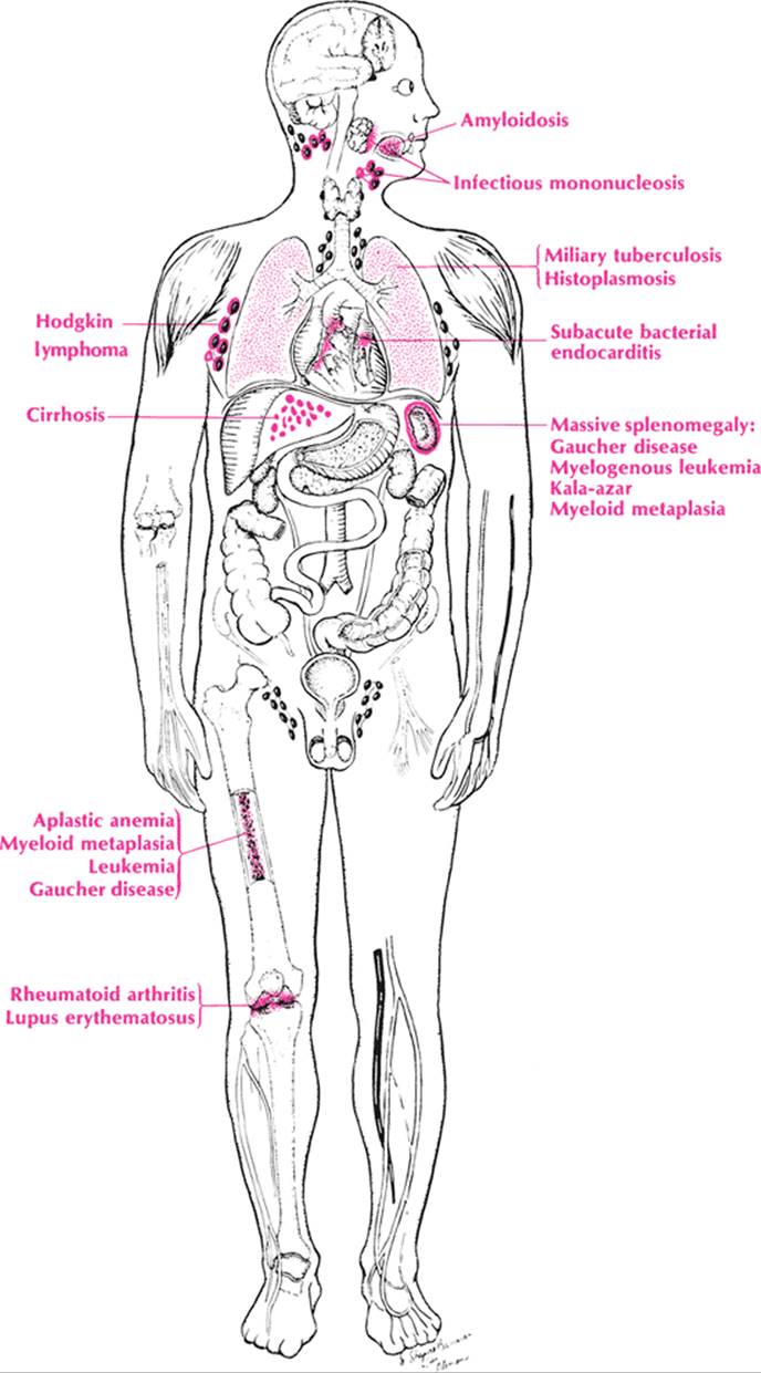
The patient is lying on the table and has a palpable mass in the left upper quadrant. The mass has a hard, smooth surface with a notch on the edge and descends on inspiration. The patient has an enlarged spleen. What can be done about it? What is causing it?
The key word is histology. Think about the histologic components: parenchyma, supporting tissue, arteries, veins, and a capsule. What is the parenchyma? It is nothing more than the components of the blood: red cells, white cells, lymph tissue, and platelets. Now it is possible to form a differential. Increased numbers of red cells recall polycythemia; increased numbers of white cells recall leukemia and infection. Increased lymph tissue suggests Hodgkin lymphoma, whereas increased supporting tissue indicates reticuloendotheliosis and acromegaly. Increased vein size occurs in obstruction of the portal vein as in cirrhosis, thrombosis of the portal vein, and CHF. If the artery has a local increase in size, an aneurysm forms, compressing the splenic veins.
A differential is at hand, but it is still incomplete. Now think of physiology. The spleen is a reserve for blood storage. It is also able to form red cells and other components of the blood when the bone marrow is atrophied, as in extramedullary erythropoiesis. More important, it is involved in the destruction of old or damaged red cells and platelets. Finally, it hypertrophies to fight infection just like the lymph glands. Extramedullary erythropoiesis recalls the splenomegaly of aplastic anemia and myeloid metaplasia, just as destruction or sequestration of cells brings to mind the splenomegaly of hemolytic anemias (e.g., hereditary spherocytosis, malaria, and lupus erythematosus) and thrombocytopenic purpura. The hypertrophy to fight infection or diffuse inflammation of the body should suggest the splenomegaly of bacterial endocarditis, kala-azar, infectious mononucleosis, miliary tuberculosis, and rheumatoid arthritis. Almost anything that causes generalized lymphadenopathy can cause splenomegaly.
Only one category of splenomegaly is not brought to mind by this approach, but it is easily remembered because it is an exception—infiltration of inert material. Thus, in gargoylism there is a foreign mucopolysaccharide in the spleen. Numerous mucopolysaccharidoses are now described in the literature. There is a buildup of lipids in the reticuloendotheliosis of Gaucher disease, Niemann–Pick disease, and Hand–Schüller–Christian disease, but these are intracellular. Amyloid may infiltrate the spleen. Metastatic carcinoma of the spleen is rare.
Table 52 summarizes the above discussion and gives additional causes of splenomegaly to consider in the differential. One final diagnosis to consider is traumatic splenomegaly.
Approach to the Diagnosis
How does one go about pinning down the diagnosis? There are several clinical clues. One looks during the physical examination for jaundice, lymphadenopathy, a rash, sore throat, hepatomegaly, and a positive Rumpel–Leede test. The combination of symptoms and signs will eliminate certain causes and make others more plausible. For example, splenomegaly with jaundice but no hepatomegaly suggests hemolytic anemia. The size of the spleen is also an important differential feature. If the spleen is very large, it should suggest myeloid metaplasia, chronic myelogenous leukemia, Gaucher disease, and kala-azar.
|
|
|
Splenomegaly |
The laboratory is the principal aid from this point on. Smears for red cell morphology, malaria, and other parasites are invaluable. Blood cultures and a lymph node and bone biopsy may be useful. If a specific disease is strongly suspected, consult Appendix A for appropriate tests.
Other Useful Tests
1. CBC and differential (anemia, leukemia)
2. Blood smear for morphology (anemia)
3. Reticulocyte count (hemolytic anemia)
4. Platelet count and clot retraction (thrombocytopenia)
5. Radioactive chromium–tagged red cell (hemolytic anemia)
|
TABLE 52. Splenomegaly |
|||||||||||||||||||||||||||||||||||
|
|||||||||||||||||||||||||||||||||||
6. Serum haptoglobins (hemolytic anemia)
7. Bone marrow examination (aplastic anemia)
8. Blood cultures (SBE)
9. Febrile agglutinins (infectious disease)
10. Heterophil antibody titer (infectious mononucleosis)
11. Brucellin agglutinins (brucellosis)
12. Blood smear for parasites (malaria, trypanosomiasis)
13. Liver function studies (cirrhosis, Banti syndrome)
14. Rheumatoid arthritis test (Felty syndrome)
15. ANA test (collagen disease)
16. Serum protein electrophoresis (lymphoma, collagen disease)
17. Hemoglobin electrophoresis (hemolytic anemia)
18. Esophagram (esophageal varices) (portal cirrhosis)
19. X-ray of long bones (Gaucher disease, metastasis)
20. Flat plate of abdomen for spleen size (splenomegaly)
21. Lymph node biopsy (Hodgkin lymphoma)
22. Liver biopsy (cirrhosis)
23. Splenic aspirate (lymphoma, leukemia)
24. Splenoportogram and splenic pulp pressure (portal cirrhosis)
25. Purified protein derivative (PPD) test, intermediate, and skin tests for various fungi (see Table 36)
26. Skin biopsy (hemochromatosis)
27. Muscle biopsy (collagen disease, trichinosis)
28. Diagnostic ultrasound (cyst, splenic aneurysm)
29. CT scan (malignancy)
30. Liver–spleen scan (splenomegaly)
|
TABLE 53. Sputum |
|||||||||||||||||||||||||||||||||||||||||||||||||||||||||||
|
|||||||||||||||||||||||||||||||||||||||||||||||||||||||||||