Abdominal Pain, Generalized
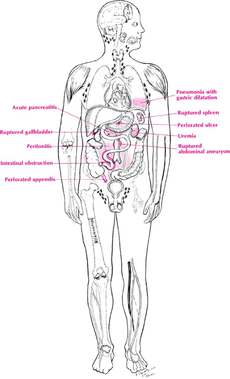
The GI tract is the only “organ” that really covers the abdomen from one end to the other. Anything that causes an irritation of all or a large portion of this “tube” may cause generalized abdominal pain. Thus, gastritis, viral and bacterial gastroenteritis, irritable bowel syndrome, ulcerative colitis, and amebic colitis fall into this category. The remainder of the causes of generalized abdominal pain can be developed by using the mnemonic ROS with the anatomy of the entire abdomen.
When faced with a patient with diffuse abdominal pain, think of R for ruptured viscus. Now take each organ and consider the possibility of its having ruptured. Thus, the stomach and duodenum suggest a ruptured peptic ulcer; the pancreas, an acute hemorrhagic pancreatitis; the gallbladder, a ruptured cholecystitis. The liver and spleen usually rupture from trauma, whereas the fallopian tube may rupture from an ectopic pregnancy. The colon ruptures from diverticulitis, ulcerative colitis, or carcinoma. What is the one thing that should make the physician suspect a ruptured viscus? Rebound tenderness is the answer. In addition, one or both testicles may be drawn up (Collins sign). If only the right testicle is drawn up, suspect a ruptured appendix or peptic ulcer. If only the left is drawn up, suspect a ruptured diverticulum. If both are drawn up, suspect pancreatitis or a generalized peritonitis.
Now take the letter O. This signifies intestinal obstruction. Think of adhesion hernia, volvulus, paralytic ileus, intussusception, fecal impaction, carcinoma, mesenteric infarction, regional ileitis, and malrotation. The best way to recall all these is with the mnemonic VINDICATE.
|
|
|
Abdominal pain, generalized |
Next take the letter S. This signifies the systemic diseases that may irritate the intestines, the peritoneum, or both. Once again the mnemonic VINDICATEwill remind one to recall the important offenders.
· V—Vascular suggests the anemias, congestive heart failure (CHF), coagulation disorders, and mesenteric artery occlusion, embolism, or thrombosis.
· I—Inflammatory includes tuberculous, gonococcal and pneumococcal peritonitis, and trichinosis.
· N—Neoplasms should suggest leukemia and metastatic carcinoma.
· D—Deficiency might suggest the gastroenteritis of pellagra.
· I—Intoxication reminds one of lead colic, uremia, and the venom of a black widow spider bite.
· C—Congenital suggests porphyria and sickle cell disease.
· A—Autoimmune brings to mind periarteritis nodosa, rheumatic fever, Henoch–Schönlein purpura, and dermatomyositis.
· T—Trauma would suggest the paralytic ileus of trauma anywhere, the crush syndrome, and hemoperitoneum.
· E—Endocrine disease suggests diabetic ketoacidosis, addisonian crisis, and hypocalcemia.
Approach to the Diagnosis
If the onset is acute, a general surgeon should be consulted at the outset. Ominous signs include boardlike rigidity, rebound tenderness, and shock with nausea and vomiting. Hyperactive bowel sounds of a high-pitched tinkling character with distention and obstipation suggest intestinal obstruction. In contrast, normal bowel sounds, little distention, good vital signs, and minimal tenderness suggest gastroenteritis or other diffuse irritation of the bowel.
It is wise to pass a nasogastric tube and attach to suction and proceed with a CBC, urinalysis, an immediate flat plate and upright of the abdomen, chest x-ray, serum amylase and lipase levels, and chemistry panel. Sometimes, lateral decubitus films are necessary to reveal the stepladder pattern of intestinal obstruction. A pregnancy test should be ordered if age and gender dictates it.
If these tests fail to confirm the clinical diagnosis and the patient's condition is deteriorating, it is probably wise to proceed immediately with an exploratory laparotomy. If the patient's condition is stable, one may order more diagnostic tests depending on the location of the pain and other symptoms and signs. For example, if the pain seems more localized to the RUQ, a gallbladder ultrasound or nuclear scan may be ordered. If it is still considered generalized, perhaps a CT scan of the abdomen and pelvis is indicated. Monitoring vital signs and doing repeated CBCs, serum amylase levels, and flat plates of the abdomen are useful in borderline cases.
Other Useful Tests
1. Quantitative urine amylase level
2. Four-quadrant peritoneal tap (peritonitis, pancreatitis, ruptured ectopic)
3. Urine porphobilinogen (porphyria)
4. IVP (renal calculus)
5. Serial cardiac enzymes (mycardial infarct)
6. Serial electrocardiograms (ECGs)
7. Double enema (intestinal obstruction)
8. Esophagoscopy (reflux esophagitis)
9. Gastroscopy (peptic ulcer)
10. Colonoscopy (diverticulitis, carcinoma)
11. Laparoscopy (ruptured viscus, PID)
12. Culdocentesis (ruptured ectopic pregnancy)
13. Pelvic sonogram (ruptured ectopic pregnancy)
14. Angiogram (mesenteric thrombosis)
15. Breath test or stool tests for Helicobacter pylori (peptic ulcer)
Right Upper Quadrant Pain
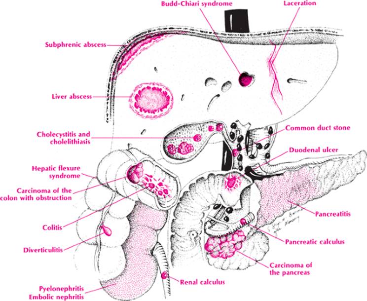
The patient is complaining of RUQ pain and you cannot just give him or her a bag of pills and send him or her home. The patient's condition may be serious. However, you are in a hurry to get out of the office because you have another important appointment. What do you do? The key is to visualize the anatomy.Imagine the liver, gallbladder, bile ducts, hepatic flexure of the colon, duodenum, and head of the pancreas. Surrounding these are the skin, fascia, ribs, and thoracic and lumbar spine, with the intercostal nerves and arteries and abdominal muscle.
Pain is usually from inflammation, trauma, or infarction. The patient gives no history of trauma, but he or she could have a contusion of the muscle from coughing hard. That is not likely, however, unless the patient has other symptoms of the respiratory tract.
The possible sources of inflammation should be narrowed down first. The liver can be inflamed from hepatitis (most likely viral), the gallbladder from cholecystitis (most likely induced by stones and bacteria), or the bile ducts from cholangitis. The colon may be involved with diverticulitis, a segment of granulomatous colitis, or perhaps there is a retrocecal appendix. The duodenum, of course, would most likely have a peptic ulcer which could cause an obstruction or a perforation if the patient is vomiting, or pallor and shock if the patient is bleeding. The pancreas could be inflamed with pancreatitis, especially if the patient drinks alcohol.
These are the most important intra-abdominal considerations, but if the mnemonic VINDICATE in Table 7 were applied one might not forget the Budd–Chiari syndrome (thrombosis of the hepatic veins), portal vein thrombosis, or pyelophlebitis; these are rare. In addition, toxic hepatitis from isoniazid, thorazine, and erythromycin estolate (Ilosone), for example, can be painful. Collagen diseases affecting the liver are another possibility.
Now let us round out the differential with extra-abdominal disorders. The skin may be involved with herpes zoster or cellulitis. A fascial rentmay cause a hernia, particularly if there was previous upper abdominal surgery. Compression of the nerve roots by a herniated disc, thoracic spondylosis, or a spinal cord tumor is possible, but unlikely. Systemic conditions, such as lead colic and porphyria, and involvement of another organ, such as the kidney, must be considered (pyelonephritis or renal colic).
|
|
|
Abdominal pain, right upper quadrant |
Approach to the Diagnosis
As in the case of generalized abdominal pain, an immediate CBC, urinalysis, chemistry profile, serum amylase and lipase levels, and flat plate and upright films of the abdomen are ordered. If cholecystitis is suspected, ultrasonography or nuclear scanning of the gallbladder (hepatoiminodiacetic acid [HIDA] scan) is ordered. If there is jaundice, a common duct stone can be ruled out by endoscopic retrograde cholangiopancreatography (ERCP).
Other Useful Tests
1. Surgery consult
2. CT scan of the abdomen
3. Quantitative urine amylase
4. Urine porphobilinogen (porphyria)
5. Gallium scan (subphrenic abscess)
6. IVP (renal stone)
7. Liver function studies (common duct stone)
8. Blood lead level
9. Pregnancy test (ruptured ectopic pregnancy)
10. X-ray of thoracolumbar spine (radiculopathy)
11. Laparoscopy (ruptured viscus)
12. Aortogram (dissecting aneurysm)
13. Lymphangiogram (Hodgkin lymphoma)
14. Exploratory laparotomy
Case Presentation #4
A 38-year-old obese white woman complained of RUQ pain, nausea and vomiting of 2 days duration.
Question #1. Utilizing the methods applied above, what is your list of possible causes at this point?
Further history reveals the pain is colicky, she is the mother of four children and had a few similar attacks in the past 5 years but never this severe. Examination reveals icteric sclera, tenderness, and rebound in the RUQ but no mass or hepatomegaly.
Question #2. What is your list of possibilities at this point?
|
TABLE 7. Right Upper Quadrant Pain |
||||||||||||||||||||||||||||||||||||||||||||||||||||||||||||||||||||||||||||||||||||||||||||||||||||||||||||||||||||||||||||||||||||||||||||||||||||||||
|
||||||||||||||||||||||||||||||||||||||||||||||||||||||||||||||||||||||||||||||||||||||||||||||||||||||||||||||||||||||||||||||||||||||||||||||||||||||||
Left Upper Quadrant Pain
Anatomy is the key to recalling the many causes of abdominal pain in the LUQ by visualizing the structures layer by layer. In the first layer are the skin, abdominal wall, and ribs; in the second layer, the spleen, colon, and stomach; and in the third layer, the pancreas, adrenal gland, kidney, aorta, and spine. Now it is possible to cross-index the organs with the various etiologies contained in the mnemonic VINDICATE (Table 8). The following discussion emphasizes the most important of these.
1. Abdominal wall and ribs. Pain will occur most commonly from herpes zoster, contusion, hernia, rib fracture, or metastatic tumor.
2. Spleen. Painful splenic infarcts are not unusual in subacute bacterial endocarditis (SBE), polycythemia, sickle cell anemia, leukemia, periarteritis nodosa, and other autoimmune disorders. A ruptured spleen is an important consideration in abdominal injuries, particularly those in children and in patients with infectious mononucleosis.
3. Stomach. Acute gaseous distention of the stomach in gastritis, pneumonia, and pyloric obstruction is a common cause of LUQ pain. Gastric carcinoma that extends beyond the wall of the stomach may cause pain. Episodic obstruction of the stomach in the “cascade stomach” should be considered in the differential diagnosis. Herniation of the stomach through the diaphragm occasionally causes LUQ pain.
|
|
|
Abdominal pain, left upper quadrant |
4. Colon. An inflamed diverticulum or an inflamed splenic flexure from granulomatous colitis may cause pain in the LUQ. Less commonly, the colon develops a perforating or constricting carcinoma in this area, which obstructs the bowel. A mesenteric infarct of the colon, as well as gas or impacted feces in the splenic flexure, may also cause LUQ pain.
5. Pancreas. Acute pancreatitis, pancreatic pseudocyst, and carcinoma of the pancreas may cause LUQ pain.
6. Adrenal gland. Adrenal infarction from emboli or Waterhouse–Friderichsen syndrome may cause pain, but neoplasms rarely do until they have become massive.
7. Kidney. Renal infarct, renal calculus, acute pyelonephritis, and nephroptosis with a Dietl crisis may cause LUQ pain. Perinephric abscess must also be considered.
8. Aorta. Dissecting or atherosclerotic aneurysms of the aorta may cause LUQ pain, especially when they occlude a feeding artery to one of the structures there.
9. Spine. Herniated disc, tuberculosis, multiple myeloma, osteoarthritis, tabes dorsalis, spinal cord tumor, and anything else that may compress or irritate the intercostal nerve roots can cause LUQ pain.
Approach to the Diagnosis
The presence or absence of other symptoms and signs will be most helpful in the diagnosis. In acute cases, a surgeon is consulted and a flat plate of the abdomen, CBC, urinalysis, and perhaps a serum amylase level should be done. If necessary, a CT scan of the abdomen is also done. Gastroscopy and colonoscopy may be desirable before other x-rays are done. In chronic cases, however, an upper GI series, barium enema, and stool examination for blood, ova, and parasites are indicated.
Other Useful Tests
1. Four-quadrant peritoneal tap (ruptured spleen)
2. Quantitative urine amylase
3. IVP (renal calculus)
4. Stool for occult blood (carcinoma, diverticulitis)
5. Gallium scan (diverticulitis, etc.)
6. X-ray of thoracolumbar spine (radiculopathy)
7. Small-bowel series (Meckel diverticulum)
8. Laparoscopy (ruptured viscus or peritonitis)
9. Aortogram (dissecting aneurysm)
10. Lymphangiogram (retroperitoneal sarcoma)
11. Exploratory laparotomy
|
|
|
Abdominal pain, right lower quadrant |
Right Lower Quadrant Pain
Most cases of acute RLQ pain are considered appendicitis until proven otherwise, but every physician has been fooled by this axiom more times than he or she would like to remember. For this reason, the astute clinician will want to have a good list of possibilities in mind. Anatomy is the key to recalling an inclusive list of causes of all RLQ pain. Visualizing the structures, layer by layer, one finds the skin and abdominal wall in the first layer; the terminal ileum, cecum, appendix, and Meckel diverticulum in the second layer; the ureters, tubes, and ovaries (in women) in the third layer; and the muscles, spine, and terminal aorta in the fourth layer. Now the organs can be cross-indexed with the various etiologies that may be encountered by using the mnemonic VINDICATE (Table 9). The following discussion emphasizes the most important diseases in the differential diagnosis.
|
TABLE 8. Left Upper Quadrant Pain |
|||||||||||||||||||||||||||||||||||||||||||||||||||||||||||||||||||||||||||||||||||||||||||||||||||||||||||||
|
|||||||||||||||||||||||||||||||||||||||||||||||||||||||||||||||||||||||||||||||||||||||||||||||||||||||||||||
1. Skin and abdominal wall. Herpes zoster, cellulitis, contusion, and especially inguinal or femoral hernias are significant causes of RLQ pain.
2. Appendix. Appendicitis is a major cause of RLQ pain.
3. Terminal ileum. Regional ileitis, tuberculosis, or typhoid and intussusceptions may involve the ileum and cause severe pain. Mesenteric adenitis and infarcts may also affect the ileum.
4. Cecum. Diverticulitis, colitis (e.g., granulomatous or amebic), and colon carcinoma are culprits that may cause RLQ pain originating in the cecum. Impacted feces are also a possible cause.
5. Meckel diverticulum. This congenital anomaly may become obstructed and inflamed, develop a pancreatitis or a perforated peptic ulcer, or communicate with a periumbilical cellulitis. All of these may cause RLQ pain.
6. Ureters. Renal calculi and hydronephrosis may cause RLQ pain.
7. Ovary and fallopian tubes. A mumps oophoritis may cause pain in the RLQ. Ovarian cysts may twist on their pedicles or rupture, causing pain, as may the rupture of a small graafian follicle in the normal cycle (mittelschmerz). Three significant lesions may involve the tube: salpingitis, endometriosis, and ectopic pregnancy. All three are painful.
8. Aorta. Dissecting aneurysms or emboli of the terminal aorta and its branches may seize the patient with acute pain.
|
TABLE 9. Right Lower Quadrant Pain |
|||||||||||||||||||||||||||||||||||||||||||||||||||||||||||||||||||||||||||||||||||||||||||||||||||||||||||||
|
|||||||||||||||||||||||||||||||||||||||||||||||||||||||||||||||||||||||||||||||||||||||||||||||||||||||||||||
9. Pelvis and spine. Osteoarthritis, ruptured disc, metastatic carcinoma, Pott disease, and rheumatoid spondylitis should be considered here.
10. Miscellaneous structures. A ruptured peptic ulcer or inflamed gallbladder may leak fluid into the right colic gutter and cause RLQ pain. Any of the numerous causes of intestinal obstruction (e.g., adhesions or volvulus) may cause pain. Omental infarcts are another miscellaneous cause. Referred pain from pneumonia or pulmonary infarct has encouraged some surgeons to insist on a chest x-ray prior to surgery.
Approach to the Diagnosis
Obviously, acute RLQ pain is suspected to be acute appendicitis until proven otherwise. However, it is wise to order flat plate and upright films of the abdomen, CBC, urinalysis, and an amylase level before surgery to dodge a curveball. Some surgeons want a chest x-ray as well, because pneumonia and other chest conditions can present with RLQ pain. A pregnancy test should be ordered for women of childbearing age to help rule out a ruptured ectopic pregnancy, but ultrasonography is even better. Surprisingly, many patients get to the operating room without a rectal or vaginal examination.
In cases of chronic RLQ pain, contrast studies such as a barium enema, IVP, upper GI series, and cholecystogram may be indicated. If these are not diagnostic, further investigation with colonoscopy, cystoscopy, culdoscopy, or laparoscopy may be needed. A CT scan of the abdomen and pelvis can often reveal the diagnosis.
Other Useful Tests
1. Stool for occult blood (mesenteric thrombosis, neoplasm)
2. Stool for ova and parasites
3. Gallium or indium scan (diverticulitis, abscess)
4. Angiogram (mesenteric thrombosis)
5. X-ray of lumbar spine (herniated disc, etc.)
6. Urine culture, sensitivity, and colony count
7. Chemistry panel
8. Sedimentation rate (inflammation)
9. Lymphangiogram (Hodgkin lymphoma)
10. Urine porphobilinogen (porphyria)
11. Small-bowel series (Meckel diverticulum)
12. Blood lead level
Left Lower Quadrant Pain
The anatomy of the LLQ, like that of the RLQ, provides a basis for recalling the causes of pain. There are fewer structures to deal with; thus, the differential diagnosis is not difficult. Visualizing the structures layer by layer, there are the skin and abdominal wall in the first layer; the sigmoid colon, omentum, and portions of small intestine in the second layer; the ureter, fallopian tubes, and ovaries (in women) in the third layer; and the aorta, pelvis, and spine beneath all these structures. Now, by using the mnemonic VINDICATE, the organs can be cross-indexed with the various etiologies that may cause pain in this area (Table 10). The following discussion emphasizes the most important diseases that must be considered in the differential diagnosis.
|
TABLE 10. Left Lower Quadrant Pain |
|||||||||||||||||||||||||||||||||||||||||||||||||||||||||||||||||||||||||||||||||||||||||
|
|||||||||||||||||||||||||||||||||||||||||||||||||||||||||||||||||||||||||||||||||||||||||
1. Skin and abdominal wall. Herpes zoster, cellulitis, contusion, and, especially, inguinal or femoral hernias are significant causes of LLQ pain.
2. Small intestine. Regional ileitis, intussusception, adhesion, volvulus, and other conditions that cause intestinal obstruction should be considered here.
3. Sigmoid colon. Diverticulitis, ischemic colitis, mesenteric adenitis and infarct, and granulomatous colitis are important causes. Carcinoma of the sigmoid may induce pain by perforating or obstructing the colon.
4. Ureters. Ureteral colic must be considered in the differential diagnosis of LLQ pain.
5. Ovary and fallopian tubes. A mumps oophoritis, ovarian cysts that twist on their pedicles or rupture, and small graafian follicles of the normal cycle that rupture are all included in the differential diagnosis of LLQ pain. The tubes may cause pain if there is an ectopic pregnancy, if they are inflamed by a salpingitis, or if they are infiltrated by endometriosis.
6. Aorta. Dissecting aneurysms and emboli of the terminal aorta may cause acute lower quadrant pain.
7. Pelvis and spine. Osteoarthritis, a ruptured disc, metastatic carcinoma, Pott disease, and rheumatoid spondylitis should be considered here.
8. Miscellaneous. Occasionally, pain in the bladder, prostate, or uterus is referred to the LLQ. A fibroid of the uterus may twist and cause pain. Impacted feces may cause severe pain. Referred pain from pneumonia, pleurisy, and myocardial infarction is uncommon but must be considered. Metabolic conditions that cause generalized abdominal pain and that should be remembered are listed on page 16.
Approach to the Diagnosis
There is no doubt about the value of a good history and physical examination, including both the rectal and pelvic areas. After this, the signs and symptoms should be summarized and grouped together; in many cases, this technique will pinpoint the diagnosis.
The laboratory workup can now proceed. In acute cases, the physician should order a flat plate of the abdomen, CBC, urinalysis (and examine it him- or herself), and serum amylase level before exploratory surgery. A pregnancy test is ordered in women of childbearing age. In chronic cases, sigmoidoscopy, barium enema, upper GI series, small-bowel follow-through, and stool examination for blood, ova, and parasites should be done before culdoscopy, peritoneoscopy, or colonoscopy is contemplated. An exploratory laparotomy remains a useful diagnostic tool even in chronic cases of LLQ pain.
Other Useful Tests
1. CT scan of the abdomen and pelvis
2. Gallium or indium scan (diverticular abscess, tuboovarian abscess)
3. Sonogram (ruptured ectopic pregnancy)
4. IVP
5. Examination of all urine for stones
6. Vaginal culture
7. Stool culture
8. Urine culture, sensitivity, and colony count
9. X-ray of lumbar spine (herniated disc, radiculopathy)
10. Peritoneal tap (ruptured ectopic pregnancy)
11. Aortogram (dissecting aneurysms)
12. Angiogram (mesenteric infarction)
13. Exploratory laparotomy
|
|
|
Abdominal pain, left lower quadrant |
Case Presentation #5
A 25-year-old white woman complained of the sudden onset of LLQ pain and occasional nausea and vomiting on the day of admission.
Question #1. Utilizing the methods described above, what are the possible causes of this patient's condition at this point?
Further history reveals she had intermittent vaginal bleeding for 2 weeks and she was treated for a vaginal discharge several months ago. Vaginal examination revealed a tender adnexal mass.
Question #2. What are the diagnostic possibilities now?
Epigastric Pain
By mental dissection of the epigastrium layer by layer from the skin to the thoracolumbar spine, one encounters all the important organs that are the sites of origin of epigastric pain (Table 11). Anatomy, therefore, is the basic science used to develop this differential diagnosis.
The skin may be the site of the pain in herpes zoster, as it is in other types of pain, although it is less likely to be midline. Cellulitis and other lesions of the skin will be readily apparent. However, muscle and fascial conditions may be missed if one does not specifically think of this layer. Thus, epigastric hernia, hiatal hernia, or contusion of the muscle will be missed, as will diaphragmatic abscesses and trichinosis of the diaphragm.
The stomach and duodenum are the next organs encountered; both are prominent causes of epigastric pain. Ulcers, especially perforated ulcers, cause severe pain. Gastritis (syphilitic, toxic, or atrophic) causes a milder form of pain. Pyloric stenosis (from whatever cause), cascade stomach, diverticula, and carcinoma or sarcoma round out the differential diagnosis here. Good collateral circulation makes vascular occlusion a less likely cause.
|
|
|
Abdominal pain, epigastric |
The colon and small intestines lie just below the stomach, so one must not forget ileitis, colitis (ulcerative or granulomatous), appendicitis, diverticulitis, Meckel diverticulum, and transverse colon carcinoma that ulcerates through the wall. Intestinal parasites and mesenteric thrombosis are additional causes that originate here. The various forms of intestinal obstruction are more important than parasites and mesenteric thrombosis.
The pancreas sits at the next layer, and acute pancreatitis is a particularly severe form of epigastric pain. Chronic pancreatitis, carcinoma, cysts of the pancreas, and mucoviscidosis cause less severe forms of epigastric pain. The lymph nodes may be involved by Hodgkin lymphoma and lymphosarcoma, leading to intestinal obstruction, but mesenteric adenitis is a much more likely cause. When the retroperitoneal nodes are involved by neoplasms (e.g., sarcoma), the pain is usually referred to the back.
The blood vessels are contained in the next layer, and one is reminded of aortic aneurysm, abdominal angina, periarteritis nodosa, and other forms of vasculitis. The sympathetic and parasympathetic nerves are involved by lead colic, porphyria, and black widow spider venom. Conditions of the thoracic spineare present in the final layer. Cord tumor, tuberculosis, herniated disc, osteoarthritis, and rheumatoid spondylitis can all lead to midepigastric pain.
Omission of the systemic diseases and diseases of other abdominal organs that sometimes cause epigastric pain is inexcusable. Pneumonia, myocardial infarction (inferior wall, particularly), rheumatic fever, epilepsy, and migraine are just a few systemic conditions that are associated with epigastric or generalized abdominal pain.
|
TABLE 11. Epigastric Pain |
|||||||||||||||||||||||||||||||||||||||||||||||||||||||||||||||||||||||||||||||||||||||||||||||||||||||||||||||||||||||||||||||||||||||||||
|
|||||||||||||||||||||||||||||||||||||||||||||||||||||||||||||||||||||||||||||||||||||||||||||||||||||||||||||||||||||||||||||||||||||||||||
Cholecystitis, hepatitis, and pyelonephritis are some local diseases that also produce midepigastric or generalized abdominal pain, which is why the target system has a useful application here. The center circle of the target is the stomach, the pancreas, and other organs in Table 11. The next circle covers the liver, kidney, gallbladder, heart, and ovaries. A further circle covers the brain and the testicles.
Approach to the Diagnosis
The approach to the diagnosis of midepigastric pain is identical to that for generalized abdominal pain (see page 16).
Hypogastric Pain
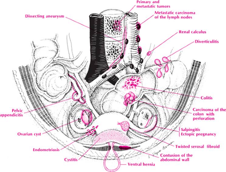
Anatomy is the basic science that will open the door to this differential diagnosis. Visualizing the structures in the hypogastrium, one sees the abdominal wall, the bladder and urinary tract, the female genital tract, the sigmoid colon and rectum, the iliac vessels, the aorta and vena cava, and the lumbosacral spine. Occasionally, other organs fall into the hypogastrium, thus they must be considered too. A pelvic kidney, visceroptosis of the transverse colon, and a pelvic appendix all may occur. Now that one has the organs in mind it is necessary only to apply the mnemonic MINT to recall the causes of hypogastric pain.
|
|
|
Abdominal pain, hypogastric |
In the abdominal wall,
· M—Malformations bring to mind ventral hernias and urachal cysts or sinuses with associated cellulitis.
· I—Inflammation includes cellulitis, carbuncle, and other skin infections.
· N—Neoplasms of the abdominal wall do not usually present with pain.
· T—Trauma suggests contusion of the rectus abdominus muscles or stab wound.
In the urinary tract, one recalls
· M—Malformations such as diverticulum, cystocele, ureterocele, bladder neck obstruction from stricture and calculus, and phimosis and paraphimosis.
· I—Inflammation suggests cystitis, prostatitis and urethritis, and Hunner ulcers.
· N—Neoplasms suggest transitional cell papilloma and carcinoma and prostate carcinoma.
· T—Trauma recalls ruptured bladder.
In the female genital tract,
· M—Malformations that may cause pain include a retroverted uterus, an ectopic pregnancy, and various congenital cysts (e.g., hydatid cyst of Morgagni) that may twist on their pedicles.
· I—Inflammation of the vagina and cervix is not usually painful except on intercourse, but endometritis and tubo-ovarian abscesses are associated with pain and fever.
· N—Neoplasms such as carcinoma of the cervix and uterus do not cause pain unless they extend beyond the uterus or obstruct the menstrual flow. However, fibroids often cause dysmenorrhea and severe pain if they twist on their pedicles, and endometriosis may spread throughout the pelvis and cause chronic or acute pain.
· T—Trauma such as perforation of the uterus during a dilatation and curettage (D & C), delivery, or by the introduction of a foreign body during sexual relations may cause abdominal pain.
The sigmoid colon and rectum may be the site of pain in:
· M—Malformations such as diverticulitis.
· I—Inflammations such as ulcerative colitis with perforation, granulomatous colitis with perforation, amebic colitis, and ischemic colitis.
· N—Neoplasms that spread beyond the lumen of the bowel or cause obstruction.
· T—Trauma from introduction of instruments or foreign bodies.
Pain in the hypogastrium may also be caused by a dissecting aneurysm of the aorta or phlebitis of the iliac veins or the inferior vena cava.The lumbosacral spine may be the site of pain in:
· M—Malformations such as spondylolisthesis and scoliosis, but these are usually associated with back pain.
· I—Inflammatory conditions of the spine such as tuberculosis and rheumatoid spondylitis are much more likely to cause hypogastric pain.
· N—Neoplasms, particularly metastatic carcinoma, multiple myeloma, and Hodgkin lymphoma, may cause hypogastric pain.
· T—Trauma of the spine may cause a herniated disc fracture or hematoma of the spine and surrounding muscles, producing hypogastric pain from a distended bladder or paralytic ileus, among other things.
The appendix and small intestine may occasionally end up in the pelvis; therefore, appendicitis and regional ileitis should not be forgotten as possible causes of hypogastric pain.
Approach to the Diagnosis
In cases of hypogastric pain, it is most important to do a good pelvic and rectal examination. Because the most common cause of hypogastric pain is cystitis or other urinary tract infection, it is essential to examine the urine (personally) and to do a culture sensitivity and colony count regardless of the findings on routine urinalysis. A vaginal culture should also be done in women on special media for Chlamydia and gonococcus to help rule out PID. Urethral smear and culture is done if there is a urethral discharge. Acute hypogastric pain may require a flat plate of the abdomen, CBC, chemistry panel, and amylase level depending on the seriousness of the patient's condition. A general surgeon may need to be consulted. Ultrasonography may be ordered to further evaluate any suspicious pelvic mass. A pregnancy test should be ordered in women of childbearing age. Three conditions that must be ruled out in the female patient are ruptured ectopic pregnancy, PID, and endometriosis. That is why a gynecologist should be consulted early if these conditions are suspected in acute cases. Chronic cases may need an IVP and cystoscopy if a urinary tract infection, stone, or neoplasm is suspected. If a problem in the lower bowel is suspected, colonoscopy or barium enema may be necessary. If there is any question about a perforated bowel or mesenteric infarction, a CT scan of the abdomen and pelvis should be done first.
Other Useful Tests
1. Stool for occult blood (mesenteric infarct, diverticulitis, neoplasm)
2. Stool for ova and parasites
3. Stool culture
4. Urine porphobilinogen (porphyria)
5. Sedimentation rate (PID)
6. Tuberculin test
7. Culdoscopy (ectopic pregnancy)
8. Laparoscopy (PID, endometriosis)
9. Exploratory laparotomy
10. Angiogram (mesenteric infarct)