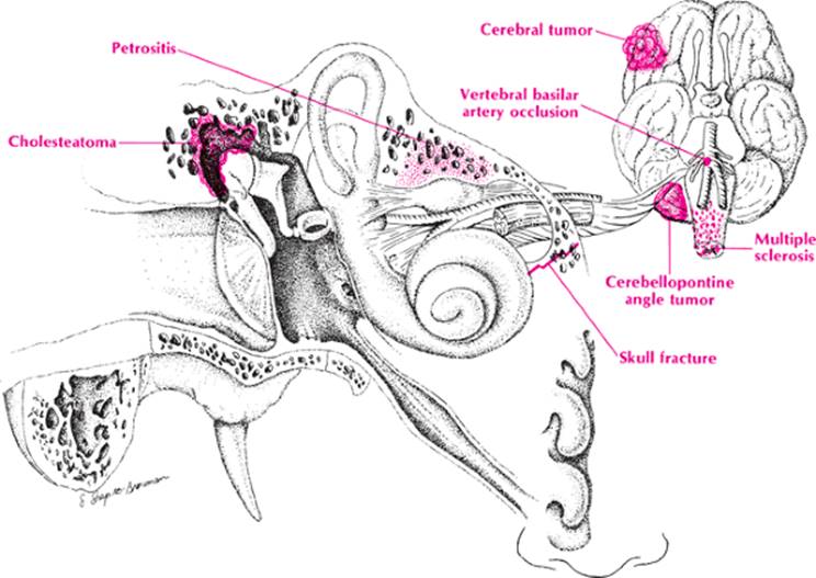
A facial palsy is usually considered to be Bell palsy and it frequently is. Nevertheless, the clinician who begins treatment without ruling out other possibilities will eventually get burned. Anatomy is the key to recalling these possibilities before the patient leaves the office. Follow the facial nerve from its origin along its pathway to its termination, and all the causes should come to mind.
|
|
|
Facial paralysis |
· Origin. Diseases of the brain and brainstem are considered here. They are usually distinguished from Bell palsy by the presence of other neurologic findings. The mnemonic ANITA will help recall them in an organized fashion.
· A—Arterial diseases include aneurysms, emboli, thromboses, and hemorrhages. Occlusion of the posterior inferior cerebellar artery will cause a peripheral facial palsy, but it can easily be distinguished from Bell palsy by the presence of a Horner syndrome, hoarseness, ataxia, and crossed hemianalgesia.
· N—Neoplasms include gliomas and the cerebellopontine angle tumor or acoustic neuroma.
· I—Inflammation suggests neurosyphilis, tuberculosis, brain abscess, and encephalitis.
· T—Trauma helps recall skull fractures and epidural and subdural hematomas.
· A—Autoimmune disease suggests multiple sclerosis, the collagen diseases, and early Guillain–Barré syndrome.
· Pathway. The facial nerve has a long pathway, and along that path it can be destroyed by the following:
· A—Arterial aneurysms
· N—Neoplasms such as acoustic neuromas and parotid gland tumors
· I—Inflammatory conditions like herpes zoster, petrositis, mastoiditis, and cholesteatomas
· T—Trauma such as basilar skull fractures and otologic surgery
· A—Autoimmune disease such as Bell palsy, or uveoparotid fever
· Termination. The site of termination of the facial nerve should suggest myasthenia gravis, muscular dystrophy, and facial hemiatrophy. These rarely present with an isolated facial palsy.
Approach to the Diagnosis
The clinical picture will frequently help to determine the cause of facial paralysis. Peripheral facial palsy as occurs in Bell palsy involves the forehead muscles and there is difficulty in closing the eyelid, whereas central facial palsy involves the face and lips and there is often associated hemiplegia or monoplegia. When there is exclusively a peripheral facial palsy without hearing loss or other neurologic signs, Bell palsy should be strongly suspected, although diabetes and myasthenia gravis need to be excluded. A bilateral peripheral nerve palsy should make one consider Guillain–Barré syndrome; be on the lookout for paralysis of the extremities as well. Bilateral facial palsy is also seen in myotonic dystrophy and myasthenia gravis. A “Bell palsy” with hearing loss and an aural discharge should prompt consideration of mastoiditis and petrositis. If there is hearing loss without a discharge, the possibility of an acoustic neuroma or cholesteatoma must be entertained. The association of a central facial palsy with hemiplegia brings up a host of possibilities including subdural hematoma, brain abscess, brain tumor, and cerebrovascular accident. The workup of these conditions is considered on page 222.
If the patient has clinical Bell palsy, one could start a therapy without a workup, but it is wise to get an x-ray of the skull and mastoids to rule out mastoiditis and petrositis and a glucose tolerance test to rule out diabetes. An acetylcholine receptor antibody titer or Tensilon test would only be ordered if the palsy were intermittent or there were other cranial nerve signs. If a middle ear infection or acoustic neuroma is suspected, the patient needs x-ray of the mastoids and petrous bones and a CT scan or MRI of the brain and auditory canal.
Other Useful Tests
1. CBC (ear infection)
2. Sedimentation rate (ear infection)
3. Venereal disease research laboratory (VDRL) test (neurosyphilis)
4. Cultures of ear discharge (otitis)
5. Audiogram and caloric tests (petrositis, acoustic neuroma)
6. Posterior fossa myelogram (acoustic neuroma)
7. Electromyogram (EMG) (Bell palsy)
8. Lyme disease antibody titer (Lyme disease)
9. Blood lead level (lead neuropathy)
10. Spinal tap (Guillain–Barré syndrome)
Case Presentation #26
A 21-year-old Hispanic man complained of acute onset of swelling of the right side of his face. Examination revealed weakness of the right facial muscles and inability to close his right eye.
Question #1. Utilizing the methods presented above, what would you consider in the differential diagnosis at this point?
A neurologist is consulted and his examination shows weakness of the left facial muscles as well. Furthermore, there is mild weakness and loss of sensation in all four extremities and diminished deep tendon reflexes.
Question #2. What is your list of possibilities now?