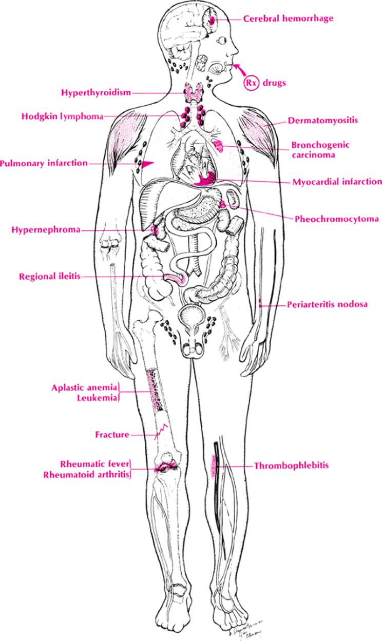
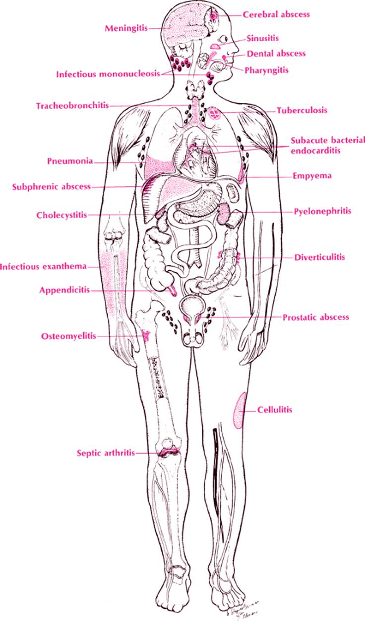
The differential diagnosis of fever is best developed using physiology first and anatomy second.
· Physiology. Increased heat in the body is caused by increased production or decreased elimination or dysfunction of the thermoregulatory system in the brain. Increased production of heat occurs in conditions with increased metabolic rate such as hyperthyroidism, pheochromocytomas, and malignant neoplasms. Poor elimination of heat may occur in congestive heart failure (CHF) (poor circulation through the skin) and conditions where the sweat glands are absent (congenital) or poorly functioning (heat stroke). Most cases of fever are caused by the effect of toxins on the thermoregulatory centers in the brain. These toxins may be exogenous from drugs, bacteria (endotoxins), parasites, fungi, rickettsiae, and virus particles, or they may be endogenous from tissue injury (trauma) and breakdown (carcinomas, leukemia, infarctions, and autoimmune disease).
· Anatomy. With the etiologies suggested by the mnemonic VINDICATE, one can apply anatomy and the various organ systems and make a useful chart (Table 28). The infections should be divided into the systemic diseases that affect more than one organ, such as typhoid, brucellosis, tuberculosis, syphilis, acquired immunodeficiency syndrome (AIDS), leptospirosis, and bacterial endocarditis, and the localized diseases that usually affect the same specific organ, such as infectious hepatitis, subacute thyroiditis, pneumococcal pneumonia, and cholera. It is wise to divide the localized infectious diseases into the “itises” (e.g., pneumonitis, hepatitis, and prostatitis), and the abscesses (dental abscess, empyema, perinephric abscess, liver abscess, and subdiaphragmatic abscess).
Also, when the physician attempts to recall the specific infections, he or she can group them into six categories beginning with the smallest organism and working up to the largest as follows: viruses, rickettsiae, bacteria, spirochetes, fungi, and parasites. Endogenous toxins released by infarctions of various organs form another convenient group. Finally, the most common neoplasms to cause fever (by tissue breakdown) are illustrated on page 172.
Approach to the Diagnosis
There are certain things to remember when a patient with fever is approached. First, a mild elevation up to 100.5°F (38°C) rectally may be normal in some people. Second, one should rule out malingering by the patient or incorrect recording by hospital personnel. Finally, psychogenic disorders must be ruled out.
The duration and severity of the fever are important. If possible, a careful chart of the fever should be made with the patient off all drugs (especially aspirin and steroids). Conditions with intermittent or relapsing fever such as brucellosis, malaria, and Mediterranean fever will be elucidated in this fashion (see Table 28).
The association with other symptoms is important. Fever, right upper quadrant pain, and jaundice suggest cholecystitis or cholangitis, whereas fever with right-sided flank pain suggests pyelonephritis. After taking a few moments to jot down the differential before launching into the history and physical examination, one can question and examine the patient more appropriately. The differential diagnosis will also lead to more appropriate use of laboratory testing.
Other Useful Tests
1. CBC (infectious disease, leukemia)
2. Urinalysis (urinary tract infection [UTI])
3. Sedimentation rate (infectious disease, collagen disease)
4. Chemistry panel (liver disease, renal disease)
5. Smear and culture of discharge from any body orifice or skin (e.g., abscess)
6. Blood cultures (septicemia, bacterial endocarditis)
7. Urine culture (pyelonephritis)
8. Bone marrow smear and culture (subacute bacterial endocarditis [SBE])
9. Stool for ova and parasites (e.g., amebiasis)
10. Blood smear for parasites and spirochetes (e.g., malaria)
|
|
|
Fever, noninfectious causes |
11. Febrile agglutinins (Salmonella, brucellosis)
12. Monospot test (infectious mononucleosis)
13. Cold agglutinins (Mycoplasma pneumoniae)
14. ANA (collagen disease)
15. Serum protein electrophoresis (multiple myeloma, collagen disease)
16. Sickle cell prep (sickle cell crisis)
17. Urine porphobilinogen (porphyria)
18. Fibrin index (Mediterranean fever)
19. Trichinella skin test or serology (trichinosis)
20. Acute- and convalescent-phase sera for viral studies
|
|
|
Fever, infectious causes |
21. Spinal fluid analysis (meningitis)
22. Urine for etiocholanolone (etiocholanolone fever)
23. Tuberculin test
24. Fungal skin test
25. Frei test (lymphogranuloma venereum)
26. Kveim test (sarcoidosis)
27. Angiotensin-converting enzyme level (sarcoidosis)
|
TABLE 28. Fever |
||||||||||||||||||||||||||||||||||||||||||||||||||||||||||||||||||||||||||||||||||||
|
||||||||||||||||||||||||||||||||||||||||||||||||||||||||||||||||||||||||||||||||||||
28. Chest x-ray (tuberculosis, pneumonia)
29. Flat plate of the abdomen (liver, spleen size, peritonitis stones)
30. X-ray of hands (sarcoidosis)
31. Gallbladder ultrasound (cholelithiasis)
32. Intravenous pyelogram (IVP) (hypernephroma, renal calculi)
33. Barium enema (neoplasm, diverticulitis)
34. CT scan of abdomen and pelvis (abscess)
35. CT scan of chest and mediastinum (abscess, neoplasm)
36. Bone scan (osteomyelitis, metastatic tumor)
37. X-ray of teeth (dental abscess)
38. Indium scan (abscess)
39. Liver biopsy (hepatic neoplasm, hepatitis, abscess)
40. Lymph node biopsy (inflammation, metastatic neoplasm)
41. Muscle biopsy (collagen disease, trichinosis)
42. Human immunodeficiency virus (HIV) antibody titer (AIDS)
43. Antistreptolysin-O (ASO) titer (rheumatic fever)
44. Epstein–Barr virus (EBV) immunoglobulins (infectious mononucleosis)
45. Transesophageal echocardiography (endocarditis)
Case Presentation #27
A 16-year-old white boy is referred to you with a history of sore throat and intermittent fever for 10 days. He was treated with penicillin by his family physician one week ago but failed to respond. On inspection, he is found to have a rash of the trunk and extremities.
Question #1. Utilizing the methods discussed above, what is your list of possibilities at this point?
Further examination discloses generalized lymphadenopathy and mild splenomegaly.
Question #2. What is your diagnosis now?