
DOORWAY INFORMATION
Opening Scenario
Gail Abbott, a 52-year-old female, comes to the office complaining of yellow eyes and skin.
Vital Signs
BP: 130/80 mm Hg Temp: 98.3°F (36.8°C)
RR: 15/minute HR: 70/minute, regular
Examinee Tasks
1. Take a focused history.
2. Perform a focused physical exam (do not perform rectal, genitourinary, or female breast exam).
3. Explain your clinical impression and workup plan to the patient.
4. Write the patient note after leaving the room.
Checklist/SP Sheet
Patient Description
Patient is a 52 yo F.
Notes for the SP
■ Sit up on the bed.
■ Show signs of scratching.
■ Exhibit RUQ tenderness on palpation.
■ If ERCP, ultrasound, or MRI is mentioned, ask for an explanation.
Challenging Questions to Ask
“My father had pancreatic cancer. Could I have it too?”
Sample Examinee Response
“It’s possible; that’s why we always rule it out in patients with yellow eyes or skin. Your family history does put you at slightly increased risk. However, we won’t know anything for certain until we run some tests.”
Examinee Checklist
Building the Doctor-Patient Relationship Entrance
□ Examinee knocked on the door before entering.
□ Examinee introduced self by name.
□ Examinee identified his/her role or position.
□ Examinee correctly used patient’s name.
□ Examinee made eye contact with the SP.
Reflective Listening
□ Examinee asked an open-ended question and actively listened to the response.
□ Examinee asked the SP to list his/her concerns and listened to the response without interrupting.
□ Examinee summarized the SP’s concerns, often using the SP’s own words.
Information Gathering
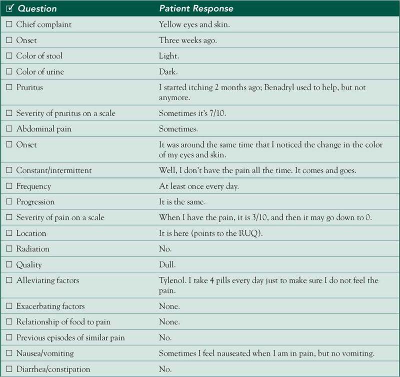
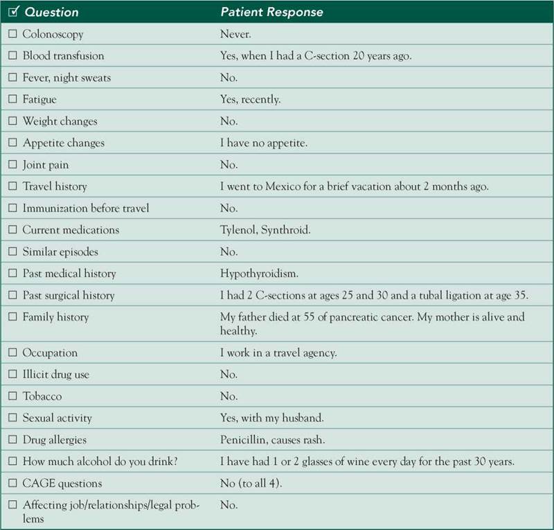
□ Examinee elicited data efficiently and accurately.
Connecting with the Patient
□ Examinee recognized the SP’s emotions and responded with PEARLS.
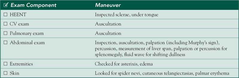
Physical Examination
□ Examinee washed his/her hands.
□ Examinee asked permission to start the exam.
□ Examinee used respectful draping.
□ Examinee did not repeat painful maneuvers.
Closure
□ Examinee discussed initial diagnostic impressions.
□ Examinee discussed initial management plans:
□ Follow-up tests.
□ Examinee asked if the SP had any other questions or concerns.
Sample Closure
Mrs. Abbott, the symptoms you describe are usually due to a disorder either in the liver itself or in the bile ducts, which are physically close to your liver. We will have to run some blood tests and conduct imaging studies such as ultrasound to get a better idea of what is going on. Once we find the cause of your problem, we can come up with an appropriate treatment plan. Until then, I recommend that you stop drinking and limit your use of Tylenol, as both may harm your liver. Do you have any questions for me?
History
HPI: 52 yo F c/o yellow skin and eyes x 3 weeks.
■ Light-colored stool and dark urine.
■ 3/10 RUQ pain, dull, intermittent (daily), no radiation, unrelated to meals, relieved by Tylenol.
■ Fatigue.
■ Anorexia.
■ Pruritus up to 7/10 in severity.
■ Nausea.
■ Recent travel to Mexico.
■ History of blood transfusion 20 years ago.
■ No diarrhea, constipation, or weight loss.
ROS: Negative except as above.
Allergies: Penicillin, causes rash.
Medications: Tylenol, Synthroid.
PMH: Hypothyroidism.
PSH: 2 C-sections, tubal ligation.
SH: No smoking, 1-2 glasses of wine/day for 30 years, CAGE 0/4, no illicit drugs. Sexually active with husband only.
FH: Father died of pancreatic cancer at age 55. No other FH of GI cancer.
Physical Examination
Patient is in no acute distress.
VS: WNL.
HEENT: Sclerae icteric.
Chest: Clear breath sounds bilaterally.
Heart: RRR; normal S1/S2; no murmurs, rubs, or gallops.
Abdomen: Soft, nondistended, C-section scar. Mild RUQ tenderness without rebound or guarding, Murphys sign,
 BS, no organomegaly or masses. No evidence of fluid wave suggestive of ascites. Skin: Jaundice, excoriations due to scratching, no spiders/telangiectasias/palmar erythema.
Extremities: No asterixis, no edema.
Differential Diagnosis
CASE DISCUSSION
Patient Note Differential Diagnoses
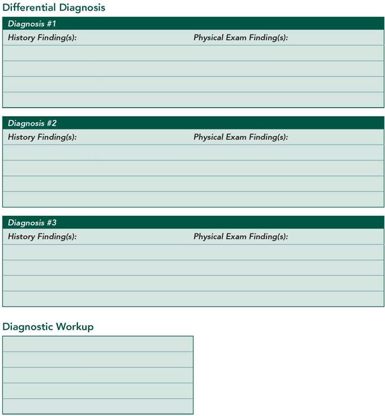
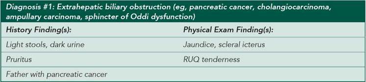
Jaundice results from hyperbilirubinemia, the cause of which may be hepatic or nonhepatic. The presence of a change in stool and urine color excludes unconjugated hyperbilirubinemia (eg, that associated with hemolysis or Gilbert’s syndrome). Thus, the predominantly conjugated hyperbilirubinemia suspected in this patient may be due to hepatocellular disease, drugs, sepsis, hereditary disorders such as Dubin-Johnson syndrome, or extrahepatic biliary obstruction. Cholangitis is ruled out by the absence of fever and chills associated with episodes of abdominal pain.
■ Extrahepatic biliary obstruction: The patient’s family history puts her at increased risk for pancreatic cancer, which classically presents with painless jaundice. However, her intermittent pain (suggesting intermittent biliary obstruction) narrows the differential to choledocholithiasis (stone in the common bile duct), cholangiocarcinoma, carcinoma of the ampulla, or sphincter of Oddi dysfunction.
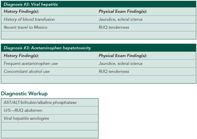
■ Viral hepatitis: The patient is at risk for hepatitis A (in light of her trip to Mexico) and chronic hepatitis C (given her remote blood transfusion). However, the intermittent nature of her RUQ pain makes acute hepatitis less likely.
■ Acetaminophen hepatotoxicity: This should be suspected in acute liver injury, as even moderate amounts of acetaminophen may overwhelm the metabolic capacity of a damaged liver (usually in alcoholics and in patients with chronic hepatitis or cirrhosis).
Additional Differential Diagnoses
■ Alcoholic hepatitis: The patient’s symptoms are consistent with this diagnosis. Hepatomegaly is often present. Although she reports drinking only one or two glasses of wine daily, patients often underreport alcohol consumption.
■ Primary biliary cirrhosis: This usually occurs in women 40-60 years of age, often with pruritus as a presenting symptom. It is commonly found in patients with other autoimmune diseases, such as hypothyroidism (as in this case). However, jaundice is usually a late finding and is not associated with RUQ pain.
Diagnostic Workup
■ AST/ALT/bilirubin/alkaline phosphatase: These levels can help differentiate a hepatocellular process (primarily associated with increased AST and ALT) from a cholestatic process (primarily associated with increased bilirubin and alkaline phosphatase).
■ U/S—RUQ abdomen: Used to diagnose biliary obstruction, stones, and intrahepatic tumors.
■ Viral hepatitis serologies: Hepatitis A IgM antibody should be checked to document recent infection. Other screening tests include hepatitis B surface antigen and hepatitis C antibody.
■ CBC: Patients with chronic liver disease often exhibit a low platelet count as a result of portal hypertension and subsequent splenomegaly.
■ PT/PTT: A coagulopathy is often seen in advanced liver disease and is attributable to synthetic dysfunction and subsequent clotting factor deficiencies.
■ Acetaminophen level: Used to diagnose acetaminophen overdose.
■ CT—abdomen: A CT scan provides information similar to that above but is more expensive.
■ MRCP/ERCP: Can identify the cause, location, and extent of biliary obstruction. ERCP is invasive but has the advantage of being both a diagnostic and a therapeutic tool in many cases. MRCP is a noninvasive MRI-based diagnostic substitute.