
DOORWAY INFORMATION
Opening Scenario
Jessica Anderson, a 21-year-old female, comes to the ED complaining of abdominal pain.
Vital Signs
BP: 120/80 mm Hg Temp: 100.5°F (38.1°C)
RR: 20/minute HR: 88/minute, regular
Examinee Tasks
1. Take a focused history.
2. Perform a focused physical exam (do not perform rectal, genitourinary, or female breast exam).
3. Explain your clinical impression and workup plan to the patient.
4. Write the patient note after leaving the room.
Checklist/SP Sheet
Patient Description
Patient is a 21 yo F, single with 1 child.
Notes for the SP
■ Exhibit right lower abdominal tenderness on palpation.
■ Show rebound tenderness (pain when the examinee removes his palpating hand).
■ Demonstrate guarding (contraction of the abdominal muscles when palpating the RLQ).
■ Experience pain in the RLQ when the examinee presses on the LLQ (Rovsing’s sign).
■ Manifest pain when the examinee extends your right hip (psoas sign).
Challenging Questions to Ask
■ “My child is in the house alone. I must leave now.”
■ “I can’t afford to stay in the hospital. Please give me a prescription for antibiotics so that I can leave.”
Sample Examinee Response
“Ms. Anderson, I understand your concern for your child’s safety. However, it is most important that we make sure your illness isn’t life threatening. Our social worker would be happy to work with you to ensure that your child is taken care of, as well as to address any financial concerns you may have.”
Examinee Checklist
Building the Doctor-Patient Relationship
Entrance
□ Examinee knocked on the door before entering.
□ Examinee introduced self by name.
□ Examinee identified his/her role or position.
□ Examinee correctly used patient’s name.
□ Examinee made eye contact with the SP.
Reflective Listening
□ Examinee asked an open-ended question and actively listened to the response.
□ Examinee asked the SP to list his/her concerns and listened to the response without interrupting.
□ Examinee summarized the SP’s concerns, often using the SP’s own words.
Information Gathering
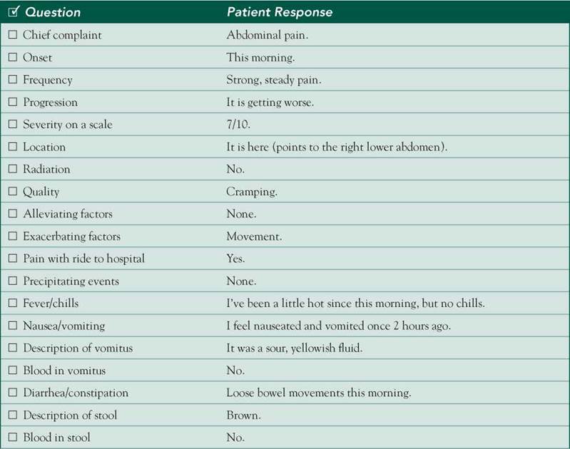
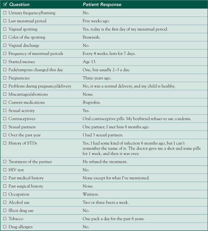
□ Examinee elicited data efficiently and accurately.
Connecting with the Patient
□ Examinee recognized the SP’s emotions and responded with PEARLS.
Physical Examination
□ Examinee washed his/her hands.
□ Examinee asked permission to start the exam.
□ Examinee used respectful draping.
□ Examinee did not repeat painful maneuvers.

Closure
□ Examinee discussed initial diagnostic impressions.
□ Examinee discussed initial management plans:
□ Follow-up tests: Examinee mentioned the need for rectal and pelvic exams.
□ Safe sex practices.
□ Help with smoking cessation.
□ Assistance of social workers to help the patient identify available financial resources.
□ Examinee asked if the SP had any other questions or concerns.
Sample Closure
Ms. Anderson, your symptoms may be due to a problem with your reproductive organs, such as an infection in your fallopian tube or a cyst on your ovary. They might also result from a complicated pregnancy, which could be indicated if your pregnancy test comes back positive. Another possibility is an infection in your appendix, which could require surgery. To ensure an accurate diagnosis, we will need to run some tests, including a blood test, a urinalysis, a pregnancy test, and possibly a CT scan of your abdomen and pelvis. I will also need to perform rectal and pelvic exams. Since cigarette smoking is associated with a variety of diseases, I advise you to quit smoking; we have many ways to help you if you are interested. I also recommend that you use a condom every time you have intercourse to prevent STDs, including HIV, and to avoid pregnancy. Our social worker can meet with you to discuss your social situation, and she can offer you a variety of resources. Do you have any questions for me?
History
HPI: 21 yo G1P1 F c/o right lower abdominal pain that started this morning. The pain is 7/10, crampy, nonradiating, and constant. It is exacerbated by movement and accompanied by fever, nausea, vomiting, and loose stools. The patient noticed some brownish spotting this morning. No urinary symptoms; no abnormal vaginal discharge.
OB/GYN: LMP 5 weeks ago. Regular periods every 4 weeks lasting 7 days. Menarche at age 13. Uncomplicated NSVD at full term 3 years ago.
ROS: Negative except as above.
Allergies: NKDA.
Medications: Ibuprofen.
PMH: STD 1 month ago, possibly treated with ceftriaxone and doxycycline.
PSH: None.
SH: 1 PPD for 6 years, 2-3 beers/week, no illicit drugs. Unprotected sex with multiple partners over the past year.
Physical Examination
Patient is in pain.
VS: WNL except for temperature of 100.5°F
Chest: No tenderness, clear breath sounds bilaterally.
Heart: RRR; normal S1/S2; no murmurs, rubs, or gallops.
Abdomen: Soft, nondistended, hypoactive BS, no hepatosplenomegaly Direct and rebound RLQ tenderness, RLQ guarding, psoas sign,
 Rovsings sign,
 obturator sign, no CVA tenderness.
Differential Diagnosis
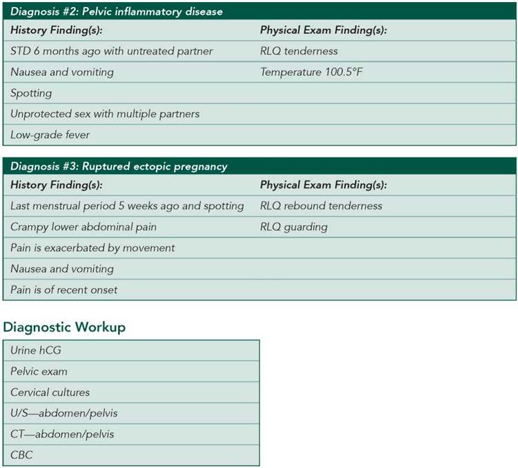
CASE DISCUSSION
Patient Note Differential Diagnoses
This case is written primarily to elicit the differential diagnosis of RLQ pain in a woman of childbearing age. The presentation of gynecologic diseases commonly mimics appendicitis.
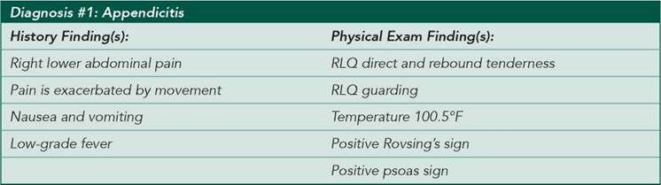
■ Appendicitis: In a patient presenting with RLQ pain, low-grade fever, nausea and vomiting, and peritoneal signs (pain exacerbated by movement), appendicitis should certainly be in the differential. The abdominal exam revealed direct and rebound RLQ tenderness, RLQ guarding, a positive psoas sign, and a positive Rovsing’s sign—all of which are associated with appendicitis. However, the onset of pain in appendicitis is usually gradual.
■ Pelvic inflammatory disease (PID): Suspicion is high for this diagnosis in a patient who presents with recent- onset lower abdominal pain and low-grade fever in the setting of a recent STD and unprotected sex with an untreated partner. The standard treatment for gonorrhea and chlamydia consists of ceftriaxone and doxycycline. Left untreated, these infections can progress to PID. Other findings suggestive of PID include abnormal menstrual bleeding, nausea and vomiting, and a history of multiple sex partners.
■ Ruptured ectopic pregnancy: Although this patient does not have previously documented PID (or a previous tubal pregnancy), the crampy lower abdominal pain, nausea and vomiting, and vaginal spotting that she is experiencing after a five-week period of amenorrhea suggest this diagnosis. However, positive psoas and Rovsing’s signs are not typical of an ectopic pregnancy.
Additional Differential Diagnoses
■ Ruptured ovarian cyst: The patient’s sudden-onset, unilateral lower abdominal pain, rebound tenderness, and guarding are consistent with this diagnosis. Rupture may occur at any time during the menstrual cycle, and symptoms may resemble a ruptured ectopic pregnancy as described above. However, this diagnosis is less common than appendicitis and PID. In addition, given the patient’s history of having her last menstrual period five weeks ago, ruptured ectopic pregnancy must be placed higher on the differential, as a ruptured ovarian cyst would not be associated with a late menstrual period.
■ Adnexal torsion: This presentation may be due to adnexal torsion, an uncommon complication that is most often associated with ovarian enlargement due to a benign mass.
■ Gastroenteritis: Viral gastroenteritis presents with crampy abdominal pain, nausea and vomiting, low-grade fever, and diarrhea. It can be difficult to distinguish from appendicitis and gynecologic etiologies but is less likely in this case given the presence of rebound tenderness.
■ Abortion: The fact that the patient’s last menstrual period was only five weeks ago makes this diagnosis less likely, but the crampy abdominal pain and vaginal spotting may signal an abortion. Furthermore, the presence of fever suggests possible septic abortion.
■ Endometriosis: This is an unlikely diagnosis, in part because the patient has no history of chronic pelvic pain, dysmenorrhea, dyspareunia, or infertility, which are often associated. In the setting of established endometriosis, this presentation in a patient with acute, severe pain, including rebound tenderness, could be due to rupture of an endometrioma (“chocolate cyst”).
Diagnostic Workup
■ Urine hCG: Positive in both ectopic and intrauterine pregnancies. Urine and serum tests are equally sensitive, but quantitative hCG levels (available only via serum test) may help diagnose and treat ectopic pregnancy.
■ Pelvic exam: Look for cervical motion tenderness and discharge, uterine size, and adnexal masses or tenderness.
■ Cervical cultures: Neisseria gonorrhoeae and Chlamydia trachomatis, the main causes of PID, are detected by means of DNA probes.
■ U/S—abdomen/pelvis: Can help diagnose appendiceal or ovarian pathology. Transvaginal ultrasound can identify an intrauterine gestational sac when the time elapsed since the last menstrual period is 35 days (this corresponds to a β-hCG of approximately 1500 mIU/mL); fluid in the cul-de-sac is nonspecific and may suggest ectopic pregnancy or a ruptured ovarian cyst.
■ CT—abdomen/pelvis: Can detect the presence of appendiceal inflammation, abscess in appendicitis, or signs of other GI or gynecologic pathology.
■ CBC: Findings are nonspecific, but leukocytosis may be seen in infection or appendicitis.
■ UA: To rule out UTI.
■ Laparoscopy: Can diagnose ectopic pregnancy (gold standard), ruptured ovarian cyst, ovarian torsion, PID ± tubo-ovarian abscess, appendicitis, and the like.