
DOORWAY INFORMATION
Opening Scenario
Charles Andrews, a 66-year-old male, comes to the clinic complaining of a tremor.
Vital Signs
BP: 135/85 mm Hg Temp: 98.6°F (37°C)
RR: 16/minute HR: 70/minute, regular
Examinee Tasks
1. Take a focused history.
2. Perform a focused physical exam (do not perform rectal, genitourinary, or female breast exam).
3. Explain your clinical impression and workup plan to the patient.
4. Write the patient note after leaving the room.
Checklist/SP Sheet
Patient Description
Patient is a 66 yo M.
Notes for the SP
■ Exhibit mild muscle rigidity in your wrists and arms—that is, when the examinee tries to move your wrists and arms, stiffen them and move them slowly.
■ Lean your back forward slightly and walk in small, shuffling steps.
■ Exhibit a resting hand tremor (pill rolling) that disappears with movement.
Challenging Questions to Ask
“Do you think I will get better?”
Sample Examinee Response
“I think your tremor will improve with medication, but I don’t know how long the improvement will last. The tremor may be a sign of a larger movement disorder called Parkinson’s disease, and we need to do some additional evaluations to explore that possibility.”
Examinee Checklist
Building the Doctor-Patient Relationship Entrance
□ Examinee knocked on the door before entering.
□ Examinee introduced self by name.
□ Examinee identified his/her role or position.
□ Examinee correctly used patient’s name.
□ Examinee made eye contact with the SP.
Reflective Listening
□ Examinee asked an open-ended question and actively listened to the response.
□ Examinee asked the SP to list his/her concerns and listened to the response without interrupting.
□ Examinee summarized the SP’s concerns, often using the SP’s own words.
Information Gathering
□ Examinee elicited data efficiently and accurately.

Connecting with the Patient
□ Examinee recognized the SP’s emotions and responded with PEARLS.
Physical Examination
□ Examinee washed his/her hands.
□ Examinee asked permission to start the exam.
□ Examinee used respectful draping.
□ Examinee did not repeat painful maneuvers.

Closure
□ Examinee discussed initial diagnostic impressions.
□ Examinee discussed initial management plans:
□ Follow-up tests.
□ Possible need to compare an old handwriting sample with a present sample.
□ Examinee offered support throughout the patient’s illness.
□ Examinee asked if the SP had any other questions or concerns.
Sample Closure
Mr. Andrews, I am sorry to have to tell you this, but on the basis of your history and physical exam, it would appear that you have Parkinson’s disease. Your symptoms may improve with medications, but eventually they will return. One indicator of disease progression involves looking closely at your handwriting. Do you think you could bring an old sample of your handwriting with you on your next visit? You should also know that about 25% of the time, patients with your symptoms do not have Parkinson’s disease. For this reason, I would like to run a few tests, including some imaging studies of your head and some blood tests. Although we won’t have those results before you leave today, I will print out a comprehensive patient pamphlet that will give you resources to help answer your questions as they come up. I want you to know that I will be here to treat you and to help you every step of the way. Do you have any questions for me?

History
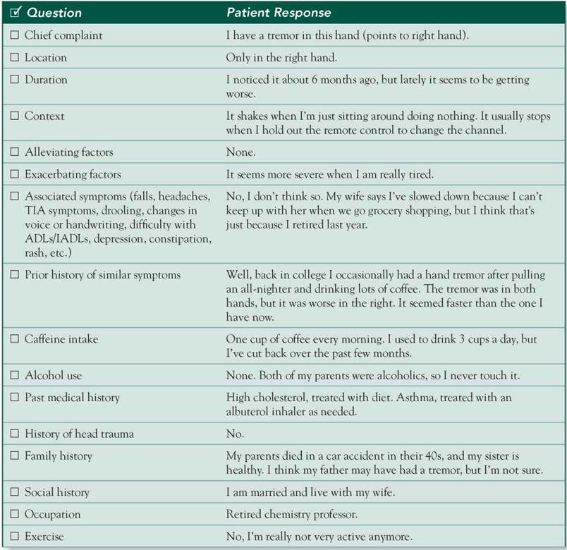
HPI: 66 yo M c/o right hand tremor for 6 months. It occurs at rest and seems to be getting worse. The tremor is exacerbated by fatigue. There are no alleviating factors (he does not drink alcohol). Reducing his caffeine intake to 1 cup of coffee daily did not seem to help. He denies associated symptoms but does say that his wife complains that he has "slowed down" since retiring last year. Specifically, he seems to be walking more slowly recently (time course unspecified, but within the past year). He had a hand tremor when very fatigued back in college, but it was bilateral and faster than his present tremor.
ROS: Negative except as above.
Allergies: NKDA.
Medications: Albuterol MDI prn (no use in past year).
PMH: High cholesterol, treated with diet. Mild asthma.
SH: No smoking, no EtOH, no illicit drugs. He is a retired chemistry professor, married and lives with his wife.
FH: Father may have had a tremor.
Physical Examination
Patient is in no acute distress.
VS: WNL.
Chest: Clear breath sounds bilaterally.
Heart: RRR; normal S1/S2; no murmurs, rubs, or gallops.
Neuro: Mental status: Alert and oriented x 3. Cranial nerves: 2-12 grossly intact. Motor: Right hand resting tremor with "pill-rolling" movement that improves or disappears during purposeful action or posture. Mild muscle rigidity in both wrists and arms, but no frank cogwheeling. Strength 5/5 throughout. DTRs: Symmetric 2+ in all extremities. Cerebellar: Romberg, rapid alternating movements and heel-to-shin test normal and symmetric. Gait: Bradykinetic, takes small steps. Walks with back slightly bent forward. Sensation: Intact to soft touch and pinprick.
Differential Diagnosis

CASE DISCUSSION
Patient Note Differential Diagnoses
■ Parkinson's disease (PD): This is the most common cause of resting tremor (ie, a tremor that is evident with the affected body part supported and completely at rest but improves or subsides with voluntary activity), although some patients with PD also have a postural/action tremor that is indistinguishable from essential tremor (ET, see below). Tremor is usually low frequency (4-6 Hz), begins in one upper extremity, and may later involve the other extremities as well. Leg tremor is more commonly due to PD than to ET. The face, lips, and jaw may be involved, but in contrast to ET, PD does not produce head tremor. Along with the tremor, the patient’s bradykinesia and rigidity suggest PD.
■ Essential tremor (ET): This is the most common neurologic cause of postural tremor (ie, tremor that is apparent when the arms are held outstretched) or action tremor (ie, tremor that increases at the end of goal- directed activity such as finger-to-nose testing). Approximately 50% of cases are familial. Tremor is usually high frequency and often asymmetrically involves the distal upper extremity. The head, voice, chin, trunk, and legs can also be involved. ET is not associated with other neurologic signs and improves following the ingestion of small amounts of alcohol. Differentiation from the classic resting tremor of PD is usually straightforward, as in this case.
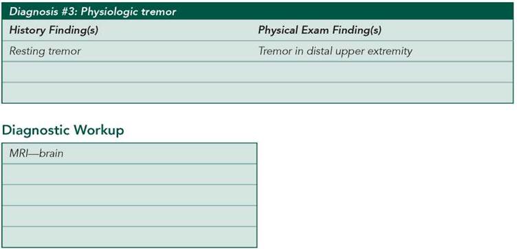
■ Physiologic tremor: This refers to a very low-amplitude, high-frequency (10- to 12-Hz) tremor present in normal individuals. The tremor is often not visible, but when enhanced by medications or other medical conditions, it is the most common cause of postural and action tremors. Conditions that can enhance physiologic tremor include anxiety, excitement, sleep deprivation/fatigue, hypoglycemia, caffeine intake, alcohol withdrawal, thyrotoxicosis, fever, and pheochromocytoma.
Additional Differential Diagnoses
■ Midbrain lesion: Midbrain injury due to stroke, trauma, or demyelinating disease is a rare cause of a solitary asymmetric resting tremor.
■ Drug-induced tremor: Many medications can enhance physiologic tremor, notably P-agonists (eg, albuterol), nicotine, theophylline, TCAs, lithium, valproic acid, and corticosteroids. Mercury and arsenic exposure may also contribute to tremor. Neuroleptics and metoclopramide can cause drug-induced parkinsonism, but tremor is often absent in these cases.
■ Psychogenic tremor: This often manifests with varying frequency and either becomes more irregular or subsides entirely when the patient is asked to perform a complex, repetitive motor task with the contralateral limb.
■ Wilson's disease: This can cause resting tremor (among other manifestations) but is not considered in patients older than 40 years of age.
■ Hyperthyroidism: This is associated with fine tremor along with a variety of other classic signs and symptoms.
Diagnostic Workup
■ MRI—brain: To rule out a structural lesion, particularly in the midbrain or basal ganglia.
■ TSH: To screen for hyperthyroidism.
■ Heavy metal screen: To screen for mercury and arsenic toxicity via urine or blood tests.
■ Ceruloplasmin, slit lamp examination for Kayser-Fleischer rings, AST/ALT, CBC, 24-hour urinary copper, liver biopsy: These tests constitute the screening tests (and diagnostic tests, in the case of liver biopsy) used to evaluate for suspected Wilson’s disease. As noted previously, the patient’s advanced age precludes consideration of Wilson’s disease.