
Mucocutaneous Signs of Systemic Cancers
ICD-9: 199.0  ICD-10: M8000/6
 Mucocutaneous findings may suggest systemic cancers in several ways:
Associations of heritable mucocutaneous disorders with systemic cancers.
By action at a distance, i.e., paraneoplastic syndromes.
Or spread of cancer to the skin or mucosal sites by direct, lymphatic, or hematogenous extension (cutaneous metastasis).
Classification of Skin Signs of Systemic Cancer1
Metastatic Cancers
Persistent Tumor. Lymphatic extension, hematogenous spread.
Direct Extension. Paget disease, extramammary Paget disease.
Lymphomas with secondary skin involvement (Section 21).
Heritable Disorders
Cowden Syndrome
Peutz–Jeghers Syndrome
Neurofibromatosis (p. 405).
Tuberous sclerosis (p. 402).
Multiple endocrine neoplasia (types 1 and 2b).
Paraneoplastic Syndromes
Acanthosis nigricans, malignant, tripe palms.
Acquired ichthyosis.
Bazex syndrome.
Carcinoid syndrome.
Dermatomyositis (p. 328).
Ectopic ACTH syndrome.
Erythema gyratum repens.
Gardner syndrome.
Glucagonoma syndrome.
Hypertrichosis lanuginosa.
Muir–Torre syndrome.
Palmar keratoses.
Paraneoplastic pemphigus.
Pruritus.
Pyoderma gangrenosum (p. 116).
Sweet syndrome (p. 120).
Vasculitis (p. 356).
Metastatic Cancer to the Skin* ICD-9: 199.0 ICD-10: M8000/6
Metastatic cancer to the skin is characterized by solitary or multiple dermal or subcutaneous nodules, occurring as metastatic cells from a distant noncontiguous primary malignant neoplasm.
They are transported to and deposited in the skin or subcutaneous tissue by one of the following routes:
Lymphatic routes.
Hematogenous spread.
Contiguous spread across the peritoneal cavity or other tissues.
Skin lesions nodule (Figs. 19-1 and 19-2), raised plaque, thickened fibrotic area. First detected when <5 mm. Fibrotic area may resemble morphea; occurring on scalp, may produce alopecia. Initially, epidermis is intact, stretched over nodule; in time, surface may become ulcerated (Fig. 19-3) or hyperkeratotic. May appear inflammatory, i.e., pink to red or hemorrhagic. Firm to indurated. May be solitary, few, or multiple. May acquire considerable size and may be mistaken for a primary skin cancer (Fig. 19-3).
*For metastatic nonmelanoma skin cancers and melanoma, see Sections 11 and 12.
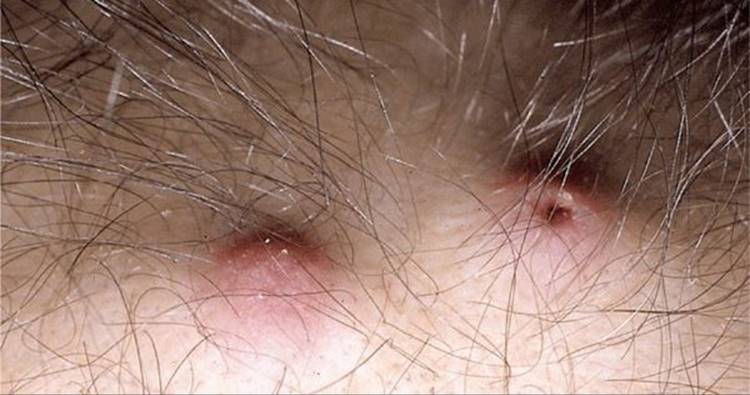
Figure 19-1. Metastatic cancer to the skin: bronchogenic cancer Dermal nodules on the scalp of a patient undergoing chemotherapy for metastatic lung cancer; the nodules were only apparent following loss of hair during chemotherapy. The nodule on the left is asymptomatic, erythematous, but noninflamed. The nodule on the right has a central depression marking a punch biopsy site.
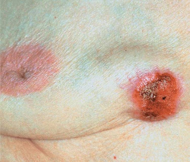
Figure 19-2. Metastatic cancer to the skin Breast cancer: Large nodule on breast in a 40-year-old woman with breast cancer, present for 4 months.

Figure 19-3. Metastatic cancer to the skin Adenocarcinoma of the GI tract. This fungating mass was just the tip of the iceberg: a much larger mass was in the subcutis.
Special Patterns of Cutaneous Involvement
Breast
Inflammatory metastatic carcinoma (carcinoma erysipelatodes): erythematous patch or plaque with an active spreading border (Fig. 19-4). Most often with breast cancer that may spread within lymphatics to the skin of involved breast, resulting in inflammatory plaques resembling erysipelas (hence, the designation carcinoma erysipelatodes). Occurs with other cancers as well [pancreas, parotid, tonsils, colon, stomach, rectum, melanoma, pelvic organs, ovary (Fig. 19-5), uterus, prostate, lung, mesothelioma (Fig. 19-6)].
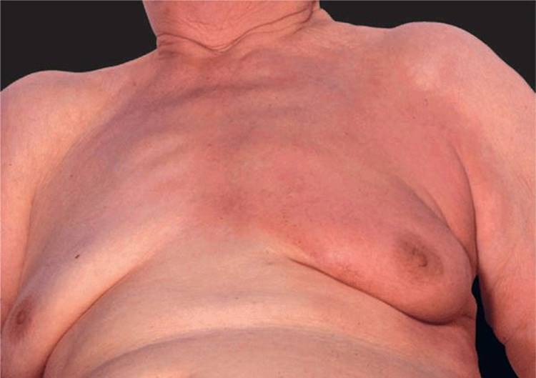
Figure 19-4. Metastatic cancer of the skin: inflammatory breast cancer (carcinoma erysipelatodes) A large erythematous and only minimally indurated lesion covering the entire breast and presternal region; the lesion is red and sharply defined and thus looks like erysipelas. There was a 2 × 2 cm lump in the breast upon palpation.
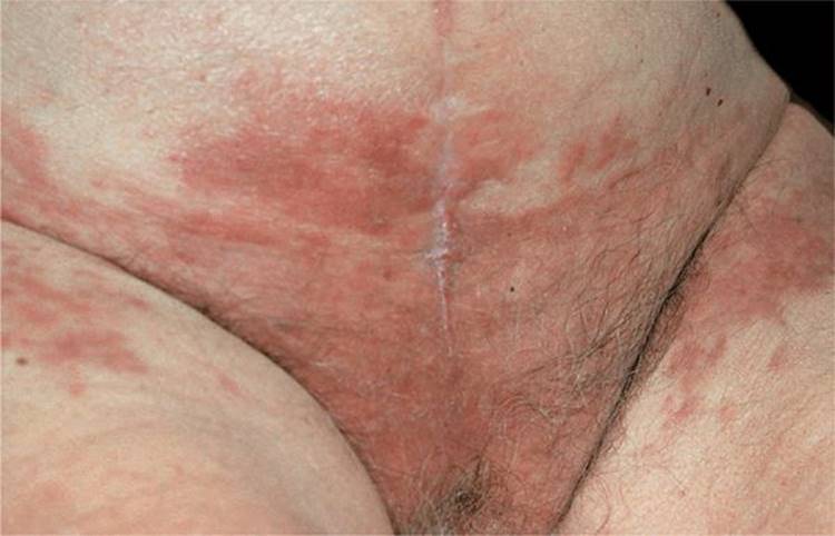
Figure 19-5. Metastatic ovarian cancer Manifesting as carcinoma erysipelatodes on the lower abdomen and inguinal region. Workup disclosed ovarian cancer with peritoneal carcinomatosis.
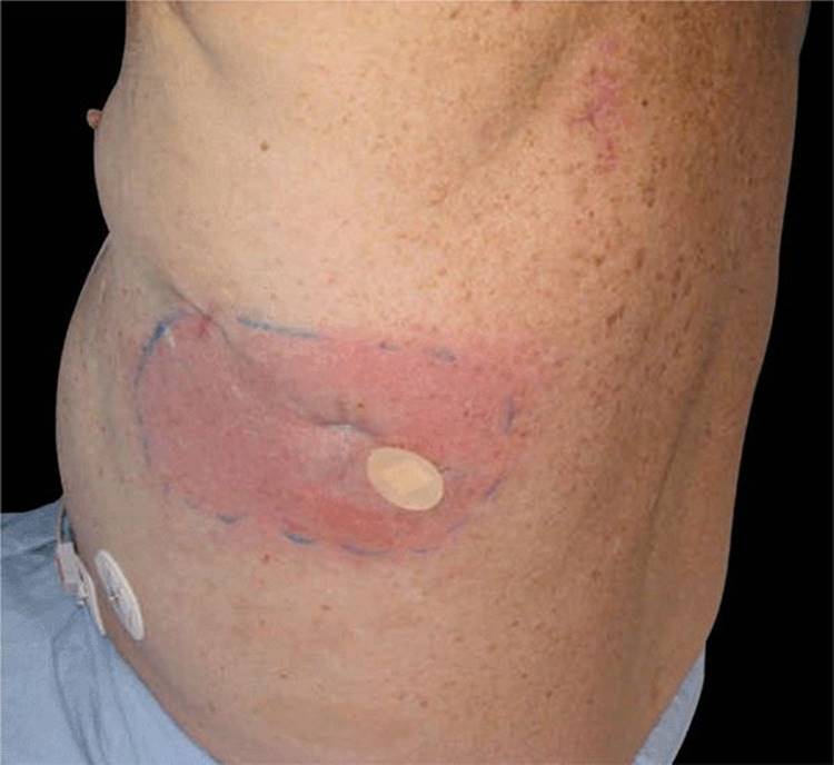
Figure 19-6. Mesothelioma An indurated erythematous patch on the lateral chest represents carcinoma erysipleatodes from mesothelioma.
Telangiectatic metastatic carcinoma (carcinoma telangiectaticum): breast cancer appearing as pinpoint telangiectases with dilated capillaries within carcinoma erysipelatodes. Violaceous papules or papulovesicles resembling lymph-angioma circumscriptum.
En cuirasse metastatic carcinoma: diffuse morphea-like induration of skin (Fig. 19-7). Usually local extension of breast cancer occurring in breast and presternal region. Sclerodermoid plaque may encase chest and resembles a metal breastplate of a cuirassier. Also occurs with primary of lung, GI tract, kidney.
Paget disease: see below.
Multiple smooth nodules on scalp: prostate adenocarcinoma, lung cancer, breast cancer (Fig. 19-1).
Alopecia neoplastica: On scalp, areas of hair loss resembling alopecia areata; well-demarcated, red-pink, smooth surface, flat.
Large Intestine. Often presents on skin of abdomen or perineal regions; also, scalp or face. Most originate in rectum. May present with metastatic inflammatory carcinoma (like carcinoma erysipelatodes) of inguinal region, supraclavicular area, or face and neck. Less commonly, sessile or pedunculated nodules on buttocks, grouped vascular nodules of groin or scrotum, or facial tumor. Rarely, cutaneous fistula after appendectomy or resembling hidradenitis suppurativa.
Lung Carcinoma. May produce a large number of metastatic nodules in a short period. Most commonly, reddish nodule(s) on scalp (Fig. 19-1). Trunk: symmetric; along direction of intercostal vessels, may be zosteriform; in scar (thoracotomy site or needle aspiration tract).
Hypernephroma. Can produce solitary lesion; also widespread. Usually appear vascular, ± pulsatile, ± pedunculated; can resemble pyogenic granuloma. Most common on head (scalp) and neck; also trunk and extremities.
Carcinoma of Bladder, Ovary. Can spread contiguously to abdominal and inguinal skin similarly to breast cancer, as described above, and look like erysipelas (Fig. 19-5).
Miscellaneous Patterns. With dilation of lymphatics and superficial hemorrhage, may resemble lymphangioma. With lymph stasis and dermal edema, resembles pigskin or orange peel.
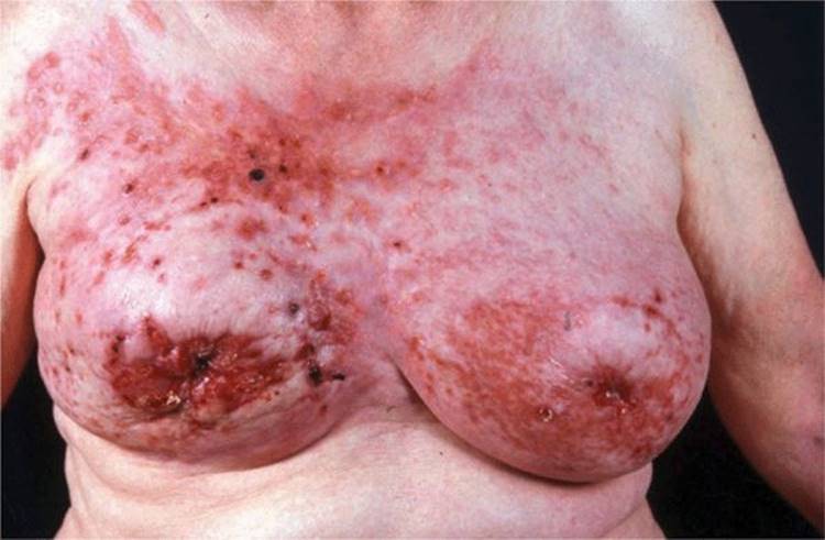
Figure 19-7. Metastatic breast cancer: cancer en cuirasse Both breasts are hard upon palpation—like an armor plate. There are multiple small and large, ulcerated nodules and there is a background of erysipelas-like erythema (carcinoma erysipelatodes).
Mammary Paget Disease ICD-9: 174.0 ICD-10: C50.01 
Mammary Paget disease (MPD) is a malignant neoplasm that unilaterally involves the nipple or areola and simulates a chronic eczematous dermatitis.
It represents contiguous spread of underlying intraductal carcinoma of the breast (1–4% of breast cancers).
Usually occurring in females (>50 years); there are rare examples in males.
Onset is insidious over several months or years. May be asymptomatic or there may be pruritus, pain, burning, discharge, bleeding, ulceration, and nipple invagination.
Skin lesion presents as red, scaling plaque, rather sharply marginated, oval with irregular borders. When scale is removed, the surface is moist and oozing (Fig. 19-8). Lesions range in size from 0.3 to 15 cm (Fig. 19-9). In early stages, there is no induration of the plaque; later, induration and infiltration develop and nodules may be palpated in breast. At initial, there is flattening or retraction of the nipple presentation, an underlying breast mass is palpable in fewer than one-half of patients. May be bilateral. Lymph node metastases occur more often when MPD is associated with an underlying palpable mass.
Differential diagnosis includes eczematous dermatitis, psoriasis, benign ductal papilloma, nipple-areola retention hyperkeratosis, impetigo, SCC in situ, familial pemphigus.
Eczematous dermatitis of the nipples is usually bilateral; it is without any induration and responds rapidly to topical glucocorticoids. Nevertheless, be suspicious of Paget disease if “eczema” persists for >3 weeks. Diagnosis verified by biopsy showing neoplastic cells in epidermis following a pathognomonic pattern of spread. Define underlying intraductal carcinoma by mammography.
Management consists of surgery, radiotherapy, and/or chemotherapy as in any other breast carcinomas. Lymph node dissection if regional nodes are palpable. Prognosis varies. When breast mass is not palpable, 92% of patients survive 5 years after excision; 82% survive 10 years. When breast mass is palpable, 38% survive 5 years; 22% survive 10 years. Prognosis worse when there is lymphadenopathy.

Figure 19-8. Mammary Paget disease A sharply demarcated red plaque mimicking eczema or psoriasis on the nipple. The plaque is slightly indurated and there is slight scaling; any red, eczema-like lesion on the nipple and areola that does not respond to topical glucocorticoids should be biopsied.

Figure 19-9. Mammary Paget disease A sharply defined psoriasiform plaque that has obliterated the areola and nipple. There was a lump in the breast and a small axillary mass.
Extramammary Paget Disease ICD-9: 709.8 ICD-10: L87.9 
Extramammary Paget disease (EPD) is a neoplasm of the anogenital and axillary skin, histologically identical and clinically similar to Paget disease of the breast.
Often representing an intraepidermal extension of a primary adenocarcinoma of underlying apocrine glands or of the lower gastrointestinal, urinary, or female genital tracts.
Often, however, it is unassociated with underlying cancer.
The histogenesis of EPD is not uniform. Occurs as an in situ upward extension of an in situ adenocarcinoma in deeper glands (25%). Alternatively, EPD may have a multifocal primary origin in the epidermis and its appendages. Primary tumors in the anorectum can arise within the rectal mucosa or intramural glands.
Insidious onset, slow spread, + itching. The lesion presents as erythematous plaque, + scaling, + erosion (Fig. 19-10), + crusting, + exudation; eczematous-appearing lesions, but borders are sharply defined (Fig. 19-10), geographic configuration. Lesions should always be biopsied.
Histopathologically, Paget cells are dispersed between keratinocytes, occur in clusters, extend down into adnexal structures (hair follicles, eccrine ducts). Adnexal adenocarcinoma is often found when carefully searched for.
In perineal/perianal EPD, underlying carcinoma should be searched for by rectal examination, proctoscopy, sigmoidoscopy, barium enema. In genital EPD, search for underlying carcinoma by cystoscopy, intravenous pyelogram; in vulvar EPD, by pelvic examination.
Differential diagnosis includes all red plaques: eczematous dermatitis, lichen simplex chronicus, lichen sclerosus et atrophicus, lichen planus, intertriginous psoriasis, Candida intertrigo, SCC in situ (erythroplasia of Queyrat), human papilloma virus–induced SCC in situ, amelanotic superficial spreading melanoma.
EPD is usually much larger than is apparent clinically. Surgical excision must be controlled histologically (Mohs micrographic surgery). If Paget cells are in dermis and regional lymph nodes are palpable, lymph node dissection may improve prognosis, which is related to underlying adenocarcinoma. EPD remains in situ in the epidermis and adnexal epithelium in >65% of cases. When no underlying neoplasm is present, there is nonetheless a high recurrence rate, even after apparently adequate excision; this is due to the multifocal origin in the epidermis and adnexal structures.
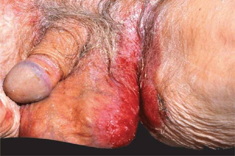
Figure 19-10. Extramammary Paget disease Moist, well-demarcated, eroded, oozing, erythematous plaque on the scrotum and inguinal fold in an older male. The lesion is commonly mistaken for Candidaintertrigo and unsuccessfully treated as such.
Cowden Syndrome (Multiple Hamartoma Syndrome) ICD-9: 759.6 ICD-10: Q85.9 
Cowden syndrome (named after the propositus) is a rare, autosomal-dominant heritable cancer syndrome with variable expressivity in a number of systems in the form of multiple hamartomatous neoplasms of ectodermal, mesodermal, and endodermal origin.
Germ-line mutations in the tumor-suppressor gene PTEN are located on chromosome 10q22–23 in most cases.
There is a special susceptibility for breast and thyroid cancers, and the skin lesions are important markers.
Skin lesions may appear first in childhood and develop over time. They consist of trichilemmomas, skin-colored, pink (Fig. 19-11B), or brown papules having the appearance of flat warts on the central area of the face, lips, and the ears; translucent punctate keratoses of the palms and soles; and hyperkeratotic, flat-topped papules on the dorsa of the hands and forearms. Mucous membranes: papules of the gingival, labial (Fig. 19-11A), and palatal surfaces that coalesce, giving a “cobblestone” appearance. Papillomas of the buccal mucosa and the tongue.
In addition to breast cancer (20%), which is often bilateral, and thyroid cancer (8%), there are various internal hamartomas:
Breast: fibrocystic disease, fibroadenomas, adenocarcinoma, gynecomastia in males.
Thyroid: goiter, adenomas, thyroglossal duct cysts, follicular adenocarcinoma.
GI tract: hamartomatous polyps throughout tract but increased in large bowel, adenocarcinoma arising in polyp.
Female genital tract: ovarian cysts, menstrual abnormalities.
Musculoskeletal: craniomegaly, kyphoscoliosis, “adenoid” facies, high-arched palate.
CNS: mental retardation, seizures, neuromas, ganglioneuromas, and meningiomas of the ear canal.
It is important to establish the diagnosis of Cowden syndrome so that these patients can be followed carefully to detect breast and thyroid cancers early.

Figure 19-11. Cowden syndrome (A) Multiple reddish, confluent papules on the oral mucosa giving a cobblestone appearance. (B) Multiple skin-colored warty papules on the face, which represent trichilemmomas.
Peutz–Jeghers Syndrome
ICD-9: 759.6 ICD-10: Q85.8 
Peutz–Jeghers syndrome is a familial (autosomal dominant, spontaneous mutation in 40%) polyposis characterized by many small, pigmented brown macules (lentigines) on the lips, oral mucous membranes (brown to bluish black), and on the bridge of the nose, palms, and soles.
The gene has been mapped to 19p13.3.
Macules on the lips may disappear over time, but not the pigmentation of the mouth; therefore, the mouth pigmentation is the sine qua non for the diagnosis (Fig. 19-12).
There are usually, but not always, multiple hamartomatous polyps in the small bowel, as well as in the large bowel and stomach, that cause abdominal symptoms such as pain, GI bleeding, anemia.
Although pigmented macules are congenital or develop in infancy and early childhood, polyps appear in late childhood or before the age of 30 years.
Adenocarcinoma may develop in polyps, and there is an increased incidence of breast, ovarian, and pancreatic cancer.
There is a normal life expectancy unless carcinoma develops in the GI tract. Malignant neoplasms may be more frequent in Japanese patients with this syndrome, and prophylactic colectomy has been recommended for these patients.
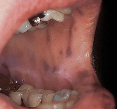
Figure 19-12. Peutz–Jeghers syndrome Multiple, dark-brown lentigines on the vermilion border of the lip and the buccal mucosa. This patient had GI bleeding due to hamartomatous polyps in the small bowel.
Glucagonoma Syndrome ICD-9: 211.7 ICD-10: M8152/0
Glucagonoma syndrome is a rare but well-described clinical entity caused by excessive production of glucagon in an α-cell tumor of the pancreas.
Characterized by superficial migratory necrolytic erythema (MNE) with erosions that crust and heal with hyperpigmentation.
Inflammatory patches and red plaques (Figs. 19-13 and 19-14) of gyrate, circinate, arcuate, or annular shape that enlarge with central clearing, resulting in geographic areas that become confluent (Fig. 19-14). Borders show vesiculation to bulla formation, crusting, and scaling.
Lesions involve perioral and perigenital regions and flexures and intertriginal areas.
Fingertips red, shining, erosive (Fig. 19-15).
There is glossitis, angular cheilitis (Fig. 19-13), blepharitis.
General examination reveals wasting, malnutrition.
Most cases are associated with glucagonoma, but the pathogenesis of MNE is not known. There exists MNE without glucagonoma.
Differential diagnosis: Includes all moist red plaque(s): acrodermatitis enteropathica, zinc deficiency, pustular psoriasis, mucocutaneous candidiasis, Hailey–Hailey disease (familial pemphigus).
Laboratory: Fasting plasma glucagon level increased to >1000 ng/L (normal 50–250 ng/L) and makes the diagnosis. There is also hyperglycemia, reduced glucose tolerance, severe malabsorption, gross hypoaminoacidemia, low serum zinc. CT scan angiography will locate tumor within pancreas and metastases in the liver.
Dermatopathology of early skin lesions shows band-like upper epidermal necrosis with retention of pyknotic nuclei and pale keratinocyte cytoplasm.
Prognosis depends on the aggressiveness of the glucagonoma. Hepatic metastases have occurred in 75% of patients at the time of diagnosis. If these are slow growing, patients may have prolonged survival, even with metastatic disease.
MNE responds poorly to all types of therapy. Some cases have responded partially to zinc replacement. MNE resolves after tumor excision. However, surgical excision of glucagonoma achieves cure in only 30% of cases because of persistent metastases (usually liver). There is poor response to chemotherapy.

Figure 19-13. Glucagonoma syndrome: migratory necrolytic erythema Inflammatory dermatosis with angular cheilitis, inflammatory, scaly, erosive, and crusted plaques and fissures around the nose and mouth.
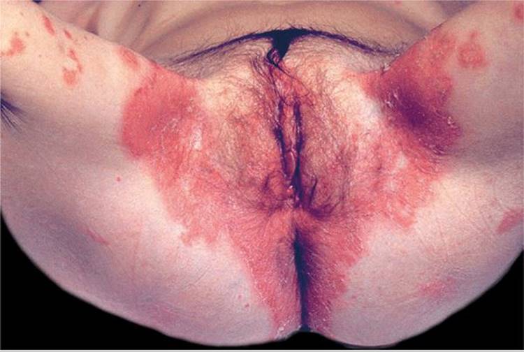
Figure 19-14. Glucagonoma syndrome: migratory necrolytic erythema Polycyclic erosions in the anogenital gluteal and sacral regions. Sharply defined with necrotic flaccid epidermis still covering part of these erosions.

Figure 19-15. Glucagonoma syndrome Fingertips are red, glistening, and partially erosive.
Malignant Acanthosis Nigricans ICD-9: 701.2 ICD-10: L83
Like other forms of acanthosis nigricans (AN) (see Section 5), malignant AN starts as a diffuse, velvety thickening and hyperpigmentation chiefly on the neck, axillae, and other body folds, as well as on the perioral and periorbital, umbilical, mamillary, and genital areas, giving the skin a dirty appearance (see Fig. 5-1).
Malignant AN differs from other forms of AN primarily because of (1) the more pronounced velvety hyperkeratosis and hyperpigmentation, (2) the pronounced mucosal involvement and involvement of the mucocutaneous junction, (3) tripe hands, and (4) weight loss and wasting due to the underlying malignancy.
AN may precede by 5 years other symptoms of a malignancy, usually adenocarcinoma of the GI or GU tract, bronchial carcinoma, or, less commonly, lymphoma. Malignant AN is a truly paraneoplastic disease, and a search for underlying malignancies is imperative. Removal of malignancy is followed by regression of AN.
See “Acanthosis Nigricans” in Section 5.
Paraneoplastic Pemphigus (PNP)
ICD-9: 694.4 ICD-10: L10.82
Mucous membranes primarily and most severely involved.
Lesions combine features of pemphigus vulgaris (page 101) and erythema multiforme (page 314), clinically, histologically, and immunopathologically.
Most prominent clinical findings consist of severe oral (Fig. 19-16) and conjunctival erosions in a patient with an underlying neoplasm.
These neoplasms are in order of frequency: non-Hodgkin lymphomas, chronic lymphatic leukemia, Castleman disease, thymoma, sarcoma, and Waldenström macroglobulinemia.
Patients with PNP may also have clinical and serologic evidence of myasthenia gravis and autoimmune cytopenia.
PNP sera contain autoantibodies to plakin antigens (in the intercellular plaque of desmosomes), envoplakin and periplakin, and to desmoplakin I and II. Less commonly patient sera may contain autoantibodies that recognize bullous pemphigoid antigen (230 kDa), plectin, and plakoglobin.
Autoantibodies of PNP cause blistering in neonatal mice and are detected by indirect immunofluorescence on rodent urinary bladder epithelium.
Treatment is directed toward elimination or suppression of malignancy but may also require systemic glucocorticoids.
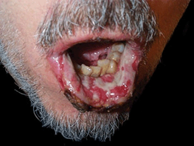
Figure 19-16. Paraneoplastic pemphigus Severe erosions covering practically the entire mucosa of the oral cavity partially covered by fibrin. Lesions are extremely painful, interfering with adequate food intake.
1Conditions covered in this section are printed in bold, conditions dealt with in other sections are in italics. Numbers in parentheses indicate page numbers. Rare conditions not discussed in this book are described in CA deWitt et al, in K Wolff et al (eds): Fitzpatrick’s Dermatology in General Medicine 7th ed. New York, McGraw-Hill, 2008, pp. 1493–1507.