
Organ transplant recipients are chronically immunosuppressed and their T cell function is impaired. Ensuing diseases are mostly infections and are similar to those occurring in other conditions associated with T cell impairment, such as AIDS. In addition, organ transplant recipients are at great risk for developing nonmelanoma skin cancer and other cancers. Bone marrow and stem cell graft recipients are candidates for graft-versus-host disease (GVHD).
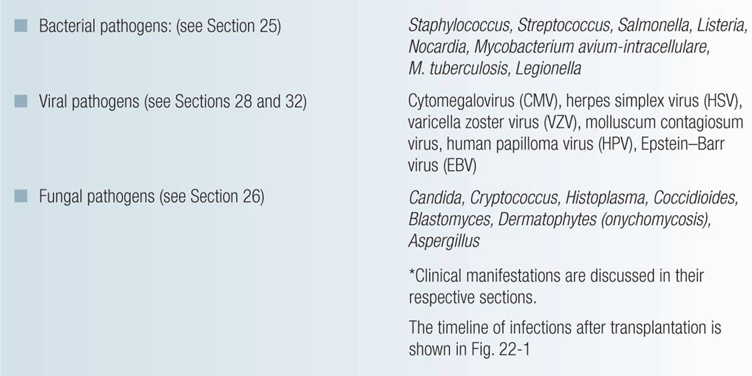
Most Common Infections Associated with Organ Transplantation* 
Timeline of common infections after transplantation
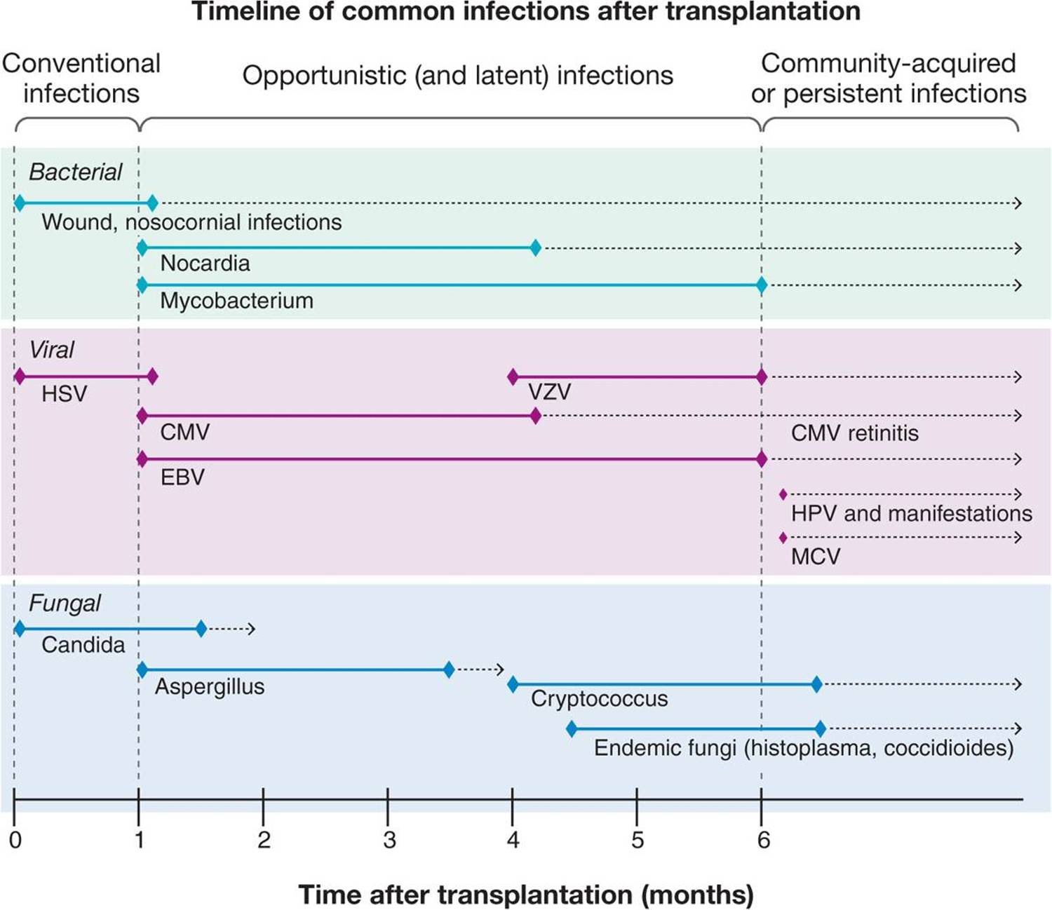
Figure 22-1. Timeline of common infections after transplantation.
Skin Cancers Associated with Organ Transplantation*
 Nonmelanoma skin cancer is the most common malignancy in adult solid organ transplant patients.
The majority are squamous cell carcinomas (SCC) (Section 11).
The risk of developing SCC increases exponentially with the length of immunosuppression.
The cumulative incidence is 80% after 20 years of immunosuppression in renal transplantation. SCC in posttransplant patients are aggressive.
HPV infection is implicated in the pathogenesis.
Other epithelial proliferative lesions are actinic keratoses, keratoacanthomas, porokeratosis, appendage tumors, and Merkel cell carcinomas (Section 11).
Children with organ transplants may also be at higher risk for the development of melanoma (Section 12).
Lymphoproliferative disorders are common in graft recipients and related to Epstein–Barr virus-mediated proliferation of B cells and most are lymphomas of B cell origin. Cutaneous T cell lymphomas account for 30% of cutaneous lymphomas in transplant patients (Section 21).
Kaposi sarcoma occurs in immunosuppressed transplant recipients with an incidence of 0.5–5%. All cases are associated with Kaposi sarcoma-associated herpesvirus (KSHV) infection (Section 21).
*Clinical manifestations are discussed in their respective sections.
Graft-Versus-Host Disease ICD:9: 996.85  ICD-10: T86.0
GVHD is the totality of organ dysfunction caused by the action of histoincompatible, immunocompetent donor cells against the tissues of an immunocompetent host.
Graft-versus-host reaction (GVHR) is the expression of GVHD in a specific organ (e.g., cutaneous GVHR).
Acute cutaneous GVHR, usually occurring 10–30 days after bone marrow transplantation (BMT). It is the earliest and most frequent GVHR. Liver and GI tract GVHR are also common.
Chronic cutaneous GVHR occurs >60 days after allogeneic BMT and manifests as lichenoid and sclerodermoid changes.
Incidence. Allogeneic BMT: 20–80% of successful engraftments. Autologous BMT: mild cutaneous GVHR occurs in 8%. Low incidence after blood transfusion in immunosuppressed patients, maternal-fetal transfer in immunodeficiency disease.
Acute Cutaneous GVHR
During the first 2 months after BMT (usually between 10 and 30 days): mild pruritus, localized/generalized; pain on pressure, palms/soles. Nausea/vomiting, abdominal pain; watery diarrhea. Jaundice; dark yellow urine.
Skin Lesions. Initially, subtle, discrete macules and/or papules on upper trunk, hands/feet (Fig. 22-2), especially palms/soles. Macules; confluent in the face, often erosive (Fig. 22-3). Painful. Mild edema with violaceous hue, periungual and on pinna. Erythema often in perifollicular array. If controlled/resolved, erythema diminishes with subsequent desquamation (Fig. 22-4) and postinflammatory hyperpigmentation. If it progresses, macules/papules become generalized, confluent, and evolve into erythroderma. Subepidermal bullae, especially over pressure/trauma sites, palms/soles. Positive Nikolsky sign. If bullae widespread with rupture/erosion, TEN-like form of acute cutaneous GVHR (see Section 8) (Fig. 22-5). For staging, see Table 22-1.
Mucosa. Lichen planus-like lesions in buccal mucosa; erosive stomatitis, oral and ocular sicca-like syndrome; esophagitis/esophageal strictures. Keratoconjunctivitis.
General Findings. Fever, jaundice, nausea, vomiting, right upper quadrant pain/tenderness, cramping, abdominal pain, diarrhea, serositis, pulmonary insufficiency, dark urine.
Chemistry. Elevated SGOT, bilirubin, alkaline phosphatase.
Dermatopathology. Focal vacuolization of basal cell layer, apoptosis of individual keratinocytes; mild perivenular mononuclear cell infiltrate. Apposition of lymphocytes to necrotic keratinocytes (satellitosis); vacuoles coalesce to form subepidermal clefts → subepidermal blister formation. Endothelial cell swelling. Immunocytochemistry: HLA-DR expression of keratinocytes precedes morphologic changes and thus represents important, early diagnostic sign.
Differential Diagnosis. Exanthematous drug reaction, viral exanthem, TEN, erythroderma.
Course and Prognosis. Mild-to-moderate GVHR responds well to treatment. Prognosis of TEN-like GVHR is grave. Severe GVHD susceptible to infections—bacterial, fungal, viral (CMV, HSV, VZV). Acute GVHD is primary or associated cause of death in 15–70% of BMT recipients.
Management Topical. Glucocorticoids. PUVA, extracorporeal photopheresis. Systemic. Methylprednisolone, tacrolimus, cyclosporine, mycophenolate mofetil, etanercept, infliximab.
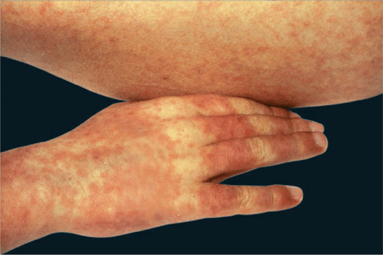
Figure 22-2. Acute cutaneous GVHR Discrete and confluent erythematous, blanching macules, and rarely elevated papules with indistinct borders involving hands and trunk. Note relative sparing over the metacarpophalangeal and proximal interphalangeal joints.
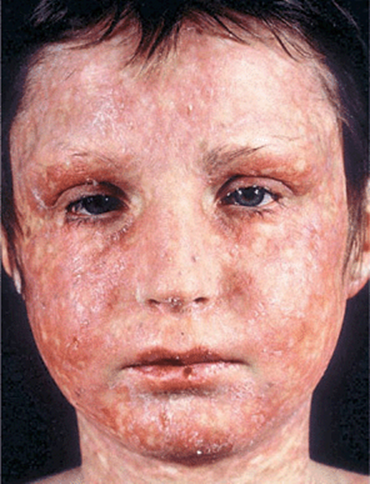
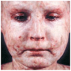
Figure 22-3. Acute cutaneous GVHR involving the face of a 10-year-old boy The individual lesions are confluent, there is slight desquamation, and there are erosions on the lips, cheeks and chin. The mucous membranes were severely involved.
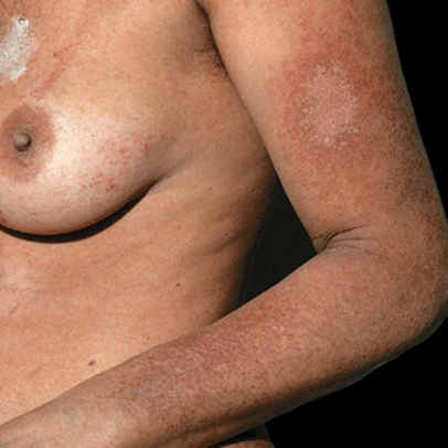
Figure 22-4. Acute cutaneous GVHR, remitting The maculopapular lesions have acquired a brownish hue and there is slight scaling.
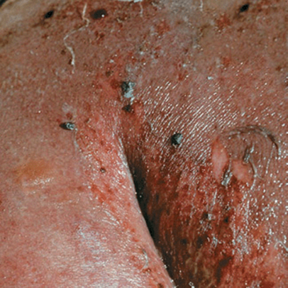
Figure 22-5. Acute GVHR, TEN-like Confluent epidermal necrosis with wrinkling and dislodgement of the necrotic epidermis, erosions, and hemorrhagic crusts. This severe reaction involved the entire skin and is indistinguishable from TEN. It occurred after allogeneic BMT and is clearly a very severe, life-threatening condition.
TABLE 22-1 CLINICAL STAGING OF ACUTE CUTANEOUS GVHR

Chronic Cutaneous GVHR 
More than 60 days after BMT. Evolving from acute GVHR or arising de novo. Acute GVHR not always followed by chronic GVHR. Clinical classification thus distinguishes between quiescent onset, progressive onset, and de novo chronic cutaneous GVHR. Chronic GVHR occurs in 25% of recipients of marrow from an HLA-identical sibling who survives > 100 days.
Skin Lesions. Flat-topped (lichen planus-like) papules of violaceous color, initially on distal extremities but later generalized (Fig. 22-6) and/or confluent areas of dermal sclerosis (Fig. 22-7A) with overlying scale resembling scleroderma mainly on trunk, buttocks, hips, and thighs. With more severe disease, severe generalized sclerodermoid changes also involving face (Fig. 22-7B) with necrosis and ulceration on acral and pressure sites. Hair loss; anhidrosis; nails: dystrophy, anonychia; vitiligo-like hypopigmentation.
Mucosa. Like erosive/ulcerative lichen planus.
General Findings. Chronic liver disease, general wasting.
Chemistry. Elevated ALT, AST, γ-glutamyltransferase.
Dermatopathology. Like lichen planus or like scleroderma.
Course and Prognosis. Sclerodermoid GVHR with tight skin/joint contracture may result in impaired mobility, ulcerations. Permanent hair loss; xerostomia, xerophthalmia, corneal ulcers, blindness. Malabsorption. Mild chronic cutaneous GVHR may resolve spontaneously. Chronic GVHR may be associated with recurrent and occasionally fatal bacterial infections.
Management. Topical glucocorticoids, PUVA, and extracorporeal photopheresis. Systemic immunosuppression with prednisone, cyclosporine, azathioprine, mycophenolate mofetil, methotrexate, tacrolimus, and thalidomide.
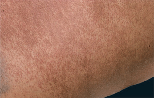
Figure 22-6. Chronic cutaneous GVHR, lichen planus-like Violaceous to brownish, lichen planus-like perifollicular papules becoming confluent on the trunk, occurring 3 months after allogeneic BMT.
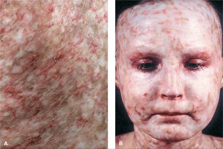
Figure 22-7. Chronic cutaneous GVHR, sclerodermoid (A) Close-up view of the back of a patient with poikilodermatous changes (hypo- and hyperpigmentation) and telangiectasias in the sclerotic skin. (B) Ebony-white bound down skin and telangiectasias in the 10-year-old boy shown in Fig. 22-3. Skin looks and feels like severe scleroderma. In this case, acute GVHR evolved directly into chronic GVHR and involved the entire skin of the head, trunk and extremities.