

Normal Nail Apparatus
 The nail apparatus is made up of:
Nail plate, the horny “dead” product.
Four specialized epithelia: proximal nail fold, nail matrix, nail bed, hyponychium.
Nail apparatus disorders can be traumatic, primary, manifestations of cutaneous disease (e.g., psoriasis), neoplastic, infectious, or manifestations of systemic diseases (e.g., lupus erythematosus).
Components of the Normal Nail Apparatus (See Fig. 32-1)
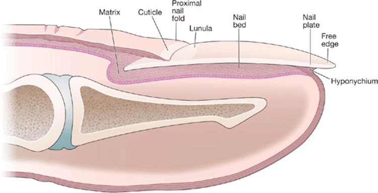
Figure 32-1. Schematic drawing of normal nail.
Local Disorders of Nail Apparatus
Local disorders affecting the nail apparatus can result in a spectrum of chronic nail diseases.
Chronic Paronychia ICD-9: 681.02  ICD-10: L03.0
Associated with damage to cuticle: mechanical or chemical.
At risk: adult women, food handlers, house cleaners.
Chronic dermatitis of proximal nail fold and matrix: chronic inflammation (eczema, psoriasis) with loss of cuticle, separation of nail plate from proximal nail fold (Fig. 32-2).

Figure 32-2. Chronic paronychia The distal fingers and periungual skin are red and scaling. The cuticle is absent; a pocket is present, formed as the proximal nail folds separate from the nail plate. The nail plates show trachonychia (rough surface with longitudinal ridging) and onychauxis (apparent nail plate thickening due to subungual hyperkeratosis of nail bed). The underlying problem is psoriasis. Candida albicans or Staphylococcus aureuscan cause space infection in the “pocket” with intermittent erythema and tenderness of the nail fold.
Predisposing factors:
Dermatosis: psoriasis, dermatitis [atopic, irritant (occupational), allergic contact], lichen planus.
Drugs: oral retinoids (isotretinoin, acitretin), indinavir.
Foreign body: hair, bristle, wood splinters.
Manifestations: first, second, and third fingers of dominant hand; proximal and lateral nail folds erythematous and swollen; cuticle absent.
Intermittently, persistent low-grade inflammation may flare into subacute painful exacerbations, resulting in discolored transverse ridging of lateral edges.
Secondary infection/colonization: Candida spp., Pseudomonas aeruginosa, or Staphylococcus aureus. Nail plate may become discolored; green undersurface with Pseudomonas. Infection associated with painful acute inflammation.
Management:
Protection.
Treat the dermatitis with glucocorticoid: topical, intralesional triamcinolone, short course of prednisone.
Treat secondary infection.
Onycholysis ICD-9: 703.8 ICD-10: L60.1
Detachment of nail from its bed at distal and/or lateral attachments (Fig. 32-3).
Onycholysis creates a subungual space that collects dirt and keratinous debris; area may be malodorous when the overlying nail plate is removed.
Etiology:
Primary: Idiopathic (fingernails in women; mechanical or chemical damage); trauma (fingernails, occupational injury; toenails, podiatric abnormalities, poorly fitting shoes).
Secondary: Vesiculobullous disorders (contact dermatitis, dyshidrotic eczema, herpes simplex); nail bed hyperkeratosis (onychomycosis, psoriasis, chronic contact dermatitis); nail bed tumors; drugs.
In psoriasis, yellowish-brown margin is visible between pink normal nail and white separated areas. In “oil spot” or “salmon-patch” variety (Fig. 32-3), nail plate–nail bed separation may start in middle of nail.
Colonization with P. aeruginosa results in a biofilm on the undersurface of the onycholytic nail plate, causing a brown or greenish discoloration (Fig. 32-4).
Other secondary pathogens that can colonize/infect the space are Candida spp., dermatophytes, and numerous environmental fungi.
Underlying disorders in fingernail onycholysis: trauma (e.g., splinter), psoriasis, photoonycholysis (e.g., doxycycline), dermatosis adjacent to nail bed (e.g., psoriasis, dermatitis, chemical exposure), congenital/hereditary.
Underlying toenail onycholysis: additional factors of onychomycosis (Trichophyton rubrum), shoe trauma.
Management: debride all nail separated from nail bed (patient should continue weekly debridement); remove debris on nail bed; treat underlying disorders.
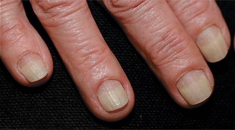
Figure 32-3. Onycholysis A 60-year-old female with distal onycholysis of fingernails, mild chronic paronychia, and loss of cuticle. Psoriasis is the likely underlying problem.
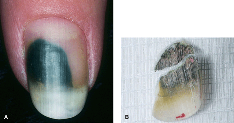
Figure 32-4. Onycholysis with Pseudomonas colonization (A) Psoriasis has resulted in distal onycholysis of the thumbnail. (B) A biofilm of Pseudomonas aeruginosa has produced the green-black discoloration of the undersurface of the onycholytic nail, which resolved following the debridement and treatment of the nail bed with glucocorticoid cream.
Green Nail Syndrome 
Usually associated with onycholysis (see above). P. aeruginosa, the most common cause, produces the green pigment pyocyanin (Fig. 32-4).
Management debride “Iytic” nail. See above.
Onychauxis and Onychogryphosis 
Onychauxis: Thickening of entire nail plate, seen in elderly.
Onychogryphosis: Onychauxis with ram’s hornlike deformity, most commonly of great toe (Fig. 32-5).
Etiology pressure from footwear in elderly; also, inherited autosomal dominant.
Keratin produced by matrix at uneven rates, with faster-growing site determining direction of deformity, without attachment to nail bed.
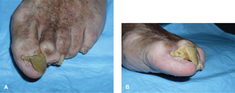
Figure 32-5. Onychauxis and onychogryphosis The great toenails appear grossly thickened with transverse ridging (onychauxis) with some medial deviation (onychogryphosis or ram’s horn deformity). (Courtesy of Dr. Nathaniel Jellinek.)
Psychiatric Disorders 
Repeated manipulation of the nail apparatus can result in changes of the paronychial skin and the nail plate.
Habit-tic Deformity. Caused by chronic, mechanical injury (Fig. 32-6). Cuticle is pushed back with inflammation and thickening of proximal nail fold. Occurs most commonly on thumbnail(s), as compulsive disorders (tic habit), caused by the index finger repeatedly picking at cuticle of thumbnail.
Obsessive Compulsive Disorder. Repeat picking at the paronychia skin can result in lichen simplex chronicus. S. aureus secondary infection is a common complication. In extreme cases, the nail plate can be destroyed (Fig. 32-7); nail biting.

Figure 32-6. Habit-tic deformity The nail plates of both thumbs are dystrophic with transverse ridging and discoloration. The cuticle is absent and the proximal nail folds excoriated. When the proximal nails and nail fold were covered with tape continually, normal nails regrew in 5 months.

Figure 32-7. Compulsive nail picking The cuticles are not formed, the proximal nail folds are inflamed and excoriated. The breaks in the integrity provide a portal of entry for S. aureus and acute paronychia.
Nail Apparatus Involvement of Cutaneous Diseases
Psoriasis 
Most common dermatosis affecting the nail apparatus.
>50% of persons with psoriasis have nail involvement at one point in time, with lifetime involvement up to 80-90%.
See also “Psoriasis” in Section 3.
Laboratory Examination
KOH preparation and/or nail clipping to pathology for PAS stain to rule out fungal colonization/infection. Onychomycosis is more common in nails with onycholysis.
Clinical Findings
Skin. Typical psoriatic lesion on nail folds (Fig. 32-8).
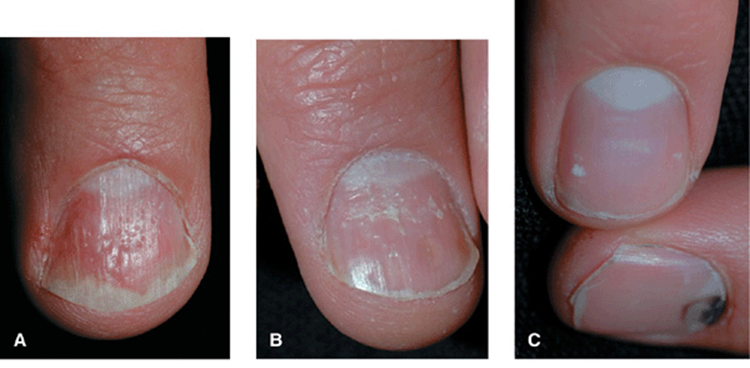

Figure 32-8. Psoriasis vulgaris (A) Multiple nail pits on the dorsal nail plate, “oil staining” of the nail bed, and distal onycholysis. (B) Trachonychia (rough surface) with oil staining and distal onycholysis. (C) Punctate leukonychia is pathognomonic for psoriasis and may be seen in only one finger. As can be seen in the nail below with traumatic subungual hemorrhage, punctate leukonychia did not occur at this site of trauma. (D) Oil staining, distal onycholysis, longitudinal ridging, adherence of the cuticle to the distal nail plate.
Matrix
• Pitting or elkonyxis: Punctate depressions; small, shallow; vary in size, depth, shape (Fig. 32-8A). May occur as regular lines (transverse; long axis) or grid-like pattern. Uncommon on toenails. Also seen in atopic dermatitis. Geometric and superficial pits seen in alopecia areata (hammered brass nails).
• Trachyonychia: Nail dull, rough, fragile (Fig. 32-8B). Twenty-nail dystrophy or sandpaper nails associated with proximal nail matrix damage: nonspecific and can also be seen in alopecia areata (see Fig. 32-10), lichen planus, atopic dermatitis. May regress spontaneously.
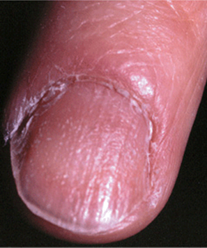
Figure 32-10. Alopecia areata: trachonychia The nail plate is rough with a “hammered brass” appearance.
• Serial transverse depressions: May mimic “washboard” nails of tic habit (pushing back cuticle).
• Longitudinal ridging: Resembles melted wax.
• Punctate leukonychia: 1- to 2-mm white spots in nail plate (mistakenly attributed to trauma) (Fig. 32-8C).
• Leukonychia: Proximal matrix involvement: surface rough and nail coarse (Fig. 32-8C).
Nail Bed
• ″Oil″ spots: Oval, salmon-colored nail beds (Fig. 32-8A, D).
• Onycholysis: Secondary to “oil” spots affecting hyponychium medially or laterally (Fig. 32-8A). May become colonized with Candida, environmental fungi (e.g., Aspergillus), Pseudomonas. Predisposes to distal/lateral onychomycosis in toenail. Up to 20% of psoriatic nails have secondary onychomycosis.
• Subungual hyperkeratosis: Nail plate becomes raised off hyponychium.
• Splinter hemorrhages.
Differential Diagnosis
Onycholysis, onychomycosis, trauma (toenails), eczema, alopecia areata.
Management
• Often unsatisfactory. See “Psoriasis” In Section 3.
• For matrix involvement, intralesional triamcinolone 3-5 mg/mL may be effective.
• For nail bed psoriasis, topical steroid (occluded) reduces hyperkeratosis.
• Systemic therapy such as methotrexate, acitretin, or “biologics” often improves nail apparatus psoriasis but may lag a few months after completion of therapy.
Lichen Planus (LP)
Nail involvement occurs in 10% of individuals with disseminated LP.
Nail apparatus involvement may be the only manifestation.
One, several, or all 20 nails may be involved (“twenty-nail syndrome,” where there is loss of all 20 nails without any other evidence of lichen planus elsewhere on the body). Onychorrhexis seen(longitudinal ridging and fissuring of the nail plate with brittleness and breakage.), though this is not a specific feature and can be seen with aging.
Similar changes are seen in lichenoid graft-versus-host disease
Course: May destroy nails.
See also “Lichen Planus” in Section 14.
Clinical Manifestations
Skin swelling with blue/red discoloration of proximal nail fold.
Matrix
• Small focus in matrix: Bulge under proximal nail fold (Fig. 32-9A).

Figure 32-9. Lichen planus (A) Middle finger: involvement of the proximal fold and matrix has caused trachonychia, longitudinal ridging, and pterygium formation. Index finger: destruction of the matrix and nail plate is complete with anonychia. Seven of ten fingernails are involved; the others are normal. (B) Involvement of the nail matrix with scarring or pterygium formation proximally dividing the nail plate in two. (C) Early involvement of the matrix with thinning of the thumbnail plates. (D) Same patient as Fig. 32-8C 2 years later, the nail plate is completely destroyed, i.e., anonychia.
• Subsequent longitudinal red line: Thinned nail plate evolving into distal split nail (onychorrhexis) (Fig. 32-9B).
• Diffuse matrix involvement: Selective atrophy of nail plate with onychorrhexis and/or transverse splitting.
• Red lunula: Focal or disseminated.
• Melanonychia, longitudinal: Transitory.
• Complete nail split.
• Pterygium formation (scar, matrix destroyed): Partial loss of the central nail plate presents as a V-shaped extension of skin of proximal nail fold adherent to nail bed (Fig. 32-9A, B).
• “Idiopathic atrophy of nails”: Acute progressive nail destruction leading to diffuse nail atrophy with and without pterygium; complete loss of nail (anonychia) (Fig. 32-9B-D).
Nail Bed. Onycholysis, distal subungual hyperkeratosis, bulla formation, permanent anonychia.
Variants
• 20-nail dystrophy of childhood: Resolves spontaneously.
• LP-like eruptions following bone marrow transplant: Graft-versus-host disease.
• Drug-induced LP-like reaction.
Management
• See “Lichen Planus,” Section 14.
• Intralesional triamcinolone.
• Systemic glucocorticoids.
Alopecia Areata (AA) 
See “Nonscarring Alopecia,” Section 31.
Manifestations:
Geometric pitting (Fig. 32-10) (small, superficial, regularly distributed).
Hammered brass appearance.
Mottled erythema of lunulae.
Trachonychia (roughness caused by excessive longitudinal striations).
Darier Disease (Darier–White Disease,
Keratosis Follicularis)
Nail changes are pathognomonic: longitudinal streaks (red and white); distal subungual hyperkeratotic papules with distal V- or wedge-shaped fissuring of nail plate (Fig. 32-11).

Figure 32-11. Darier disease Red and white longitudinal streaks on the fingernails with V-nicking in distal portion of plate. [From Goldsmith LA et al. (eds.). Fitzpatrick’s Dermatology in General Medicine, 8th ed. New York: McGraw-Hill, 2012.]
Chemical Irritant or Allergic Damage or Dermatitis 
Chemicals in nail polish and adhesive for paste- on nails can cause damage to the nail plate, i.e., discoloration, onychoschizia (splitting or lamination of the nail plate, usually in the horizontal plane at free edge; Fig. 32-12). Irritant or allergic contact dermatitis can also occur on the paronychial skin.

Figure 32-12. Chemically damaged nail False nail glued to the fingernail has chemically damaged the nail plate with leukonychia and onychoschizia (splitting and lamination of nail plate).
Neoplasms of the Nail Apparatus ICD-9: 703.8 ICD-10: L60
Benign tumors: Fibroma/fibrokeratoma, subungual exostosis, myxoid cyst, glomus tumor (painful red nail bed patch), onychomatricoma, nail matrix nevi.
Malignant tumors: Squamous cell carcinoma, melanoma, Merkel cell tumor.
Myxoid Cysts of Digits (See “Digital Myxoid Cyst” in Section 9)
Pseudocyst or ganglion originates in distal interphalangeal joint, associated with osteoarthritis (Heberden nodes).
Lesions can present on the proximal nail fold (Fig. 32-13), above and compressing the matrix, resulting in a longitudinal depressed groove in the nail plate.
When cysts expand between the periosteum and matrix, nail becomes dystrophic with a dusky red lunula.

Figure 32-13. Myxoid cysts (A) Dermal erythema and swelling of the proximal nail folds with associated longitudinal groove of the nail plate. (B) Clear gelatinous fluid has drained from the index finger on the right (crusted site). Degenerative joint disease is present in both distal interphalangeal joints.
Longitudinal Melanonychia
Manifestations: Tan, brown, or black longitudinal streak within nail plate (Fig. 32-14).
Pathogenesis: (1) Increased melanin synthesis in normally nonfunctional matrix melanocytes, (2) increase in total number of melanocytes synthesizing melanin, (3) nevomelanocytic nevus (junctional, Fig. 32-14).
. Onset Congenital or acquired. Most originate in distal matrix.
Differential diagnosis: Focal activation of nail matrix (e.g., trauma), hyperplasia of nail matrix melanocytes, nevomelanocytic nevus (junctional), drug-induced [e.g., zidovudine (AZT)] hydroxychloroquine, or melanoma of nail matrix.

Figure 32-14. Junctional nevomelanocytic nevus of the nail matrix A junctional nevus is present in the nail matrix resulting in a longitudinal brown stripe in the nail bed. The proximal nail fold/cuticle is not pigmented.
Nail Matrix Nevi 
Appear as longitudinal melanonychia (Fig. 32-14).
Onset: childhood.
Course: color and width change with aging.
Acrolentiginous Melanoma (ALM) (See Section 12) 
Mean age: 55–60 years. Incidence: 2–3% of melanomas in whites; 15-20% in blacks, Asian, Native Americans. Usually asymptomatic; most patients notice pigmented lesion, usually after trauma.
Dermatopathology: In situ or invasive.
Findings: Arises subungually or periungually, presenting with longitudinal melanonychia and/or nail plate dystrophy (Fig. 32-15). Matrix lesions usually present as ALM in whites or broadening of an existing ALM in blacks.
Hutchinson sign: Periungual extension of brown-black pigmentation onto proximal and lateral nail folds (Fig. 32-15A).
25% of ALM may be amelanotic (pigmentation not obvious or prominent).
Distribution: Thumbs, great toes (hallux).
Differential diagnosis: Subungual hemorrhage (Fig. 32-158).
Indications for biopsy: Periungual pigmentation, adult age, change in color/width of band, hyperpigmented lines within the band, proximal portion of band wider than distal; thumb, index finger, or toe involvement; blurred margins, history of trauma.
Prognosis: 5-year survival rates from 35% to 50%.

Figure 32-15. Acrolentiginous melanoma versus subungual hemorrhage (A) Melanoma arose in the nail matrix of the thumb with resultant nail plate dystrophy, subungual melanosis, and extension into the proximal nail fold and beyond it (Hutchinson sign). (B) Trauma to the proximal nail resulted in hemorrhage and a transverse depression across the nail plate. Hemorrhage extends to the longitudinal dermal ridges.
Squamous Cell Carcinoma (See Section 11)
SCC in situ (SCCIS) occurring periungually is usually caused by the oncogenic human papillomavirus types 16 and 18.
Findings: Skin-colored or hyperpigmented, keratotic, hyperkeratotic, or warty papules/plaques; onycholysis; failure of nail formation.
Distribution: Proximal and lateral nails, matrix, hyponychium (Fig. 32-16).
Invasive SCC arises within SCCIS.
Symptoms: Pain if periosteal invasion has occurred.
Findings: Solitary nodule is most common, often destroying the nail.
Distribution: Much more common on fingers (thumb and index finger most often) than toes; multiple fingers may be involved in the immunocompromised host.
Management Mohs surgery or amputation of digit for more deeply invasive lesions involving periosteum.

Figure 32-16. HPV-induced in situ and invasive squamous cell carcinoma (A) The right index fingernail bed shows hyperkeratotic failure of nail plate formation. Biopsy of the nail bed reported SCCIS with HPV-induced changes (koilocytosis). (B) Progression into invasive squamous cell carcinoma may present as hyperkeratotic papules or (C) complete obliteration of the nail unit. (Parts B and C courtesy of Dr. Nathaniel Jellinek.)
Infections of the Nail Apparatus ICD-9: 681.9 ICD-10: L03.019
Dermatophytes are the most common pathogens infecting the nail apparatus.
S. aureus and group A streptococcus cause acute soft-tissue infection of the nail fold.
Candida and S. aureus can cause secondary infection of chronic paronychia.
Recurrent herpes simplex virus infection.
Bacterial Infections
• S. aureus is the most common cause of acute paronychia.
• Felon is an acute infection of the finger tip.
• Management: See “Antimicrobial Therapy” in Section 25.
Acute Paronychia ICD-10 L03.01
Acute infection of lateral or proximal nail fold.
Usually associated with break in integrity of epidermis (e.g., hang nail), trauma.
Findings: Throbbing pain, erythema, swelling, pain, ± abscess formation (Fig. 32-17).
Infection may extend deeper, forming a felon (Fig. 32-18).

Figure 32-17. Acute paronychia The proximal nail fold is red and edematous (cellulitis) with pus formation.
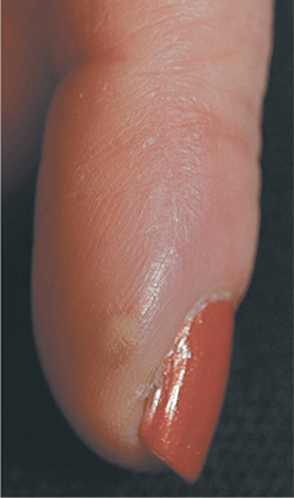
Figure 32-18. Felon An abscess is seen on the fingertip with surrounding erythema and swelling. Methicillin-sensitive S. aureus (MSSA) was isolated on culture of the pus.
Felon ICD-9: 681.01 ICD-10: L03.0 
Soft-tissue infection of pulp space of distal phalanx (Fig. 32-18); closed space infection of multiple compartments created by fibrous septa passing between the skin and periosteum.
History Penetrating injury, splint, paronychia.
Findings: Pain, erythema, swelling, abscess (Fig. 32-18).
Distribution: Thumb, index finger.
Complications: Osteitis, osteomyelitis of distal phalanx, sequestration of diaphysis of the phalanx; rupture into distal interphalangeal joint with septic arthritis; extension into distal end of flexor tendon sheath, producing tenosynovitis.
Course: May be rapid and severe; contained by unyielding skin of fingertip, infection creates tension with microvascular compromise, necrosis, and abscess formation.
Fungal Infections and Onychomycosis
• Candida spp. usually cause “space” infections of chronic paronychia or onycholytic nail and can cause destruction of the nail in the immunocompromised host.
• Dermatophytes infect the skin around the nail apparatus and cause superficial destruction of nail.
• Onychomycosis: Chronic progressive fungal infection of nail apparatus, most commonly caused by dermatophytes, less often by Candida spp.; molds and environmental fungi can be cultured from diseased nails but are not usually primary pathogens.
Candida Onychia
Candida albicans infections of the nail apparatus occur most often on fingers, commonly as secondary infection of chronic paronychia. Onychia describes inflammation of the matrix of the nail resulting in shedding of nail.
Invasion of nail plate usually occurs only in the immunocompromised host, i.e., chronic mucocutaneous candidiasis (CMC) or HIVIAIDS disease.
Etiology and Epidemiology
Etiology. C. albicans and other species. Normal flora, which causes infection if local ecology is changed in favor of yeast or in association with altered immune status. See “Candidiasis,” Section 26.
Classification
• Subungual infection associated with onycholysis.
• Intermittent flares of chronic paronychia.
• Colonization in tinea unguium.
• Total nail dystrophy (Fig. 32-19): chronic CMC and HIV/AIDS disease.

Figure 32-19. Candida onychomycosis: total dystrophic type The entire fingernail plate is thickened and dystrophic and is associated with a paronychial infection; both findings were caused by C. albicansin an individual with advanced HIV/AIDS disease.
Chronic CMC. See “Candidiasis,” Section 26.
Clinical Findings
See “Candidiasis,” Section 26.
HIV/AIDS. Candidal onychia and paronychia are common in children with HIV/AIDS, often associated with mucosal candidiasis.
Nail Apparatus. Chronic Paronychia with Acute Candidal Flare. Candida spp. can cause painful chronic infection with pain, tenderness, erythema, ± pus. Nail may become dystrophic with areas of opacification; white, yellow, green, or black discoloration; with transverse furrowing. Colonization in Tinea Unguium. Secondary pathogen in distal/lateral onychomycosis.
Total Nail Dystrophy. Proximal/lateral nail folds are inflamed and thickened. Fingertips appear bulbous. Nail is invaded and may eventually become totally dystrophic (Fig. 32-19). HIV/AIDS: one nail may be involved. CMC: 20 nails may be involved in time.
Other Findings. See “Candidiasis,” Section 26.
Differential Diagnosis
Tinea unguium, psoriasis, eczema, chronic paronychia, lichen planus.
Management
See “Candidiasis,” Section 26.
Tinea Unguium/Onychomycosis
Symptoms: nails lose protective and manipulative function.
Complications:
Pain in toenail with pressure from shoes.
Predispose to secondary bacterial infections.
Ulcerations of the underling nail bed.
Complications occur more commonly in the growing population of immunocompromised individuals and diabetic patients.
See also Section 26.
Classification by Anatomic Site Involved
Distal and Lateral Subungual Onychomycosis (DLSO) (Fig. 32-20). Infection begins in hyponychial area or nail fold, extending subungually. May be either primary, i.e., involving a healthy nail apparatus, or secondary (e.g., psoriasis) associated with onycholysis. Always associated with tinea pedis.

Figure 32-20. Onychomycosis of toenails: distal and lateral subungual type (DLSO) The toenails are white, caused by onycholysis and subungual hyperkeratosis. The dorsum of the feet shows erythema and scaling, i.e., tinea pedis. T. rubrum was detected on culture.
Superficial White Onychomycosis (SWO). Pathogen invades surface of dorsal nail (Fig. 32-21). Etiology: Trichophyton mentagrophytes or T. rubrum (children). Much less commonly, mold: Acremonium, Fusarium, Aspergillus terreus.
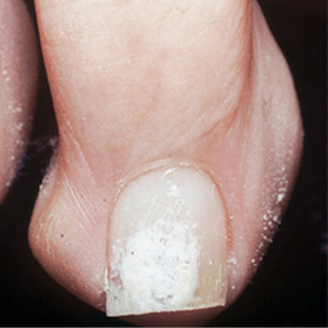
Figure 32-21. Onychomycosis of toenails: superficial white type (SWO) The dorsal nail plate is chalky white. White nail dystrophy can easily be treated by curettage; KOH preparation of the curetting shows hyphae.
Proximal Subungual Onychomycosis (PSO). Pathogen enters by way of the posterior nail fold-cuticle area and then migrates along the proximal nail groove to involve the underlying matrix, proximal to the nail bed, and finally the underlying nail (Fig. 32-22). Etiology: T. rubrum. Findings: Leukonychia that extends distally from under proximal nail fold. Usually one or two nails involved. Always associated with immunocompromised states.

Figure 32-22. Tinea unguium: proximal subungual onychomycosis type The proximal nail plate is a chalky white color due to invasion from the undersurface of the nail matrix. The patient had advanced HIV/AIDS disease.
Etiology and Epidemiology
Age of Onset. Children or adults. Once acquired, usually does not remit spontaneously. Therefore, the incidence increases with advancing age; 1% of individuals <18 years affected; almost 50% of those >70 years.
Sex. Somewhat more common in men.
Etiologic Agents. Between 95% and 97% caused by T. rubrum and T. mentagrophytes.
Molds. Acremonium, Fusarium, and Aspergillus spp. can rarely cause SWO. Dermatosis such as psoriasis, which results in onycholysis and subungual hyperkeratosis, or dermatophytic onychomycosis can be secondarily colonized/infected by molds.
Geographic Distribution. Worldwide. Etiologic agent varies in different geographic areas. More common in urban than in rural areas (associated with wearing occlusive footwear).
Prevalence. Incidence varies in different geographic regions. In the United States and Europe, up to 10% of adult population affected (related to occlusive footwear). In developing nations where open footwear is worn, uncommon.
Transmission. Dermatophytes. Anthropophilic dermatophyte infections are transmitted from one individual to another, by fomite or direct contact, commonly among family members. Some spore forms (arthroconidia) remain viable and infective in the environment for up to 5 years.
Molds. Ubiquitous in environment; not transmitted between humans.
Risk Factors. Atopics are at increased risk for T. rubrum infections. Diabetes mellitus, treatment with immunosuppressive drugs, HIV/AIDS. For toenail onychomycosis, most important factor is wearing of occlusive footwear.
Pathogenesis
Primary Onychomycosis/Tinea Unguium. The probability of nail invasion by fungi increases with defective vascular supply (i.e., with increasing age, chronic venous insufficiency, peripheral arterial disease), in posttraumatic states (lower leg fractures), or disturbance of innervation (e.g., injury to brachial plexus, trauma of spine).
Secondary Onychomycosis. Infection occurs in already altered nail apparatus, such as psoriatic or traumatized nail.
DLSO (Fig. 32-20). Nail bed produces soft keratin stimulated by fungal infection that accumulates under the nail plate, thereby raising it. Matrix is usually not invaded, and production of normal nail plate remains unimpaired despite fungal infection.
Clinical Manifestation
Approximately 80% of onychomycosis occurs on the feet, especially on the big toes; simultaneous occurrence on toe- and fingernails is not common.
DLSO. White patch is noted on the distal or lateral undersurface of the nail and nail bed, usually with sharply demarcated borders. With progressive infection, the nail becomes opaque, thickened, cracked, friable, raised by underlying hyperkeratotic debris in hyponychium (Fig. 32-20). When fingernails are involved, pattern is usually two feet and one hand.
SWO. A white chalky plaque is seen on the proximal nail plate, which may become eroded with loss of the nail plate (Fig. 32-21). SWO may coexist with DLSO. Occurs almost exclusively on the toenails, rarely on the fingernails.
PSG (Fig. 32-22). A white spot appears from beneath proximal nail fold. In time, white discoloration fills lunula, eventually moving distally to involve much of undersurface of the nail. Occurs more commonly on toenails.
Differential Diagnosis
DLSO. Psoriatic nails (“oil drop” staining of the distal nail bed and nail pits is seen in psoriasis but not onychomycosis), eczema, Reiter syndrome and keratoderma blennorrhagicum, onychogryphosis, pincer nails, congenital nail dystrophies.
SWO. Traumatic or chemical injury to nail, psoriasis with leukonychia.
Laboratory Examinations
All clinical diagnoses of onychomycosis should be confirmed by laboratory testing (see “Dermatophytoses,” Section 26).
Nail Samples. For DLSO: distal portion of involved nail bed; SWO: involved nail surface; PSO: punch biopsy through nail plate to involved nail bed.
Direct Microscopy. Specific identification of pathogen is usually not possible by microscopy, but, in most cases, yeasts can be differentiated from dermatophytes by morphology.
Fungal Culture. Isolation of the pathogen permits better use of oral antifungal agents.
Histology of Nail Clipping. Indicated if clinical findings suggest onychomycosis after negative KOH wet mounts. PAS stain is used to detect fungal elements in the nail. Most reliable technique for diagnosing onychomycosis.
Diagnosis
Clinical diagnosis is never adequate. Clinical findings confirmed by finding fungal forms in KOH preparation, nail clipping, and/or isolation of pathogenic fungus on culture.
Course and Prognosis
Without effective therapy, onychomycosis does not resolve spontaneously; progressive involvement of multiple toenails is the rule. DLSO persists after topical treatment of tinea pedis and often results in repeated episodes of epidermal dermatophytosis of feet, groin, and other sites. Tinea pedis and/or DLSO provide portal of entry for recurrent bacterial infections (S. aureus, group A streptococcus), especially cellulitis of lower leg after venous harvesting. Prevalence in diabetic patients estimated to be 32%; Diabetic patients need early intervention and should be screened regularly by a dermatologist and/or podiatrist. Untreated HIV/AIDS is associated with increased prevalence of dermatophytoses. Long-term relapse rate with newer oral agents such as terbinafine or itraconazole reported to be 15-21% 2 years after successful therapy; mycologic cultures may be positive without any clinically apparent disease.
Management
See Section 26 and Table 32-1.
TABLE 32-1 MANAGEMENT OF TINEA UNGUIUM

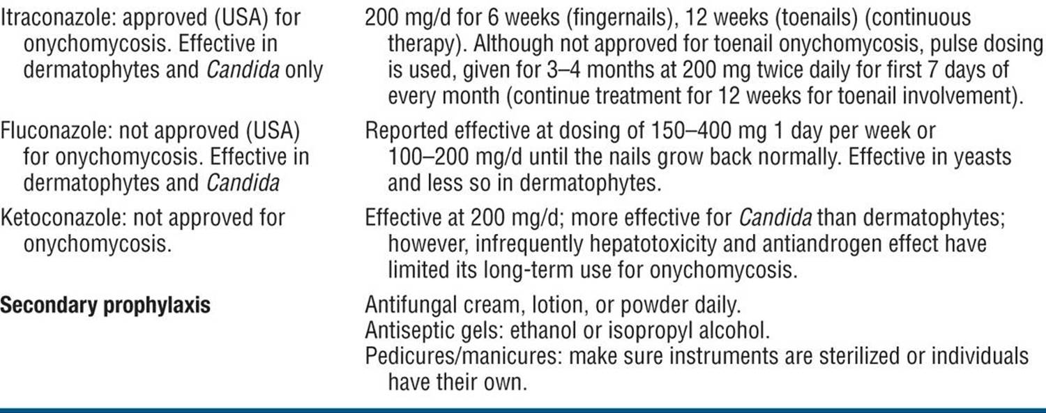
Indications for Systemic Therapy. Fingernail involvement, limitation of function, pain (thickened great toenails with pressure on nail bed, ingrowing toe nails), physical disability, potential for secondary bacterial infection, source of recurrent epidermal dermatophytosis, quality-of-life issues (poorer perceptions of general and mental health, social functioning, physical appearance, difficulty in trimming nails, discomfort in wearing shoes). Early onychomycosis is easier to cure in younger, healthier individuals than in older individuals with more extensive involvement and associated medical conditions.
Nail Signs of Multisystem Diseases ICD-9: 703.8 ICD-10: L60.0
A wide spectrum of systemic disorders can affect the nail apparatus.
Transverse or Beau Lines
Systemic disease implicated if all 20 nails involved. Single nail involvement is usually traumatic, compulsive picking, or tearing at the nails (onychotillomania). Pathogenesis: Occur after any severe, sudden, acute, particularly febrile illness; damage to matrix. Etiology High fever, postnatal, cytotoxic drugs, severe adverse cutaneous drug reaction, dermatologic disease (eczema, erythroderma, paronychia), viral infection (hand-foot-and-mouth disease, measles), Kawasaki syndrome, peripheral ischemia. Findings: Transverse, bandlike depressions in nail, extending from one lateral edge to the other, affecting all nails at corresponding levels (Fig. 32-23). If duration of disease completely inhibits matrix activity for 7–14 days, transverse depression results in total division of nail plate (onychomadesis). Multiple parallel lines with chemotherapy. Duration: Thumbnails (lines present for 6-9 months) and large nails (lines present for up to 2 years) are most reliable markers.

Figure 32-23. Cancer chemotherapy: Beau lines Multiple transverse ridging of multiple fingernails was associated with chemotherapy for breast cancer.
Leukonychia
True Leukonychia. Attributable to matrix dysfunction:
Total leukonychia: Usually inherited.
Subtotal leukonychia: Distal nail pink.
Transverse leukonychia: 1- to 2-mm wide arcuate bands.
Punctate leukonychia: Psoriasis, trauma.
Longitudinal leukonychia: Darier disease (Fig. 32-11).
Pseudoleukonychia. SWO (Fig. 32-21), chemical damage to nail keratin.
Apparent Leukonychia. Due to alteration of matrix and/or nail bed (e.g., apparent macrolunula); may involve all fingernails:
Terry-type leukonychia
Association: Hepatic disorders.
Findings: Opaque white plate obscuring lunula and extending to within 1-2 mm from distal edge of nail (Fig. 32-24). Involves all nails evenly.
Uremic Half-and-Half Nail of Lindsay
Association: Renal disorders.
Findings: Proximal nail dull white obscuring lunula (20-60% of nail); distal nail pink/reddish.
Banded nails (Muehrcke lines) (see Fig. 32-34)
Paired, narrow, white transverse bands.
Association: Cancer antineoplastic chemotherapy, hypoalbuminemia; unilateral following trauma.
Findings: Bands are parallel to lunula, separated from one another, and from lunula, by strips of pink nail.

Figure 32-24. Apparent leukonychia: Terrytype nails The proximal two-thirds of the nail plate is white, whereas the distal third shows the red color of the nail bed.
Yellow Nail Syndrome
Symptoms: Nails stop growing. Association: Lymphedema, respiratory tract disease (bronchiectasis, chronic bronchitis, malignant neoplasms), rheumatoid arthritis, internal malignancies. Pathogenesis:Arrest in nail growth. Findings: Nails hard, excessively curved from side to side; diffuse pale yellow to dark yellow-green discoloration (Fig. 32-25). Cuticles absent. Secondary onycholysis common. Distribution: 20 nails.
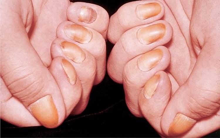
Figure 32-25. Yellow nail syndrome Diffuse yellow-to-green color of the fingernails, nail thickening, slowed growth, and excessive curvature from side to side of all 10 fingernails.
Periungual Fibroma
Synonym: Koenen tumors. Association: Tuberous sclerosis (see “Tuberous Sclerosis,” Section 16); occur in 50% of individuals. Onset. Puberty. Findings: Usually multiple, small to large, elongated to nodular tumors; produce a longitudinal groove in nail plate due to matrix compression (Fig. 32-26).
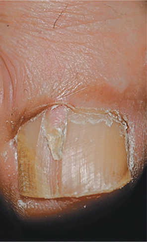
Figure 32-26. Tuberous sclerosis: periungual fibroma A skin-colored tumor is seen emerging from beneath the proximal nail fold associated with a longitudinal groove in the nail plate.
Splinter Hemorrhages 
Distal splinter hemorrhages seen with minor trauma (most common cause, occurring in up to 20% of normal population); psoriasis, atopic dermatitis. Proximal splinter hemorrhages: trauma (Fig. 32-15B), sideropenic anemia, bacterial endocarditis (Fig. 32-27), trichinosis, antiphospholipid antibody syndrome, altitude sickness. Findings: Tiny linear structures, usually 2-3 mm long, arranged in the long axis of nail; plum colored when formed, darkening to brown or black within 1-2 days; they subsequently move superficially and distally with nail growth.

Figure 32-27. Infective endocarditis: splinter hemorrhage Subungual hemorrhage in the mid-portion of the fingernail bed in a 60-year-old female with enterococcal endocarditis; subconjunctival hemorrhage was also present.
Nail Fold/Periungual Erythema And Telangiectasia
Associated with connective tissue (collagen-vascular) disease.
Periungual Erythema. Association: Systemic lupus erythematosus (SLE), dermatomyositis (DM). HIV/AIDS or hepatitis C virus infection, rhinophyma, scleroderma, hypertrophic pulmonary osteodystrophy, Kawasaki disease, hand and foot syndrome, microvasculitis. Findings: Periungual erythema, edema, alterations of cuticle, secondary nail changes.
Telangiectasia. Association: Scleroderma, SLE, DM; rheumatoid arthritis. Findings: Linear wiry vessels perpendicular to nail base overlie proximal nail folds (Fig. 32-28); usually bright red; may be black if thrombosed. SLE and DM: arise within erythema. Scleroderma and DM: enlarged capillary loops with reduced capillary density and avascular areas.
Cuticle Hyperkeratosis and Hemorrhages. SLE and DM.
Discoid LE. See Fig. 32-29.

Figure 32-28. Systemic lupus erythematosus: Nail fold erythema and telangiectasia A 64-year-old female with systemic LE with arthritis, fatigue, and photosensitivity for decades. Proximal nail folds are enlarged with erythema, telangiectasia, and thromboses. The cuticle is elongated.

Figure 32-29. Discoid lupus erythematosus: Nail fold and matrix involvement and nail dystrophy Proximal nail folds show erythema, scarring, and depigmentation associated with nail matrix inflammation.
Pterygium Inversum Unguium
Nail plate adheres to fingertip skin in scleroderma.
Systemic Amyloidosis
Nail dystrophy resembling lichen planus with severe onychodystrophy (nail plate thinned, longitudinally fissured with subungual hemorrhages) can precede diagnosis of primary systemic amyloidosis. Biopsy of nail apparatus confirms the diagnosis of amyloidosis with amyloid deposits in the superficial dermis of the nail matrix (Fig. 32-30).

Figure 32-30. Systemic amyloidosis Nail findings preceded the diagnosis of systemic amyloidosis. The matrix is inflamed with resultant thinning of the proximal nail plate and disintegration distally.
Koilonychia
Spoon-shaped nails (Fig. 32-31). Etiology (more often due to local rather than systemic factors): hereditary and congenital; Plummer-Vinson syndrome (iron-deficiency anemia, dysphagia, glossitis). Findings: In early stages, nail plate becomes flattened; later, edges become everted upward and nail appears concave.

Figure 32-31. Koilonychia The fingernail plate is concave; no other nails were involved. There were no associated systemic factors.
Clubbed Nails
Angle between proximal nail fold and nail plate is >180°. May occur with or without cyanosis. Pathogenesis: Hypertrophy of soft-tissue components of digital pulp; hyperplasia of fibrovascular tissue at base of nail (nail can be “rocked"); local cyanosis. Etiology
Cardiovascular disorders: Aortic aneurysm, congenital, and acquired cardiovascular disease.
Bronchopulmonary disorders: Intrathoracic neoplasms, chronic intrathoracic suppurative disorders.
Gastrointestinal disorders: Inflammatory bowel disease, GI neoplasms, hepatic disorders, multiple polyposis, bacillary dysentery, amoebic dysentery.
Chronic methemoglobinemia.
Findings: Digit is bulbous; nail plate enlarged and excessively curved (Fig. 32-32). Increased curvature usually affects all 20 nails.

Figure 32-32. Lung cancer: clubbed fingers Bulbous enlargement and broadening of the fingertips in a smoker with lung cancer. The tissue between the nail and underlying bone has a spongy quality giving a “floating” sensation when pressure is applied downward and forward at the junction between the plate and proximal fold. Cigarette smoke has stained the left middle finger.
Drug-Induced Nail Changes
Drugs causing adverse nail changes are similar to those causing adverse changes in cutaneous and mucosal sites.
Antimalarials: Discoloration (Fig. 32-33).
Chemotherapy: Beau lines (Fig. 32-23), onychomadesis, Muehrcke lines (Fig. 32-34), hemorrhagic onycholysis, pyogenic granulomas, melanonychia.
Antiretrovirals: Melanonychia [zidovudine (AZT)]; pyogenic granuloma (indinavir).
Beta-blockers: Digital ischemia.
Bleomycin: Digital ischemia.
PUVA: Photo-onycholysis, melanonychia.
Retinoids: Nail fragility, pyogenic granuloma, paronychia.
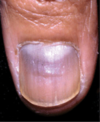
Figure 32-33. Nail discoloration: quinacrine Bluish discoloration of the nai I i n a patient with SLE treated with quinacrine.
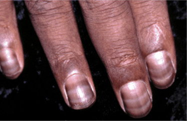
Figure 32-34. Nail discoloration and transverse bands (Muehrcke lines): Period transverse bands on the fingernail in a patient with breast cancer being treated with chemotherapy (5-fluorouracil).