
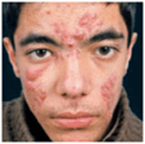
Acne Vulgaris (Common Acne) and Cystic
Acne ICD-9: 706.1  ICD-10: L70.0
 An inflammation of pilosebaceous units, very common.
Appears in certain body areas (face, trunk, rarely buttocks).
Most frequently in adolescents.
Manifests as comedones, papulopustules, nodules, and cysts.
Results in pitted, depressed, or hypertrophic scars.
Epidemiology
Occurrence. Very common, affecting approximately 85% of young people.
Age of Onset. Puberty; may appear first at 25 years or older.
Sex. More severe in males than in females.
Race. Lower incidence in Asians and Africans.
Genetic Aspects. There is a multifactorial genetic background and familial predisposition. Most individuals with cystic acne have parent(s) with a history of severe acne. Severe acne may be associated with XYY syndrome (rare).
Pathogenesis
Key factors are follicular keratinization, androgens, and Propionibacterium acnes (see Fig. 1-3).
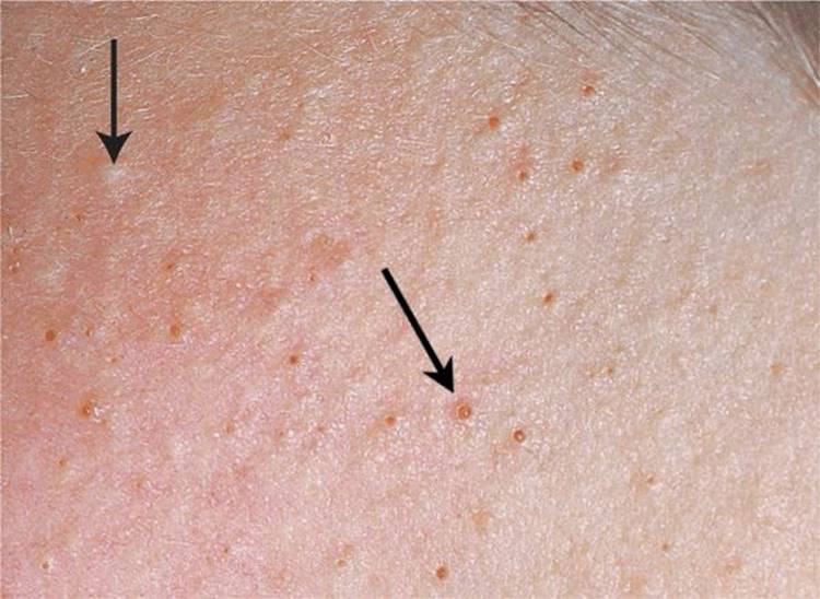
Figure 1-1. Acne vulgaris: comedones Comedones are keratin plugs that form within follicular ostia, frequently associated with surrounding erythema and pustule formation. Comedones associated with small ostia are referred to as closed comedones or “white heads” (upper arrow); those associated with large ostia are referred to as open comedones or “black heads” (lower arrow). Comedones are best treated with topical retinoids.
Follicular plugging (comedone) prevents drainage of sebum; androgens (quantitatively and qualitatively normal in serum) stimulate sebaceous glands to produce more sebum. Bacterial (p. acnes) lipase converts lipids to fatty acids and produce proinflammatory mediators (IL-I, TNF-α) that lead to an inflammatory response. Distended follicle walls break, sebum, lipids, fatty acids, keratin, bacteria enter the dermis, provoking an inflammatory and foreign-body response. Intense inflammation leads to scars.
Contributory Factors. Acnegenic mineral oils, rarely dioxin, and others.
Drugs. Lithium, hydantoin, isoniazid, glucocorticoids, oral contraceptives, iodides, bromides and androgens (e.g., testosterone), danazol.
Others. Emotional stress can cause exacerbations. Occlusion and pressure on the skin, such as by leaning face on hands is a very important and often unrecognized exacerbating factor (acne mechanica). Acne is not caused by any kind of food.
Clinical Manifestation
Duration of Lesions. Weeks to months.
Season. Often worse in fall and winter.
Symptoms. Pain in lesions (especially nodulocystic type).
Skin Lesions. Comedones—open (blackheads) or closed (whiteheads); comedonal acne (Fig. 1-1). Papules and papulopustules—i.e., a papule topped by a pustule; papulopustular acne (Fig. 1-2). Nodulesor cysts—1–4 cm in diameter (Fig. 1-4); nodulocystic acne. Soft nodules result from repeated follicular ruptures and reencapsulations with inflammation, abscess formation (cysts), and foreign-body reaction (Fig. 1-3). Round isolated single nodules and cysts coalesce to linear mounds and sinus tracts (Fig. 1-4). Sinuses: draining epithelial-lined tracts, usually with nodular acne. Scars: atrophic depressed (often pitted) or hypertrophic (at times, keloidal). Seborrhea of the face and scalp often present and sometimes severe.
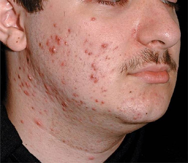
Figure 1-2. 20-year-old male In this case of papulopustular acne, some inflammatory papules become nodular and thus represent early stages of nodulocystic acne.

Figure 1-3. Acne pathogenesis [From Zaenglein AL et al. Acne vulgaris and acneiform eruptions. In: Goldsmith LA et al. eds. Fitzpatrick's Dermatology in General Medicine. 8th ed. New York, NY: McGraw-Hill; 2012.]

Figure 1-4. Nodulocystic acne A symmetric distribution in the face of a teenage boy. This image clearly shows that even nodulocystic acne starts with comedones—both open and closed comedones can be seen in this face—that then transform into papulopustular lesions, which enlarge and coalesce eventually to lead to nodulocystic acne. It is not surprising that these lesions are very painful, and it is understandable that nodulocystic acne also severely impacts the social life of these adolescents.
Sites of Predilection. Face, neck, trunk, upper arms, buttocks.
Special Forms
Neonatal Acne. On nose and cheeks in newborns or infants, related to glandular development; transient, self-healing.
Acne Excoriée. Usually in young women, associated with extensive excoriations and scarring due to emotional and psychological problems (obsessive compulsive disorder).
Acne Mechanica. Flares of acne on cheeks, chin, forehead, because of leaning face on hands, or on forehead, also from pressure of football helmet.
Acne Conglobata. Severe cystic acne (Figs. 1-5 and 1-6) with more involvement of the trunk than the face. Coalescing nodules, cysts, abscesses, and ulceration; occurs also on buttocks. Spontaneous remission rare. Rarely in XYY genotype or polycystic ovary syndrome.

Figure 1-5. Acne conglobata In this severe nodulocystic acne, there are large confluent nodules and cysts forming linear mounds that correspond to interconnecting channels. There is pustulation, crusting, and scarring and lesions are very painful.
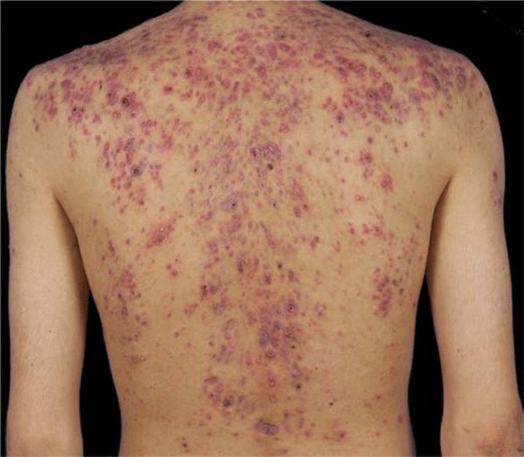
Figure 1-6. Acne conglobata on the trunk Inflammatory nodules and cysts have coalesced, forming abscesses and even leading to ulceration. There are many recent red scars following resolution of inflammatory lesions on the entire back but also on the chest.
Acne Fulminans. Teenage boys. Acute onset, severe cystic acne with suppuration and ulceration; malaise, fatigue, fever, generalized arthralgias, leukocytosis, elevated ESR.
Tropical Acne. With severe folliculitis, inflammatory nodules, draining cysts on trunk and buttocks in tropical climates; secondary infection with Staphylococcus aureus.
Occupational Acne. Due to exposure to tar derivatives, cutting oils, chlorinated hydrocarbons (see “Chloracne,” below). Not restricted to predilection sites, can appear on other (covered) body sites, like arms, legs, buttocks.
Chloracne. Due to exposure to chlorinated aromatic hydrocarbons in electrical conductors, insecticides, and herbicides. Sometimes very severe due to industrial accidents or intended poisoning (e.g., dioxin).
Acne Cosmetica. Due to comedogenic cosmetics. Pomade Acne. On the forehead, usually in Africans applying pomade to hair.
SAPHO Syndrome. Synovitis, acne fulminans, palmoplantar pustulosis, hidradenitis suppurativa, hyperkeratosis, and osteitis; very rare.
PAPA Syndrome. Sterile pyogenic arthritis, pyoderma gangrenosum acne. An inherited autoinflammatory disorder; very rare.
Acne-Like Conditions Which Are Not Acne
Steroid Acne. No comedones. Following systemic or topical glucocorticoids. Monomorphous folliculitis—small erythematous papules and pustules on chest and back.
Drug-Induced Acne. No comedones. Monomorphous acne-like eruption due to phenytoin, lithium, isoniazid, high-dose vitamin B complex, epidermal growth factor inhibitors (see Section 23), halogenated compounds.
Acne Aestivalis. No comedones. Papular eruption after sun exposure. Usually on forehead, shoulders, arms, neck, and chest.
Gram-Negative Folliculitis. Multiple tiny yellow pustules on top of acne vulgaris in long-term antibiotic administration.
Diagnosis and Differential Diagnosis
Note: Comedones are required for diagnosis of any type of acne. Comedones are not a feature of acne-like conditions (above), and the following conditions: Face S. aureus folliculitis, pseudofolliculitis barbae, rosacea, perioral dermatitis. Trunk Malassezia folliculitis, “hot-tub” pseudomonas folliculitis, S. aureus folliculitis, and acne-like conditions (see above).
Laboratory Examination
No laboratory examinations are required. In the overwhelming majority of acne patients, hormone levels are normal. If an endocrine disorder is suspected, determine free testosterone, follicle-stimulating hormone, luteinizing hormone, and DHEAS to exclude hyperandrogenism and polycystic ovary syndrome. Recalcitrant acne can also be related to congenital adrenal hyperplasia (11β or 21β hydroxylase deficiency). If systemic isotretinoin treatment is planned, determine transaminase (ALT, AST), triglyceride, and cholesterol levels.
Course
Often clears spontaneously by the early twenties but can persist to the fourth decade or older. Flares occur in the winter and with the onset of menses. The sequela of acne is scarring that may be avoided by treatment, especially with oral isotretinoin early in the course of the disease (see below).
Management
The goal of therapy is to remove the plugging of the pilar drainage, reduce sebum production, and treat bacterial colonization. Long-term goal is prevention of scarring.
Mild Acne
Usetopicalantibiotics (clindamycinanderythromycin) and benzoyl peroxide gels (2%, 5%, or 10%). Topical retinoids (retinoic acid, adapalene, tazarotene) require detailed instructions regarding gradual increases in concentration from 0.01% to 0.025% to 0.05% cream/gel or liquid.
Best combined with benzoyl peroxide-erythromycin gels.
Note: Acne surgery (extractions of comedones) is helpful only when properly done and after pretreatment with topical retinoids.
Moderate Acne. Add oral antibiotics to the above regimen. Minocycline is most effective, 50-100 mg/d, or doxycycline, 50-100 mg twice daily, tapered to 50 mg/d as acne lessens. Use of oral isotretinoin in moderate acne to prevent scarring has become much more common and is effective.
Severe Acne. In addition to topical treatment, systemic treatment with isotretinoin is indicated for cystic or conglobate acne or for any other acne refractory to treatment. This retinoid inhibits sebaceous gland function and keratinization and is very effective. Oral isotretinoin leads to complete remission in almost all cases, which last for months to years in the majority of patients.
Indications for Oral Isotretinoin. Moderate, recalcitrant, nodular acne.
Contraindications. Isotretinoin is teratogenic and effective contraception is imperative. Concurrent tetracycline and isotretinoin may cause pseudotumor cerebri (benign intracranial swelling); therefore, the two medications should never be used together.
Warnings. Determine blood lipids, transaminases (ALT, AST) before therapy. About 25% of patients can develop increased plasma triglycerides. Patients may develop mild-to-moderate elevation of transaminase levels that normalize with reduction in the dose of the drug. Eyes: Night blindness has been reported, and patients may have decreased tolerance to contact lenses. Skin: An eczema-like rash due to drug-induced dryness can occur and responds dramatically to low potency (class III) topical glucocorticoids. Dry lips and cheilitis almost always occur and must be treated. Reversible thinning of hair may occur very rarely, as may paronychia. Nose: Dryness of nasal mucosa and nosebleeds occur rarely. Other systems: Rarely, depression, headaches, arthritis, and muscular pain, pancreatitis occur. For additional rare possible complications, consult the package insert.
Dosage. Isotretinoin, 0.5-1 mg/kg given in divided doses with food. Most patients clear within 20 weeks with 1 mg/kg. Recent studies suggest that 0.5 mg/kg is equally effective.
Other Systemic Treatments for Severe Acne. Adjunctive systemic glucocorticoids may be required in severe acne conglobata, acne fulminans, and the SAPHO and PAPA syndromes. The TNF-α inhibitor infliximab and anakinra are investigational drugs in these severe forms and show promising effects. Note: For inflammatory cysts and nodules, intralesional triamcinolone (0.05 mL of a 3-5 mg/mL solution) is helpful. Web site: http://www.aad.org/pamphlets/acnepamp.html
Rosacea ICD-9: 695.3 ICD-10: L71 
A common chronic inflammatory acneiform disorder of the facial pilosebaceous units.
It is coupled with an increased reactivity of capillaries leading to flushing and telangiectasia.
 May result in rubbery thickening of nose, cheeks, forehead, or chin due to sebaceous hyperplasia, edema, and fibrosis.
Epidemiology
Occurrence. Common, affecting approximately 10% of fair-skinned people.
Age of Onset. 30-50 years; peak incidence between 40 and 50 years.
Sex. Females predominantly, but rhinophyma occurs mostly in males.
Ethnicity. Celtic persons (skin phototypes I and II) but also southern Mediterraneans; less frequent in pigmented persons (skin phototypes V and VI, i.e., brown and black).
Staging (Plewig and Kligman Classification)
The rosacea diathesis: episodic erythema, “flushing and blushing.”
Stage I: Persistent erythema with telangiectases.
Stage II: Persistent erythema, telangiectases, papules, tiny pustules.
Stage III: Persistent deep erythema, dense telangiectases, papules, pustules, nodules; rarely persistent “solid” edema of the central part of the face.
Note: Progression from one stage to another does not always occur. Rosacea may start with stage II or III and stages may overlap.
Clinical Manifestation
History of episodic reddening of the face (flushing) in response to hot liquids; spicy foods; alcohol; exposure to sun and heat. Acne may have preceded the onset of rosacea by years but rosacea usually arises de novo.
Duration of Lesions. Days, weeks, months.
Skin Symptoms. Concern about cosmetic facial appearance.
Skin Lesions. Early. Pathognomonic flushing—“red face” (Fig. 1-7); tiny papules and papulopustules (2-3 mm), pustule often small (<1 mm) and on the apex of the papule (Figs. 1-8 and 1-9). No comedones.
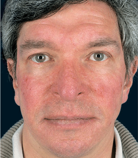
Figure 1-7. Erythematous rosacea (stage I) The early stages of rosacea often present by episodic erythema, “flushing and blushing,” which is followed by persistent erythema, which is due to multiple tiny telangiectasias, resulting in a red face.
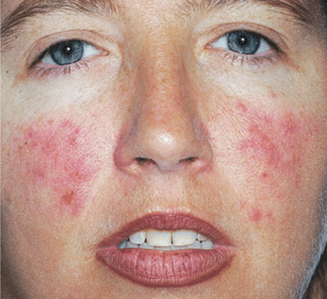
Figure 1-8. Rosacea Moderately severe rosacea in a 29-year-old female with persistent erythema, telangiectasia, red papules (stage II), and tiny pustules.
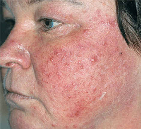
Figure 1-9. Rosacea, stages II-III Telangiectasia, papules and pustules, and some swelling in a 50-year-old woman. There are no comedones.
Late. Red facies and dusky-red papules and nodules (Figs. 1-8-1-11) Scattered, discrete lesions. Telangiectases. Marked sebaceous hyperplasia and lymphedema in chronic rosacea, causing disfigurement of the nose, forehead, eyelids, ears, and chin (Fig. 1-11).
Distribution. Symmetric localization on the face (Fig. 1-10). Rarely, neck, chest (V-shaped area), back, and scalp.
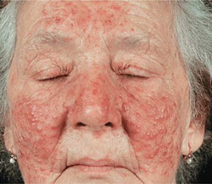
Figure 1-10. Papulopustular rosacea (early stage III) In this 65-year-old female, rosacea involves almost the entire face, sparing only the upper lip and chin. Papules and pustules have coalesced—again no comedones—and have already led to some swelling of the cheeks, which present “solid” edema.
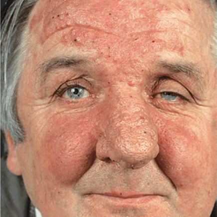
Figure 1-11. Rosacea (stage III) Here the persistent “solid” edema of the nose, forehead, and parts of the cheeks is the leading symptom. Papules, pustules, and crusted pustules are superimposed on this persistent edema. The enlarged nose feels rubbery and already represents rhinophyma.
Special Lesions
Rhinophyma (enlarged nose), metophyma (enlarged cushion-like swelling of the forehead), blepharophyma (swelling of the eyelids), otophyma (cauliflower-like swelling of the ear-lobes), and gnathophyma(swelling of the chin) result from marked sebaceous gland hyperplasia (Fig. 1-11) and fibrosis. Upon palpation: soft, rubber-like.
Eye Involvement
“Red” eyes as a result of chronic blepharitis, conjunctivitis, and episcleritis. Rosacea keratitis, albeit rare, is a serious problem because corneal ulcers may develop.
Laboratory Examinations
Bacterial Culture. Rule out S. aureus infection. Scrapings may reveal massive concurrent Demodex folliculorum infestation.
Dermatopathology. Nonspecific perifollicular and pericapillary inflammation with occasional foci of “tuberculoid” granulomatous areas; dilated capillaries. Later stages: diffuse hypertrophy of the connective tissue, sebaceous gland hyperplasia, and epithelioid granuloma without caseation.
Differential Diagnosis
Facial Papules/Pustules. Acne (in rosacea there are no comedones), perioral dermatitis, S. aureus folliculitis, gram-negative folliculitis, D. folliculorum infestation.
Facial Flushing/Erythema. Seborrheic dermatitis, prolonged use of topical glucocorticoids, systemic lupus erythematosus; dermatomyositis.
Course
Prolonged. Recurrences are common. After a few years, the disease may disappear spontaneously; usually it is for lifetime. Men and very rarely women may develop rhinophyma, gnathophyma, etc.
Management
Prevention. Marked reduction or elimination of alcohol may be helpful in some patients.
Topical
Metronidazole gel or cream, 0.75% or 1%, once or twice daily.
Topical antibiotics (e.g., erythromycin gel) less effective.
Systemic. Oral antibiotics are more effective than topical treatment.
Minocycline or doxycycline, 50-100 mg once or twice daily, first-line antibiotics; very effective. Tetracycline, 1-1.5 g/d in divided doses until clear; then gradually reduce to once-daily doses of 250-500 mg; oral metronidazole 500 mg bid, effective.
Maintenance Treatment. Minocycline or doxycycline 50 mg/d of 50 mg on alternate days or 250-500 g tetracycline.
Oral Isotretinoin. For severe disease (especially stage III) not responding to antibiotics and topical treatments. A low-dose regimen of 0.5 mg/kg body weight per day is effective in most patients, but occasionally 1 mg/kg may be required.
Ivermectin. Single dose of 12 mg po in case of massive demodex infestation.
Rhinophyma and Telangiectasia. Treated by surgery or laser surgery with excellent cosmetic results. Web site http://www.aad.org/pamphlets/rosacea.html. The ß-blocker carvedilol 6.5 mg bid has been reported to reduce erythema and telangiectasia.
Perioral Dermatitis ICD-9: 695.3  ICD-10: L71.0
  *
*
 Discrete erythematous micropapules and microvesicles.
Often confluent in the perioral and periorbital skin.
Occurs mainly in young women; can occur in children and the old.
*Rarely
Epidemiology and Etiology
Age of Onset. 16-45 years; can occur in children and the old.
Sex. Females predominantly.
Etiology. Unknown but may be markedly aggravated by potent topical (fluorinated) glucocorticoids.
Clinical Manifestation
Duration of Lesions. Weeks to months. Skin symptoms perceived as cosmetic disfigurement; occasional itching or burning, feeling of tightness.
Skin Lesions. 1- to 2-mm erythematous papulopustules on an erythematous background (Fig. 1-12) irregularly grouped, symmetric. Lesions increase in number with central confluence and satellites (Fig. 1-13); confluent plaques may appear eczematous with tiny scales. There are no comedones.
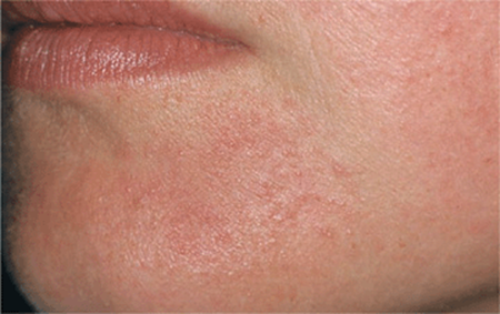
Figure 1-12. Perioral dermatitis Moderate involvement with early confluence of tiny papules and a few pustules in a perioral distribution in a young woman. Note typical sparing of the vermilion border (mucocutaneous junction).
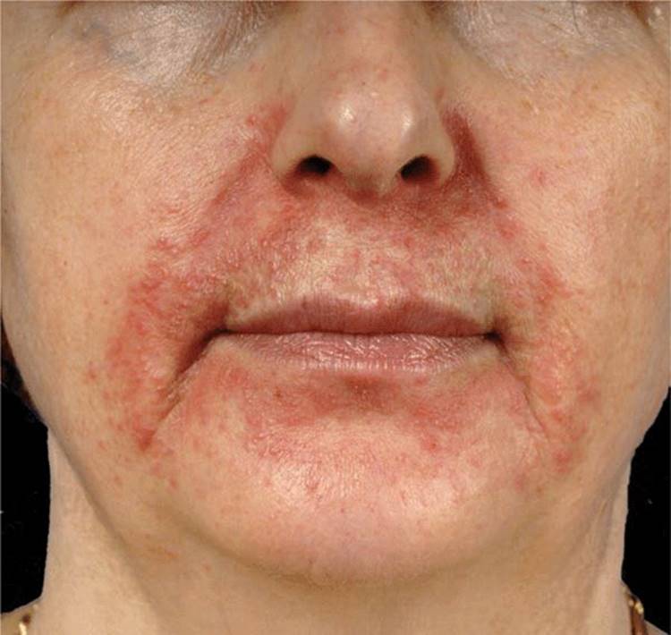
Figure 1-13. Perioral dermatitis Preferential location around the mouth and nasolabial folds and cheeks. This 38-year-old woman has been treated with fluorinated corticosteroids that led to a worsening of the condition.
Distribution. Initially perioral. Rim of sparing around the vermilion border of lips (Figs. 1-12 and 1-13) nasiolabial; at times, in the periorbital area (Fig. 1-14). Uncommonly, only periorbital.
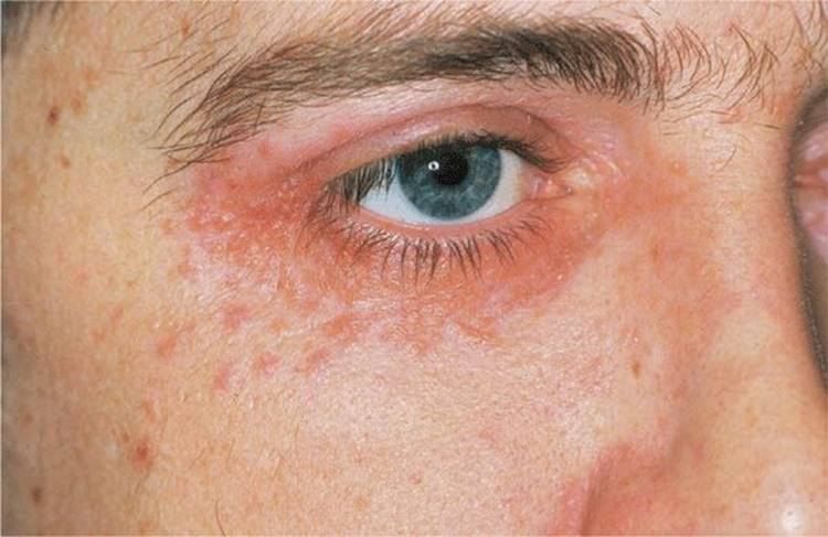
Figure 1-14. Periorbital dermatitis Note presence of tiny papules and a few pustules around the eye. This is a much less common site than the lesions around the mouth.
Laboratory Examinations
Culture. Rule out S. aureus infection.
Differential Diagnosis
Allergic contact dermatitis, atopic dermatitis, seborrheic dermatitis, rosacea, acne vulgaris, steroid acne.
Course
Appearance of lesions is usually subacute over weeks to months. At times, it is misdiagnosed as an eczematous or a seborrheic dermatitis and treated with a potent topical glucocorticoid preparation, aggravating perioral dermatitis, or inducing steroid acne.
Management
Topical
Avoid topical glucocorticoids; metronidazole, 0.75% gel two times daily or 1% once daily; erythromycin, 2% gel applied twice daily.
Systemic
Minocycline or doxycycline, 100 mg daily until clear, then 50 mg daily for another 2 months (caution, doxycycline is a photosensitizing drug) or Tetracycline, 500 mg twice daily until clear, then 500 mg daily for 1 month, then 250 mg daily for an additional month.
Hidradenitis Suppurativa ICD-9: 705.83 ICD-10: L73.2 
A chronic, suppurative, often cicatricial disease of apocrine gland-bearing skin.
Involves the axillae, the anogenital region, and, rarely, the scalp (called cicatrizing perifolliculitis).
May be associated with severe nodulocystic acne and pilonidal sinuses (also termed follicular occlusion syndrome).
Synonyms: Apocrinitis, hidradenitis axillaris, abscess of the apocrine sweat glands, acne inversa.
Epidemiology
Laboratory Examinations
Age of Onset. From puberty to climacteric.
Sex. Affects more females than males; estimated to be 4% of female population. Males more often have anogenital and females axillary involvement.
Race. All races.
Heredity. Mother-daughter transmission has been observed. Families give a history of nodulocystic acne and hidradenitis suppurativa occurring separately or together in blood relatives.
Etiology and Pathogenesis
Etiology. Unknown. Predisposing factors: obesity, smoking, and genetic predisposition to acne.
Pathogenesis. Keratinous plugging of the hair follicle → dilatation hair follicle and secondarily of the apocrine duct → inflammation → bacterial growth in dilated follicle and duct → rupture → extension of suppuration/tissue destruction → ulceration and fibrosis, sinus tract formation.
Clinical Manifestation
Symptoms. Intermittent pain and marked point tenderness related to abscess.
Skin Lesions. Open comedones, double comedones → very tender, red inflammatory nodules/abscesses (Fig. 1-15) → resolve or drain purulent/seropurulent material → moderately to exquisitely tender sinus tracts; → fibrosis, “bridge” scars, hypertrophic and keloidal scars, contractures (Figs. 1-16 and 1-17). Rarely, lymphedema of the associated limb.
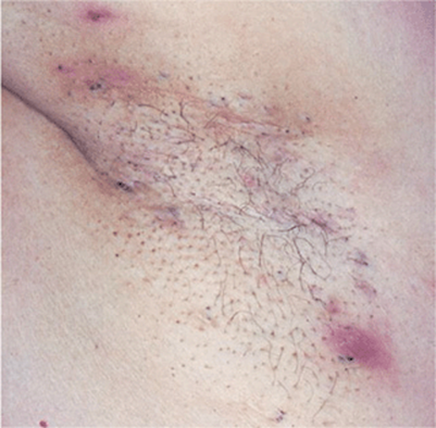
Figure 1-15. Hidradenitis suppurativa Many black comedones, some of which are paired, are a characteristic finding, associated with deep, exquisitely painful abscesses and old scars in the axilla.
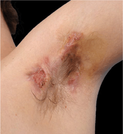
Figure 1-16. Hidradenitis suppurativa Multiple bulging and depressed scars, draining sinuses and larger ulcer in the axilla of a 24-year-old male.
Distribution. Axillae, breasts, anogenital area, groin. Often bilateral; may extend over entire back, buttocks, perineum involving scrotum or vulva (Figs. 17-17 and 1-18), and scalp.
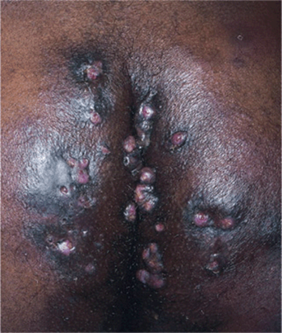
Figure 1-17. Hidradenitis suppurativa Severe scarring on the buttocks, inflammatory painful nodules with fistulas, and draining sinuses. When the patient sits down, pus will squirt from the sinus openings.
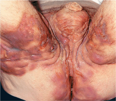
Figure 1-18. Hidradenitis suppurativa The entire perigenital and perianal skin as well as the buttocks and inner aspects of the thighs are involved in this 50-year-old male. There is considerable inflammation, and pressure releases purulent exudate from multiple sinuses. The patient had to wear a large diaper, because whenever he was seated, secretions would squirt from the sinuses.
Associated Findings. Cystic acne, pilonidal sinus. Often obesity.
Laboratory Examinations
Bacteriology. Various pathogens may secondarily colonize or “infect” lesions. These include S. aureus, streptococci, Escherichia coli, Proteus mirabilis, and Pseudomonas aeruginosa.
Dermatopathology. Keratin occlusion of hair follicle, ductal/tubular dilatation, inflammatory changes limited to follicular apparatus → destruction of apocrine/eccrine/pilosebaceous apparatus, fibrosis, pseudoepitheliomatous hyperplasia in sinuses.
Differential Diagnosis
Painful papule, nodule, abscess in groin and axilla: furuncle, carbuncle, lymphadenitis, ruptured inclusion cyst, painful lymphadenopathy in lymphogranuloma venereum, or cat-scratch disease. Also:lymphogranuloma venereum, donovanosis, scrofuloderma, actinomycosis, sinus tracts, and fistulas associated with ulcerative colitis and regional enteritis.
Course and Prognosis
The severity varies considerably. Patients with mild involvement with recurrent, self-healing, tender red nodules often do not seek therapy. Usually a spontaneous remission with age (>35 years). Some course in relentlessly progressive, with marked morbidity related to chronic pain, draining sinuses, and scarring, with restricted mobility (Figs. 1-17 and 1-18). Complications (rare): fistulas to urethra, bladder, and/or rectum; anemia, amyloidosis.
Management
Hidradenitis suppurativa is not simply an infection, and systemic antibiotics are only part of the treatment program. Combinations of (1) intralesional glucocorticoids, (2) surgery, (3) oral antibiotics, and (4) isotretinoin are used.
Medical Management
Acute Painful Nodule and Abscess. Intralesional triamcinolone (3-5 mg/mL) into the wall followed by incision and drainage of abscess.
Chronic Low-Grade Disease. Oral antibiotics: erythromycin (250-500 mg qid), tetracycline (250-500 mg qid), or minocycline (100 mg bid); or a combination of clindamycin, 300 mg bid, with rifampin (300 mg bid); may take weeks or months.
Prednisone. Concurrently, if pain and inflammation are severe: 70 mg daily for 2-3 days, tapered over 14 days.
Oral Isotretinoin. Not useful in severe disease, but useful in early disease to prevent follicular plugging and when combined with surgical excision of individual lesions. TNF-α inhibitors (e.g., infliximab) show promising results in severe cases.
Surgical Management
• Incise and drain acute abscesses.
• Excise chronic recurrent, fibrotic nodules, or sinus tracts.
• With extensive, chronic disease, complete excision of axilla or involved anogenital area extending down to fascia, requires split skin grafting.
Psychological Management
Patients become very depressed because of pain, soiling of clothing by draining pus, odor, and the site of occurrence (anogenital area). Therefore, every effort should be made to deal with the disease, using every modality possible.
Fox Fordyce Disease 
Rare, mostly in females, after puberty.
Eruption consists of skin-colored or slightly erythematous papules localized to axillae and/or genitofemoral region.
Very pruritic.
Results from plugging of follicular infundibula → rupture → inflammation.
Treatment: topical clindamycin, electrocoagulation, liposuction/curettage.