EQUIPMENT
 Centimeter ruler (flexible, clear)
Flashlight with transilluminator
Wood’s lamp (to view fluorescing lesions)
Handheld magnifying lens (optional)
EXAMINATION
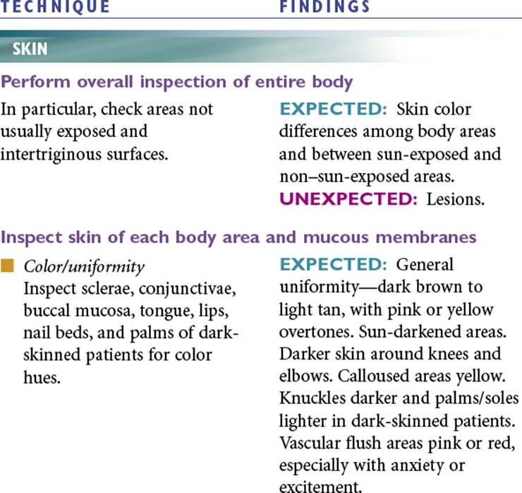

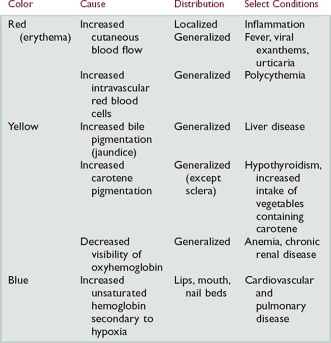
Cutaneous Color Changes

|
TECHNIQUE |
FINDINGS |
|
Pigmented nevi. Nonpigmented striae. Freckles. Birthmarks. |
|
|
UNEXPECTED:Dysplastic, precancerous, or cancerous nevi. Chloasma. Unpigmented skin. Generalized or localized color changes. Vascular skin lesions. Vascular changes. |
|
|
|
EXPECTED:Thickness variations, with eyelids thinnest, areas of rubbing thickest. Calluses on hands and feet. |
|
UNEXPECTED:Atrophy. Hyperkeratosis. Corns. |
|
|
|
EXPECTED:Bilateral symmetry. |
|
|
EXPECTED:Clean. |
|
Palpate skin |
|
|
|
EXPECTED:Minimal perspiration or oiliness. Increased perspiration (associated with activity, environment, obesity, anxiety, excitement) noticeable on palms, scalp, forehead, axillae. |
|
UNEXPECTED:Damp intertriginous areas. |
|
|
Palpate with dorsal surface of hand or fingers. |
EXPECTED:Cool to warm. Bilateral symmetry. |
|
|
EXPECTED:Smooth, soft, and even. Roughness resulting from heavy clothing, cold weather, or soap. |
|
UNEXPECTED:Extensive or widespread roughness. |
|
|
|
EXPECTED:Resilience. |
|
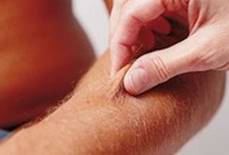
Gently pinch skin on forearm or in sternal area, and release. |
UNEXPECTED:Failure of skin to return to place quickly. |
|
|
|
|
Inspect and palpate lesions |
|
|
Measure all dimensions.
Use Wood’s lamp to distinguish fluorescing lesions.
Transilluminate to determine presence of fluid.
Note color, odor, amount, and consistency of lesion.
Check lesion for annular, grouped, linear, arciform, or diffuse arrangement.
Check lesion for generalized/localized, body region, patterns, or discrete/confluent. |
UNEXPECTED:See table on pp. 36–39 |
Primary Skin Lesions
|
Description |
Examples |
|
Macule |
|
|
Flat, circumscribed area that is a change in skin color; less than 1 cm in diameter |
Freckles, flat moles (nevi), petechiae, measles, scarlet fever |
|
Measles. From Habif, 2004.
|
|
|
Papule |
|
|
Elevated, firm, circumscribed area less than 1 cm in diameter |
Wart (verruca), elevated moles, lichen planus |
|
Lichen planus. From Weston et al, 1996.
|
|
|
Patch |
|
|
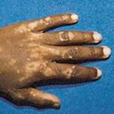
Flat, nonpalpable, irregular-shaped macule more than 1 cm in diameter |
Vitiligo, port-wine stains, mongolian spots, café au lait spots |
|
Vitiligo. From Weston et al, 1991.
|
|
|
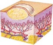
Plaque |
|
|
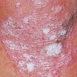
Elevated, firm, and rough lesion with flat top surface greater than 1 cm in diameter |
Psoriasis, seborrheic and actinic keratoses |
|
Plaque. From Habif, 2004.
|
|
|
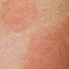
Wheal |
|
|
Elevated irregular-shaped area of cutaneous edema; solid, transient; variable diameter |
Insect bites, urticaria, allergic reaction |
|
Wheal. From Farrar et al, 1992.
|
|
|
Nodule |
|
|
Elevated, firm, circumscribed lesion; deeper in dermis than a papule; 1 to 2 cm in diameter |
Erythema nodosum, lipomas |
|
Hypertrophic nodule. From Goldman and Fitzpatrick, 1999.
|
|
|
Tumor |
|
|
Elevated and solid lesion; may or may not be clearly demarcated; deeper in dermis; greater than 2 cm in diameter |
Neoplasms, benign tumor, lipoma, hemangioma |
|
Hemangioma. From Weston et al, 1996.
|
|
|
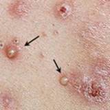
Vesicle |
|
|
Elevated, circumscribed, superficial, not into dermis; filled with serous fluid; less than 1 cm in diameter |
Varicella (chickenpox), herpes zoster (shingles) |
|
Vesicles caused by varicella. From Farrar et al, 1992.
|
|
|
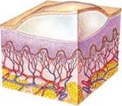
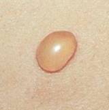
Bulla |
|
|
Vesicle greater than 1 cm in diameter |
Blister, pemphigus vulgaris |
|
Blister. From White, 1994.
|
|
|
Pustule |
|
|
Elevated, superficial lesion; similar to a vesicle but filled with purulent fluid |
Impetigo, acne |
|
Acne. From Weston et al, 1996.
|
|
|
Cyst |
|
|
Elevated, circumscribed, encapsulated lesion; in dermis or subcutaneous layer; filled with liquid or semisolid material |
Sebaceous cyst, cystic acne |
|
Sebaceous cyst. From Weston et al, 1996.
|
|
|
Telangiectasia |
|
|
Fine, irregular red lines produced by capillary dilation |
Telangiectasia in rosacea |
|
Telangiectasia. From Lemmi and Lemmi, 2000.
|
|
|
TECHNIQUE |
FINDINGS |
|
HAIR |
|
|
Inspect hair over entire body |
|
|
|
EXPECTED:Light blond to black and gray, with alterations caused by rinses, dyes, and permanents. |
|
|
EXPECTED:Hair present on scalp, lower face, neck, nares, ears, chest, axillae, back and shoulders, arms, legs, pubic area, and around nipples. Scalp hair loss in adult men, adrenal androgenic female-pattern alopecia in adult women. |
|
UNEXPECTED:Localized or generalized hair loss, inflammation, or scarring. Broken/absent hair shafts. Hirsutism in women. |
|
|
Palpate for texture |
|
|
EXPECTED:Coarse or fine, curly or straight, shiny, smooth, and resilient. Fine vellus covering body; coarse terminal hair on scalp, on pubis, on axillary areas, and in male beard. |
|
|
UNEXPECTED:Dryness and brittleness. |
|
|
NAILS |
|
|
Inspect nails |
|
|
|
EXPECTED:Variations of pink with varying opacity. Pigment deposits in persons with dark skin. White spots. |
|
UNEXPECTED:Yellow or green-black discoloration. Diffuse darkening. Pigment deposits in persons with light skin. Longitudinal red, brown, or white streaks or white bands. White, yellow, or green tinge. Blue nail beds. Blue-black discoloration. |
|
|
|
EXPECTED:Varying shape, smooth and flat/slightly convex, with edges smooth and rounded. |
|
UNEXPECTED:Jagged, broken, or bitten edges or cuticles. Peeling. Absence of nail. |
|
|
|
EXPECTED:Clean and neat. |
|
UNEXPECTED:Unkempt. |
|
|
|
EXPECTED:Longitudinal ridging and beading. |
|
UNEXPECTED:Longitudinal ridging and grooving with lichen planus. Transverse grooving, rippling, and depressions. Pitting. |
|
|
Palpate nail plate |
|
|
|
EXPECTED:Hard and smooth with uniform thickness. |
|
UNEXPECTED:Thickening or thinning. |
|
|
|
EXPECTED:Firmness. |
|
Gently squeeze between thumb and finger. |
UNEXPECTED:Separation. Boggy nail base. |
|
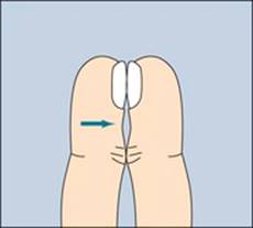
Measure nail base angle |
|
|
Inspect fingers when patient places dorsal surfaces of fingertips together. |
EXPECTED:160-degree angle. |
|
UNEXPECTED:Clubbing. |
|
|
Expected finding.
|
Clubbing.
|
|
Inspect and palpate proximal and lateral nail folds |
|
|
UNEXPECTED:Redness, swelling, pus, warts, cysts, tumors, and pain. |
|
AIDS TO DIFFERENTIAL DIAGNOSIS
|
ABNORMALITY |
DESCRIPTION |
|
Eczematous dermatitis |
Subjective Data:Itching may or may not be present; those with atopic dermatitis often report allergy history (allergic rhinitis, asthma). |
|
Objective Data:Acute: erythematous, pruritic, weeping vesicles; subacute: erythema and scaling; chronic: thick, lichenified, pruritic plaques. Atopic dermatis: during childhood lesions involve flexures, the nape, and the dorsal aspects of the limbs. In adolescence and adulthood lichenified plaques affect the flexures, head, and neck. |
|
|
Folliculitis |
Subjective Data:Acute onset of papules and pustules associated with pruritus or mild discomfort; may have pain with with deep folliculitis. |
|
Objective Data:Primary lesion small pustule 1 to 2 cm in diameter that is located over a pilosebaceous orifice and may be perforated by a hair. Pustule may be surrounded by inflammation or nodular lesions. After the pustule ruptures, a crust forms. |
|
|
Tinea (dermatophytosis) |
Subjective Data:Pruritus. |
|
Objective Data:Papular, pustular, vesicular, erythematous, or scaling lesions. Possible secondary bacterial infection. Hyphae on microscopic examination of skin scraping with KOH solution. |
|
|
Basal cell carcinoma |
Subjective Data:Persistent sore or lesions that has not healed; crusting; itching. |
|
Objective Data:Shiny nodule that is pearly or translucent; may be pink, red, white, tan, black, or brown growth with a slightly elevated rolled border and a crusted indentation in the center. |
|
|
Squamous cell carcinoma |
Subjective Data:Persistent sore or lesion that has not healed or that has grown in size; crusting and/or bleeding. |
|
Objective Data:Elevated-growth with a central depression wartlike growth; scaly red patch with irregular borders; open sore. |
|
|
Malignant melanoma |
Subjective Data:New mole or preexisting mole that has changed or is changing; history of melanoma, dysplastic or atypical nevi; family history of melanoma. |
|
Objective Data:Characteristic asymmetry, irregular borders, variegated colors, and is growing or larger than 6 mm (see ABCDs of Melanoma on p. 49). |
|
|
Hirsutism |
Subjective Data:Growth of terminal hair in women in the male distribution pattern on the face, body, and pubic areas. |
|
Objective Data:Presence of thick, dark terminal hairs in androgen-sensitive sites: face, chest, areola, external genitalia, upper and lower back, buttock, inner thigh, linea. |
|
|
Paronychia |
Subjective Data:Acute: history of nail trauma or manipulation; acute onset. Chronic: History of repeated exposure to moisture, e.g., through hand-washing. Evolves slowly initially with tenderness and mild swelling. |
|
Objective Data:Redness, swelling, tenderness at lateral and proximal nail folds. Possible purulent drainage under cuticle. Acute or chronic (with nail rippling). |
|
|
Oncychomycosis |
Subjective Data:Yellow, crumbling nail. |
|
Objective Data:Distal nail plate turns yellow or white as hyperkeratotic debris accumulates, causing the nail to separate from the nail bed. |
|
|
Redness and swelling where nail pierces the lateral nail fold and grows into the dermis. |
AIDS TO DIFFERENTIAL DIAGNOSIS
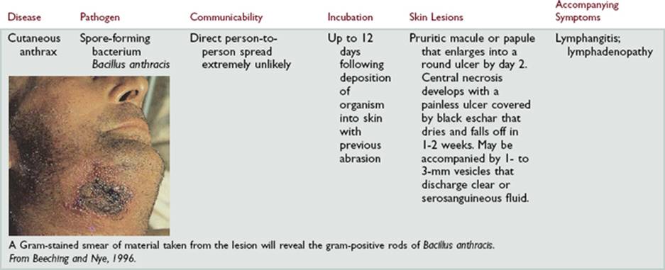
Cutaneous Manifestations of Two Pathogens That May Be Used in Biologic Warfare

Pediatric Variations
EXAMINATION
|
TECHNIQUE |
FINDINGS |
|
SKIN |
|
|
Inspect hands and feet of newborns for skin creases |
|
|
EXPECTED:Number of creases is indication of maturity of newborn; the older the gestational age, the more creases. |
|
|
UNEXPECTED:Single transverse crease across palm frequently seen in infants with Down syndrome. |
|
AIDS TO DIFFERENTIAL DIAGNOSIS
|
ABNORMALITY |
DESCRIPTION |
|
Seborrheic dermatitis |
Subjective Data:Thick greasy scalp scales or body rash. |
|
Objective Data:Thick, yellow, adherent crusted scalp, ear, or neck lesions. |
|
|
Impetigo |
Subjective Data:Lesion, typically on the face, that itches and burns. |
|
Objective Data:Honey-colored crusted or ruptured vesicles. |
|
|
Miliaria (“prickly heat”) |
Subjective Data:Parent reports rash noted while undressing infant. |
|
Objective Data:Irregular, red, macular rash on covered areas. |
|
|
Chickenpox (varicella) |
Subjective Data:Fever, headache, sore throat, malaise. |
|
Objective Data:Pruritic maculopapular skin eruption that becomes vesicular in a matter of hours. |
|
|
German measles (rubella) |
Subjective Data:Fever, coryza, sore throat, cough. |
|
Objective Data:Koplik spots on buccal muscosa; generalized light pink to red maculopapular rash. |
SAMPLE DOCUMENTATION
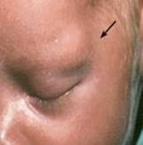
Objective.Skin: Dark pink maculopapular lesions on face, torso, extremities; large urticarial wheal on right cheek. No excoriation or secondary infection. Turgor resilient. Skin uniformly warm and dry. No edema.
Hair: Curly, black, thick with female distribution pattern. Texture coarse.
Nails: Opaque, short, well-groomed, uniform, and without deformities. Nail bed pink. Nail base angle 160 degrees. No redness, exudates, or swelling in surrounding folds and no tenderness to palpation.
From American Academy of Dermatology, 2005.
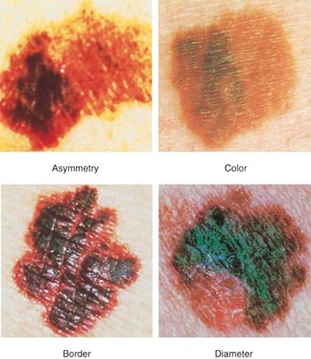
The ABCDs of Melanoma
Characteristics that should alert you to the possibility of malignant melanoma:
A Asymmetry of lesion. One half of a mole or birthmark does not match the other.
B Borders. Edges are irregular, ragged, notched, or blurred. Pigment may be streaming from the border.
C Color. The color is not the same all over and may have differing shades of brown or black, sometimes with patches of red, white, or blue.
D Diameter. The diameter is larger than 6 mm (about the size of a pencil eraser) or is growing larger.
 Thickness