
RESPIRATORY FUNCTION
The major classes of lung diseases include obstructive lung diseases (e.g., asthma, chronic obstructive pulmonary disease, and bronchiectasis), restrictive lung diseases (e.g., interstitial lung diseases, chest wall abnormalities, and neuromuscular diseases), and vascular abnormalities (e.g., pulmonary thromboembolism and pulmonary arterial hypertension). The respiratory system includes not only the lungs but also the chest wall, pulmonary circulation, and central nervous system. There are three key types of respiratory system physiologic disturbances that occur in varying combinations in different lung diseases: ventilatory function, pulmonary circulation, and gas exchange.
Disturbances in Ventilatory Function
Ventilation involves the delivery of gas to the alveoli. Pulmonary function tests are used to assess ventilatory function. The classification of lung volumes, which are measured with pulmonary function testing, is shown in Fig. 137-1. Spirometry involves forced exhalation from total lung capacity (TLC) to residual volume (RV); key measurements from a spirogram are the forced expiratory volume in 1 s (FEV1) and the forced vital capacity (FVC). Expiratory flow rates may be plotted against lung volumes to yield a flow-volume curve. Plateau of the inspiratory curve on the flow-volume loop suggests extrathoracic large airway obstruction, while plateau of the expiratory curve suggests intrathoracic large airway obstruction. Other lung volumes, including TLC and RV, are measured under static conditions using either helium dilution or body plethysmography. Lung volumes and flow rates are typically compared with population-based normal values that adjust for the age, height, sex, and race of the pt.

FIGURE 137-1 Spirogram of a slow vital capacity maneuver demonstrating various lung volumes.
There are two major patterns of abnormal ventilatory function detected by pulmonary function testing: restrictive and obstructive (Tables 137-1 and 137-2). The presence of obstruction is determined by a reduced ratio of FEV1/FVC (with abnormal often defined using a threshold of <0.7), and the severity of airflow obstruction is determined by the level of reduction of FEV1. With airflow obstruction, TLC may be normal or increased, and RV is typically elevated. With severe airflow obstruction, the FVC is often reduced.
TABLE 137-1 COMMON RESPIRATORY DISEASES BY DIAGNOSTIC CATEGORIES


TABLE 137-2 ALTERATIONS IN VENTILATORY FUNCTION IN DIFFERENT PULMONARY DISEASE CATEGORIES
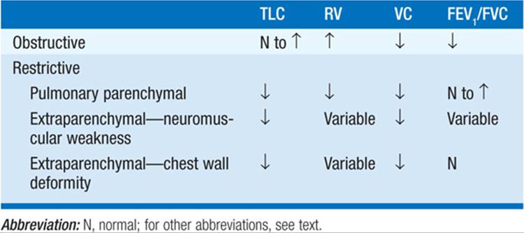
The presence of a restrictive pattern is determined by a reduction in lung volumes, especially TLC. When pulmonary parenchymal processes cause restriction, RV is also decreased, but the FEV1/FVC is normal. With extraparenchymal etiologies of restrictive ventilatory defects, such as neuromuscular weakness or chest wall abnormalities, the impact on RV and FEV1/FVC is more variable. Weakness of the respiratory muscles can be assessed by measuring maximal inspiratory and expiratory pressures.
Disturbances in Pulmonary Circulation
The pulmonary vasculature normally handles the right ventricular output (~5 L/min) at a low pressure. Normal mean pulmonary artery pressure (PAP) is 15 mmHg. When cardiac output increases, pulmonary vascular resistance (PVR) normally falls, leading to only small increases in mean PAP.
Assessment of the pulmonary vasculature requires measuring pulmonary vascular pressures and cardiac output to derive PVR. PVR rises with hypoxemia (due to vasoconstriction), intraluminal thrombi (due to diminished cross-sectional area from obstruction), or destruction of small pulmonary vessels (due to scarring or loss of the alveolar walls).
All diseases of the respiratory system causing hypoxemia are capable of causing pulmonary hypertension. However, pts with prolonged hypoxemia related to chronic obstructive pulmonary disease, interstitial lung disease, chest wall disease, and obesity-hypoventilation/obstructive sleep apnea are particularly likely to develop pulmonary hypertension. When pulmonary vessels are directly affected, as with recurrent pulmonary emboli, the decrease in cross-sectional area of the pulmonary vasculature is the primary mechanism for increased PVR, rather than hypoxemia.
Disturbances in Gas Exchange
The primary functions of the respiratory system are to remove CO2 from blood entering the pulmonary circulation and to provide O2 to blood leaving the pulmonary circulation. Normal tidal volume is approximately 500 mL and normal respiratory rate is approximately 15 breaths/min, leading to a total minute ventilation of approximately 7.5 L/min. Because of anatomic dead space, alveolar ventilation is approximately 5 L/min. Gas exchange depends on alveolar ventilation rather than total minute ventilation.
Partial pressure of CO2 in arterial blood (Paco2) is directly proportional to the amount of CO2 produced each minute ( co2) and inversely proportional to alveolar ventilation (A)

Adequate movement of gas between alveoli and pulmonary capillaries by diffusion is required for normal gas exchange. Diffusion can be tested by measuring the diffusing capacity of the lung for a low (and safe) concentration of carbon monoxide (DLCO) during a 10-second breath-hold. DLCO measurement is typically corrected for the pt’s hemoglobin level. Diffusion abnormalities rarely result in arterial hypoxemia at rest but can cause hypoxemia with exercise. A restrictive ventilatory defect with reduced DLCO suggests parenchymal lung disease. The pattern of normal spirometry, normal lung volumes, and reduced DLCO is consistent with pulmonary vascular disease. Gas exchange is critically dependent on proper matching of ventilation and perfusion.
Assessment of gas exchange is commonly performed with arterial blood gases, which provide measurements of the partial pressures of O2 and CO2 The actual content of O2 in blood is determined by both Po2and hemoglobin concentration. The alveolar-arterial O2 difference [(A – a) gradient] can provide useful information when assessing abnormalities in gas exchange. The normal (A – a) gradient is <15 mmHg under age 30 but increases with aging. In order to calculate the (A – a) gradient, the alveolar Po2 (Paco2) must be calculated:

where FIO2 = fractional concentration of inspired O2 (0.21 while breathing room air), PB = barometric pressure (760 mmHg at sea level), PH2O = water vapor pressure (47 mmHg when air is saturated at 37°C), and R = respiratory quotient (the ratio of CO2 production to O2 consumption, usually assumed to be 0.8). Severe arterial hypoxemia rarely occurs purely due to alveolar hypoventilation while breathing air at sea level. The (A – a) gradient is calculated by subtracting the measured Pao2 from the calculated PAO2.
Adequacy of CO2 removal is reflected in the partial pressure of CO2 measured in an arterial blood gas. Pulse oximetry is a valuable, widely used, and noninvasive tool to assess O2 saturation, but it provides no information about Paco2. Other limitations of pulse oximetry include relative insensitivity to oxygenation changes when Pao2 is >60 mmHg, problems with obtaining an adequate signal when cutaneous perfusion is decreased, and inability to distinguish oxyhemoglobin from other forms of hemoglobin, such as carboxyhemoglobin and methemoglobin.
Mechanisms of Abnormal Respiratory Function
The four basic mechanisms of hypoxemia are (1) decrease in inspired Po2, (2) hypoventilation, (3) shunt, and (4) ventilation/perfusion mismatch. Decrease in inspired Po2(e.g., at high altitude) and hypoventilation (characterized by an increased Paco2) both lower arterial oxygenation by reducing alveolar oxygenation; thus, the (A – a) gradient is normal. Shunting (e.g., intracardiac shunt) causes hypoxemia by bypassing the alveolar capillaries. Shunting is characterized by an elevated (A – a) gradient and is relatively refractory to oxygenation improvement with supplemental O2. Ventilation/perfusion mismatch is the most common cause of hypoxemia; it is associated with an elevated (A – a) gradient, but supplemental O2 corrects the hypoxemia by raising the O2 content of blood from regions with low ventilation/perfusion ratios. An algorithm for approaching the hypoxemic pt is shown in Fig. 137-2.

FIGURE 137-2 Flow diagram outlining the diagnostic approach to the pt with hypoxemia. COPD, chronic obstructive pulmonary disease. (From SE Weinberger: Principles of Pulmonary Medicine, 4th ed. Philadelphia, Saunders, 2004; with permission.)
Hypercapnia is caused by inadequate alveolar ventilation. Potential contributing factors include (1) increased CO2 production, (2) decreased ventilatory drive, (3) malfunction of the respiratory pump or increased airway resistance, and (4) inefficiency of gas exchange (increased dead space or ventilation/perfusion mismatch).
Although diffusion abnormalities rarely cause hypoxemia at rest, assessment of DLCO can be used to determine the functional integrity of the alveolar-capillary membrane. Diseases that solely affect the airways typically do not reduce the DLCO. DLCO is reduced in interstitial lung disease, emphysema, and pulmonary vascular disease. DLCO can be elevated in alveolar hemorrhage, asthma, and congestive heart failure.
DIAGNOSTIC PROCEDURES
NONINVASIVE PROCEDURES
Radiographic Studies
The chest x-ray (CXR), generally including both posteroanterior and lateral views, is often the first diagnostic study in pts presenting with respiratory symptoms. With some exceptions (e.g., pneumothorax), the CXR pattern is usually not sufficiently specific to establish a diagnosis; instead, the CXR serves to detect disease, assess magnitude, and guide further diagnostic investigation. With diffuse lung disease, CXR can detect an alveolar, interstitial, or nodular pattern. CXR can also detect pleural effusion and pneumothorax, as well as abnormalities in the hila and mediastinum. Lateral decubitus views can be used to estimate the size of freely flowing pleural effusions.
Chest CT is widely used to clarify radiographic abnormalities detected by CXR. Advantages of chest CT compared with CXR include (1) ability to distinguish superimposed structures due to cross-sectional imaging; (2) superior assessment of tissue density, permitting accurate assessment of the size and density of pulmonary nodules and improved identification of abnormalities adjacent to the chest wall, such as pleural disease; (3) with the use of IV contrast, ability to distinguish vascular from nonvascular structures, which is especially useful in assessing hilar and mediastinal abnormalities; (4) with CT angiography, ability to detect pulmonary emboli; and (5) due to superior visible detail, improved recognition of parenchymal and airway diseases, including emphysema, bronchiectasis, lymphangitic carcinoma, and interstitial lung disease.
A variety of other imaging techniques are used less commonly to assess respiratory disease. Magnetic resonance imaging (MRI) is generally less useful than CT for evaluation of the respiratory system but can be helpful as a nonradioactive tool to assist in the evaluation of intrathoracic cardiovascular pathology and to distinguish vascular and nonvascular structures without IV contrast. Ultrasound is not useful for assessing the pulmonary parenchyma, but it can detect pleural abnormalities and guide thoracentesis of a pleural effusion. Pulmonary angiography can assess the pulmonary arterial system for venous thromboembolism but has largely been replaced by CT angiography.
Nuclear Medicine Imaging
Ventilation-perfusion lung scans can be used to assess for pulmonary thromboembolism but have also largely been replaced by CT angiography. Positron emission tomographic (PET) scanning assesses the uptake and metabolism of a radiolabeled glucose analogue. Because malignant lesions usually have increased metabolic activity, PET scanning, especially when combined with CT images in PET/CT, is useful to assess pulmonary nodules for potential malignancy and to stage lung cancer. PET studies are limited in assessing lesions <1 cm in diameter; false-negative screening for malignancy can result from lesions with low metabolic activity, such as bronchioloalveolar cell carcinoma. False-positive PET signals can be observed in inflammatory conditions such as pneumonia.
Sputum Exam
Sputum can be obtained by spontaneous expectoration or induced by inhalation of an irritating aerosol like hypertonic saline. Sputum is distinguished from saliva by the presence of bronchial epithelial cells and alveolar macrophages as opposed to squamous epithelial cells. Sputum exam should include gross inspection for blood, color, and odor, as well as Gram’s stain and routine bacterial culture. Bacterial culture of expectorated sputum may be misleading due to contamination with oropharyngeal flora. Sputum samples can also be assessed for a variety of other pathogens, including mycobacteria, fungi, and viruses. Sputum samples induced by hypertonic saline can be stained for the presence of Pneumocystis jiroveci. Cytologic examination of sputum samples can be used as an initial screen for malignancy.
INVASIVE PROCEDURES
Bronchoscopy
Bronchoscopy is a procedure that provides direct visualization of the tracheobronchial tree, typically to the subsegmental level. The fiberoptic bronchoscope is used in most cases, but rigid bronchoscopy is valuable in specific circumstances, including massive hemorrhage and foreign body removal. Flexible fiberoptic bronchoscopy allows visualization of the airways; identification of endobronchial abnormalities, including tumors and sites of bleeding; and collection of diagnostic specimens by washing, brushing, biopsy, or lavage. Washing involves instilling sterile saline through the bronchoscope channel onto the surface of a lesion; part of the saline is suctioned back through the bronchoscope and processed for cytology and microorganisms. Bronchial brushings can be obtained from the surface of an endobronchial lesion or from a more distal mass or infiltrate (potentially with fluoroscopic guidance) for cytologic and microbiologic studies. Biopsy forceps can be used to obtain biopsies of endobronchial lesions or passed into peribronchial alveolar tissue (often with fluoroscopic guidance) to obtain transbronchial biopsies of more distal lung tissue. Transbronchial biopsy is particularly useful in diagnosing diffuse infectious processes, lymphangitic spread of cancer, and granulomatous diseases. Complications of transbronchial biopsy include hemorrhage and pneumothorax.
Bronchoalveolar lavage (BAL) is an adjunct to fiberoptic bronchoscopy, permitting collection of cells and fluid from distal air spaces. After wedging the bronchoscope in a subsegmental airway, saline is instilled and then suctioned back through the bronchoscope for analyses, which can include cytology, microbiology, and cell counts. BAL is especially useful in the diagnosis of P. jiroveci pneumonia and some other infections.
Additional bronchoscopic approaches to obtain tissue samples from locations adjacent to the trachea or large bronchi for cytologic assessment of malignancy include transbronchial needle aspiration (TBNA). TBNA can be supplemented with endobronchial ultrasound (EBUS), which can allow guided aspiration of hilar and mediastinal lymph nodes.
Percutaneous Needle Aspiration of the Lung
A needle can be inserted through the chest wall and into a pulmonary lesion to aspirate material for cytologic and microbiologic studies. Percutaneous needle aspiration is usually performed under CT guidance. Owing to the small size of the sample obtained, sampling error is a limitation of the procedure.
Thoracentesis
Thoracentesis should be performed as an early step in the evaluation of a pleural effusion of uncertain etiology. Analysis of pleural fluid can determine the etiology of the effusion (Chap. 144). Large-volume thoracentesis can be therapeutic by palliating dyspnea.
Mediastinoscopy
Tissue biopsy of mediastinal masses or lymph nodes is often required for cancer diagnosis and staging. Mediastinoscopy is performed from a suprasternal approach, and a rigid mediastinoscope is inserted—from which biopsies can be obtained. Lymph nodes in the aortopulmonary location typically require a parasternal mediastinotomy to provide access for biopsy.
Video-Assisted Thoracic Surgery
Video-assisted thoracic surgery (VATS), also known as thoracoscopy, is widely used for the diagnosis of pleural lesions as well as peripheral parenchymal infiltrates and nodules. VATS, which requires that the pt tolerate single lung ventilation during the procedure, involves passing a rigid scope with a camera through a trocar and into the pleural space; instruments can be inserted and manipulated through separate intercostal incisions. VATS has largely replaced “open biopsy,” which requires a thoracotomy.

For a more detailed discussion, see Kritek P, Choi AMK: Approach to the Patient With Disease of the Respiratory System, Chap. 251, p. 2084, in HPIM-18; Naureckas ET, Solway J: Disturbances of Respiratory Function, Chap. 252, p. 2087, in HPIM-18; and Fuhlbrigge AL, Choi AMK: Diagnostic Procedures in Respiratory Disease, Chap. 253, p. 2094, in HPIM-18.