
CHOLELITHIASIS
There are two major types of gallstones: cholesterol and pigment stones. Cholesterol gallstones contain >50% cholesterol monohydrate. Pigment stones have <20% cholesterol and are composed primarily of calcium bilirubinate. In the United States, 80% of stones are cholesterol and 20% are pigment.
EPIDEMIOLOGY
One million new cases of cholelithiasis per year in the United States. Predisposing factors include demographic/genetics (increased prevalence in North American Indians), obesity, weight loss, female sex hormones, age, ileal disease, pregnancy, type IV hyperlipidemia, and cirrhosis.
SYMPTOMS AND SIGNS
Many gallstones are “silent,” i.e., present in asymptomatic pts. Symptoms occur when stones trigger inflammation or cause obstruction of the cystic or common bile ducts. Major symptoms: (1) biliary colic—a severe steady ache in the RUQ or epigastrium that begins suddenly; often occurs 30–90 min after meals, lasts for several hours, and occasionally radiates to the right scapula or back; (2) nausea, vomiting. Physical exam may be normal or show epigastric or RUQ tenderness.
LABORATORY
Occasionally, mild and transient elevations in bilirubin [<85 μmol/L (<5 mg/dL)] accompany biliary colic.
IMAGING
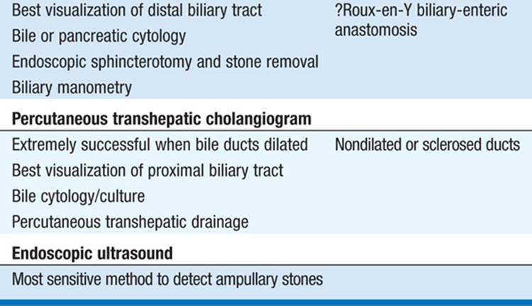
Only 10% of cholesterol gallstones are radiopaque. Ultrasonography is best diagnostic test. The oral cholecystogram has been largely replaced by ultrasound, but may be used to assess the patency of the cystic duct and gallbladder emptying function (Table 161-1).
TABLE 161-1 DIAGNOSTIC EVALUATION OF THE BILE DUCTS


DIFFERENTIAL DIAGNOSIS
Includes peptic ulcer disease (PUD), gastroesophageal reflux, irritable bowel syndrome, and hepatitis.
COMPLICATIONS
Cholecystitis, pancreatitis, cholangitis.
TREATMENT Cholelithiasis
In asymptomatic pts, risk of developing complications requiring surgery is small. Elective cholecystectomy should be reserved for: (1) symptomatic pts (i.e., biliary colic despite low-fat diet); (2) persons with previous complications of cholelithiasis (see below); and (3) presence of an underlying condition predisposing to an increased risk of complications (calcified or porcelain gallbladder). Pts with gallstones >3 cm or with an anomalous gallbladder containing stones should also be considered for surgery. Laparoscopic cholecystectomy is minimally invasive and is the procedure of choice for most pts undergoing elective cholecystectomy. Oral dissolution agents (ursodeoxycholic acid) partially or completely dissolve small radiolucent stones in 50% of selected pts within 6–24 months. Because of the frequency of stone recurrence and the effectiveness of laparoscopic surgery, the role of oral dissolution therapy has been largely confined to pts who are not candidates for elective cholecystectomy.
ACUTE CHOLECYSTITIS
Acute inflammation of the gallbladder is usually caused by cystic duct obstruction by an impacted stone. Inflammatory response is evoked by: (1) mechanical inflammation from increased intraluminal pressure; (2) chemical inflammation from release of lysolecithin; (3) bacterial inflammation, which plays a role in 50–85% of pts with acute cholecystitis.
ETIOLOGY
90% calculous; 10% acalculous. Acalculous cholecystitis is associated with higher complication rate and acute illness (i.e., burns, trauma, major surgery), fasting, hyperalimentation leading to gallbladder stasis, vasculitis, carcinoma of gallbladder or common bile duct, some gallbladder infections (Leptospira, Streptococcus, Salmonella, or Vibrio cholerae), but in >50% of cases an underlying explanation is not found.
SYMPTOMS AND SIGNS
(1) Biliary colic (RUQ or epigastric pain) that progressively worsens; (2) nausea, vomiting, anorexia; and (3) fever. Examination typically reveals RUQ tenderness; palpable RUQ mass found in 20% of pts. Murphy’s sign is present when deep inspiration or cough during palpation of the RUQ produces increased pain or inspiratory arrest.
LABORATORY
Mild leukocytosis; serum bilirubin, alkaline phosphatase, and AST may be mildly elevated.
IMAGING
Ultrasonography is useful for demonstrating gallstones and occasionally a phlegmonous mass surrounding the gallbladder. Radionuclide scans (HIDA, DIDA, DISIDA, etc.) may identify cystic duct obstruction.
DIFFERENTIAL DIAGNOSIS
Includes acute pancreatitis, appendicitis, pyelonephritis, peptic ulcer disease, hepatitis, and hepatic abscess.
COMPLICATIONS
Empyema, hydrops, gangrene, perforation, fistulization, gallstone ileus, porcelain gallbladder.
TREATMENT Acute Cholecystitis
No oral intake, nasogastric suction, IV fluids and electrolytes, analgesia (meperidine or NSAIDs), and antibiotics (ureidopenicillins, ampicillin sulbactam, ciprofloxacin, third-generation cephalosporins; anaerobic coverage should be added if gangrenous or emphysematous cholecystitis is suspected; imipenem/meropenem cover the spectrum of bacteria causing ascending cholangitis but should be reserved for the most life-threatening infections when other antibiotics have failed). Acute symptoms will resolve in 70% of pts. Optimal timing of surgery depends on pt stabilization and should be performed as soon as feasible. Urgent cholecystectomy is appropriate in most pts with a suspected or confirmed complication. Delayed surgery is reserved for pts with high risk of emergent surgery and where the diagnosis is in doubt.
CHRONIC CHOLECYSTITIS
ETIOLOGY
Chronic inflammation of the gallbladder; almost always associated with gallstones. Results from repeated acute/subacute cholecystitis or prolonged mechanical irritation of gallbladder wall.
SYMPTOMS AND SIGNS
May be asymptomatic for years, may progress to symptomatic gallbladder disease or to acute cholecystitis, or present with complications.
LABORATORY
Tests are usually normal.
IMAGING
Ultrasonography preferred; usually shows gallstones within a contracted gallbladder (Table 161-1).
DIFFERENTIAL DIAGNOSIS
Peptic ulcer disease, esophagitis, irritable bowel syndrome.
TREATMENT Chronic Cholecystitis
Surgery indicated if pt is symptomatic.
CHOLEDOCHOLITHIASIS/CHOLANGITIS
ETIOLOGY
In pts with cholelithiasis, passage of gallstones into common bile duct (CBD) occurs in 10–15%; increases with age. At cholecystectomy, undetected stones are left behind in 1–5% of pts.
SYMPTOMS AND SIGNS
Choledocholithiasis may present as an incidental finding, biliary colic, obstructive jaundice, cholangitis, or pancreatitis. Cholangitis usually presents as fever, RUQ pain, and jaundice (Charcot’s triad).
LABORATORY
Elevations in serum bilirubin, alkaline phosphatase, and aminotransferases. Leukocytosis usually accompanies cholangitis; blood cultures are frequently positive. Amylase is elevated in 15% of cases.
IMAGING
Diagnosis usually made by cholangiography either preoperatively by endoscopic retrograde cholangiopancreatography (ERCP) or intraoperatively at the time of cholecystectomy. Ultrasonography may reveal dilated bile ducts but is not sensitive for detecting CBD stones (Table 161-1).
DIFFERENTIAL DIAGNOSIS
Acute cholecystitis, renal colic, perforated viscus, pancreatitis.
COMPLICATIONS
Cholangitis, obstructive jaundice, gallstone-induced pancreatitis, and secondary biliary cirrhosis.
TREATMENT Choledocholithiasis/Cholangitis
Laparoscopic cholecystectomy and ERCP have decreased the need for choledocholithotomy and T-tube drainage of the bile ducts. When CBD stones are suspected prior to laparoscopic cholecystectomy, preoperative ERCP with endoscopic papillotomy and stone extraction is the preferred approach. CBD stones should be suspected in gallstone pts with (1) history of jaundice or pancreatitis, (2) abnormal LFT, and (3) ultrasound evidence of a dilated common bile duct or stones in the duct. Cholangitis treated like acute cholecystitis; no oral intake, hydration, analgesia, and antibiotics are the mainstays; stones should be removed surgically or endoscopically.
PRIMARY SCLEROSING CHOLANGITIS (PSC)
PSC is a sclerosing, inflammatory, and obliterative process involving the biliary tree.
ETIOLOGY
Associations: inflammatory bowel disease (75% of cases of PSC—especially ulcerative colitis), AIDS, rarely retroperitoneal fibrosis.
SYMPTOMS AND SIGNS
Pruritus, RUQ pain, jaundice, fever, weight loss, and malaise. 44% may be asymptomatic at diagnosis. May progress to cirrhosis with portal hypertension.
LABORATORY
Evidence of cholestasis (elevated bilirubin and alkaline phosphatase) common.
RADIOLOGY/ENDOSCOPY
Transhepatic or endoscopic cholangiograms reveal stenosis and dilation of the intra- and extrahepatic bile ducts.
DIFFERENTIAL DIAGNOSIS
Cholangiocarcinoma, Caroli disease (cystic dilation of bile ducts), Fasciola hepatica infection, echinococcosis, and ascariasis.
TREATMENT Primary Sclerosing Cholangitis
No satisfactory therapy. Cholangitis should be treated as outlined above. Cholestyramine may control pruritus. Supplemental vitamin D and calcium may retard bone loss. Glucocorticoids, methotrexate, and cyclosporine have not been shown to be effective. Ursodeoxycholic acid improves liver tests but has not been shown to affect survival. Surgical relief of biliary obstruction may be appropriate but has a high complication rate. Liver transplantation should be considered in pts with end-stage cirrhosis. Median survival: 9–12 years after diagnosis, with age, bilirubin level, histologic stage, and splenomegaly being predictors of survival.

For a more detailed discussion, see Greenberger NJ, Paumgartner G: Diseases of the Gallbladder and Bile Ducts, Chap. 311, p. 2615, in HPIM-18.