
DEFINITION
These diseases result from IgE-dependent release of mediators from sensitized basophils and mast cells upon contact with an offending antigen (allergen). Associated disorders include anaphylaxis, allergic rhinitis, urticaria, asthma, and eczematous (atopic) dermatitis. Atopic allergy implies a familial tendency to the development of these disorders singly or in combination.
PATHOPHYSIOLOGY
IgE binds to the surface of mast cells and basophils through a high-affinity receptor. Cross-linking of this IgE by antigen causes cellular activation with subsequent release of preformed and newly synthesized mediators including histamine, prostaglandins, leukotrienes (C4, D4, and E4, collectively known as slow-reacting substance of anaphylaxis—SRS-A), acid hydrolases, neutral proteases, proteoglycans, and cytokines (Fig. 167-1). These mediators have been implicated in many pathophysiologic events associated with immediate-type hypersensitivity, such as vasodilatation, increased vasopermeability, smooth-muscle contraction, and chemotaxis of neutrophils and other inflammatory cells. The clinical manifestations of each allergic reaction depend largely on the anatomic site(s) and time course of mediator release.

FIGURE 167-1 Bioactive mediators of three categories generated by IgE-dependent activation of murine mast cells can elicit common but sequential target cell effects leading to acute and sustained inflammatory responses. LT, leukotriene; PAF, platelet-activating factor; PGD,2, prostaglandin D2; IL, interleukin; GM-CSF, granulocytemacrophage colony-stimulating factor; INF, interferon; TNF, tumor necrosis factor.
URTICARIA AND ANGIOEDEMA
DEFINITION
May occur together or separately. Urticaria involves only the superficial dermis and presents as circumscribed wheals with raised serpiginous borders and blanched centers; wheals may coalesce. Angioedemainvolves deeper layers of skin and may include subcutaneous tissue. The classification of urticaria-angioedema focuses on mechanisms that elicit clinical disease and can be useful for differential diagnosis (see Table 167-1).
TABLE 167-1 CLASSIFICATION OF URTICARIA AND/OR ANGIOEDEMA


PATHOPHYSIOLOGY
Characterized by massive edema formation in the dermis (and subcutaneous tissue in angioedema). Presumably the edema is due to increased vasopermeability caused by mediator release from mast cells or other cell populations.
DIAGNOSIS
History, with special attention to possible offending exposures and/or ingestion as well as the duration of lesions. Vasculitic urticaria typically persists >72 h, whereas conventional urticaria often lasts <48 h.
• Skin testing to food and/or inhalant antigens.
• Physical provocation, e.g., challenge with vibratory or cold stimuli.
• Laboratory exam: complement levels, ESR (neither an elevated ESR nor hypocomplementemia is observed in IgE-mediated urticaria or angioedema); C1 inhibitor (C1INH) testing for deficiency of C1INH antigen (type 1) or a nonfunctional protein (type 2) if history suggests hereditary angioedema; cryoglobulins, hepatitis B antigen, and antibody studies; autoantibody screen.
• Skin biopsy may be necessary.
DIFFERENTIAL DIAGNOSIS
Atopic dermatitis, contact sensitivity, cutaneous mastocytosis (urticaria pigmentosa), systemic mastocytosis.
PREVENTION
Identification and avoidance of offending agent(s), if possible.
TREATMENT Urticaria and Angioedema
• H1 antihistamines may be helpful: e.g., chlorpheniramine up to 24 mg PO daily; diphenhydramine 25–50 mg PO qid; hydroxyzine 40–200 mg PO daily; cyproheptadine 8–32 mg PO daily; or the low or nonsedating class, e.g., loratidine 10 mg PO daily; desloratidine 5mg PO daily; fexofenadine up to 180 mg PO daily; cetirizine 5–10 mg PO daily; levocetirizine 5 mg PO daily.
• H2 antihistamines: e.g., ranitidine 150 mg PO bid may add benefit.
• Leukotriene receptor antagonists can be add-on therapy: e.g., montelukast 10 mg daily or zafirlukast 20 mg bid.
• Topical glucocorticoids are of no value in the management of urticaria and/or angioedema. Systemic glucocorticoids should not be used in the treatment of idiopathic, allergen-induced, or physical urticaria because of their long-term toxicity.
ALLERGIC RHINITIS
DEFINITION
An inflammatory condition of the nose characterized by sneezing, rhinor-rhea, and obstruction of nasal passages; may be associated with conjunctival and pharyngeal itching, lacrimation, and sinusitis. Seasonal allergic rhinitis is commonly caused by exposure to pollens, especially from grasses, trees, weeds, and molds. Perennial allergic rhinitis is frequently due to contact with house dust (containing dust mite antigens) and animal danders.
PATHOPHYSIOLOGY
Deposition of pollens and other allergens on nasal mucosa of sensitized individuals results in IgE-dependent triggering of mast cells with subsequent release of mediators that cause development of mucosal hyperemia, swelling, and fluid transudation. Inflammation of nasal mucosal surface probably allows penetration of allergens deeper into tissue, where they contact perivenular mast cells. Obstruction of sinus ostia may result in development of secondary sinusitis, with or without bacterial infection.
DIAGNOSIS
Accurate history of symptoms correlated with time of seasonal pollination of plants in a given locale; special attention must be paid to other potentially sensitizing antigens such as materials associated with pets, e.g., dander.
• Physical examination: nasal mucosa may be boggy or erythematous; nasal polyps may be present; conjunctivae may be inflamed or edematous; manifestations of other allergic conditions (e.g., asthma, eczema) may be present.
• Skin tests to inhalant and/or food antigens.
• Nasal smear may reveal large numbers of eosinophils; presence of neutrophils may suggest infection.
• Total and specific serum IgE (as assessed by immunoassay) may be elevated.
DIFFERENTIAL DIAGNOSIS
Vasomotor rhinitis, URI, irritant exposure, pregnancy with nasal mucosal edema, rhinitis medicamentosa, non-allergic rhinitis with eosinophilia, rhinitis due to α-adrenergic agents.
PREVENTION
Identification and avoidance of offending antigen(s).
TREATMENT Allergic Rhinitis
• Older antihistamines (e.g., chlorpheniramine, diphenhydramine) are effective but cause sedation and psychomotor impairment including reduced hand-eye coordination and impaired automobile driving skills. Newer anti-histamines (e.g., fexofenadine, loratadine, desloratadine, cetirizine, levocetirizine, olopatadine, bilastine, and azelastine) are equally effective but are less sedating and more H1 specific.
• Oral sympathomimetics, e.g., pseudoephedrine 30–60 mg PO qid; may aggravate hypertension; combination antihistamine/decongestant preparations may balance side effects and provide improved pt convenience.
• Topical vasoconstrictors—should be used sparingly due to rebound congestion and chronic rhinitis associated with prolonged use.
• Topical nasal glucocorticoids, e.g., beclomethasone, 2 sprays in each nostril bid, or fluticasone, 2 sprays in each nostril once daily.
• Topical nasal cromolyn sodium, 1–2 sprays in each nostril qid.
• Montelukast 10 mg PO daily is approved for seasonal and perennial rhinitis.
• Hyposensitization therapy, if more conservative therapy is unsuccessful.
SYSTEMIC MASTOCYTOSIS
DEFINITION
A systemic disorder characterized by mast cell hyperplasia; generally involves bone marrow, skin, GI mucosa, liver, and spleen. Classified as (1) indolent, (2) associated with concomitant hematologic disorder, (3) aggressive, (4) mastocytic leukemia, and (5) mast cell sarcoma.
PATHOPHYSIOLOGY AND CLINICAL MANIFESTATIONS
The clinical manifestations of systemic mastocytosis are due to tissue occupancy by the mast cell mass, the tissue response to that mass (fibrosis), and the release of bioactive substances acting both locally (urticaria pigmentosa, crampy abdominal pain, gastritis, peptic ulcer) and at distal sites (headache, pruritus, flushing, vascular collapse). Clinical manifestations may be aggravated by alcohol, use of narcotics (e.g., codeine), ingestion of NSAIDs.
DIAGNOSIS
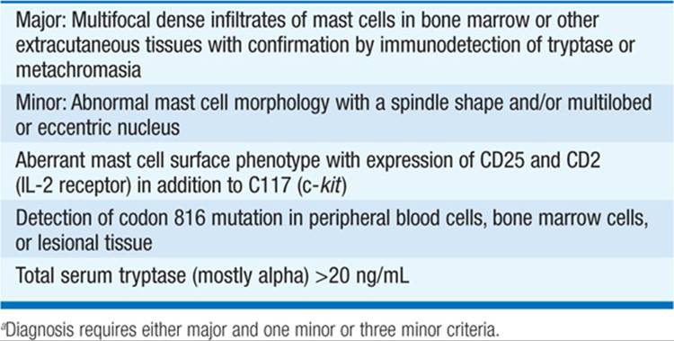
Although the diagnosis of mastocytosis may be suspected on the basis of clinical and laboratory findings, it can be established only by tissue biopsy (usually bone marrow biopsy). The diagnostic criteria for systemic mastocytosis are shown in Table 167-2. Laboratory studies that can help support a diagnosis of systemic mastocytosis include measurement of urinary or blood levels of mast cell products such as histamine, histamine metabolites, prostaglandin D2 (PGD2) metabolites, or mast cell tryptase. Other studies including bone scan, skeletal survey, GI contrast studies may be helpful. Other flushing disorders (e.g., carcinoid syndrome, pheochromocytoma) should be excluded.
TABLE 167-2 DIAGNOSTIC CRITERIA FOR SYSTEMIC MASTOCYTOSISa
TREATMENT Systemic Mastocystosis
• H1 and H2 antihistamines.
• Proton pump inhibitors for gastric hypersecretion.
• Oral cromolyn sodium for diarrhea and abdominal pain.
• NSAIDs (in nonsensitive pts) may help by blocking PGD2 production.
• Systemic glucocorticoids may help, but frequently are associated with complications.
• Hydroxyurea to reduce mast cell lineage progenitors may have merit in aggressive systemic mastocytosis.
• Chemotherapy for frank leukemias.

For a more detailed discussion, see Austen KF: Allergies, Anaphylaxis, and Systemic Mastocytosis, Chap. 317, p. 2707, in HPIM-18.