
DEFINITION AND PATHOGENESIS
A clinicopathologic process characterized by inflammation of and damage to blood vessels, compromise of vessel lumen, and resulting ischemia. Clinical manifestations depend on size and location of affected vessel. Most vasculitic syndromes appear to be mediated by immune mechanisms. May be primary or sole manifestation of a disease or secondary to another disease process. Unique vasculitic syndromes can differ greatly with regards to clinical features, disease severity, histology, and treatment.
PRIMARY VASCULITIS SYNDROMES
Granulomatosis with Polyangiitis (Wegener’s)
Granulomatous vasculitis of upper and lower respiratory tracts together with glomerulonephritis; upper airway lesions affecting the nose and sinuses can cause purulent or bloody nasal discharge, mucosal ulceration, septal perforation, and cartilaginous destruction (saddlenose deformity). Lung involvement may be asymptomatic or cause cough, hemoptysis, dyspnea; eye involvement may occur; glomerulonephritis can be rapidly progressive and asymptomatic and can lead to renal failure.
Churg-Strauss Syndrome
Granulomatous vasculitis of multiple organ systems, particularly the lung; characterized by asthma, peripheral eosinophilia, eosinophilic tissue infiltration; glomerulonephritis can occur.
Polyarteritis Nodosa (PAN)
Medium-sized muscular arteries involved; frequently associated with arteriographic aneurysms; commonly affects renal arteries, liver, GI tract, peripheral nerves, skin, heart; can be associated with hepatitis B.
Microscopic Polyangiitis
Small-vessel vasculitis that can affect the glomerulus and lungs; mediumsized vessels also may be affected.
Giant Cell Arteritis
Inflammation of medium- and large-sized arteries; primarily involves temporal artery but systemic and large vessel involvement may occur; symptoms include headache, jaw/tongue claudication, scalp tenderness, fever, musculoskeletal symptoms (polymyalgia rheumatica); sudden blindness from involvement of optic vessels is a dreaded complication.
Takayasu’s Arteritis
Vasculitis of the large arteries with strong predilection for aortic arch and its branches; most common in young women; presents with inflammatory or ischemic symptoms in arms and neck, systemic inflammatory symptoms, aortic regurgitation.
Henoch-Schönlein Purpura
Characterized by involvement of skin, GI tract, kidneys; more common in children; may recur after initial remission.
Cryoglobulinemic Vasculitis
Majority of cases are associated with hepatitis C where an aberrant immune response leads to formation of cryoglobulin; characterized by cutaneous vasculitis, arthritis, peripheral neuropathy, and glomerulonephritis.
Idiopathic Cutaneous Vasculitis
Cutaneous vasculitis is defined broadly as inflammation of the blood vessels of the dermis; due to underlying disease in >70% of cases (see “Secondary Vasculitis Syndromes,” below) with 30% occurring idiopathically.
Miscellaneous Vasculitic Syndromes
• Kawasaki disease (mucocutaneous lymph node syndrome)
• Isolated vasculitis of the central nervous system
• Behçet’s syndrome
• Cogan’s syndrome
• Polyangiitis overlap syndrome
SECONDARY VASCULITIS SYNDROMES
• Drug-induced vasculitis
• Serum sickness
• Vasculitis associated with infection, malignancy, rheumatic disease
EVALUATION (SEE FIG. 170-1)
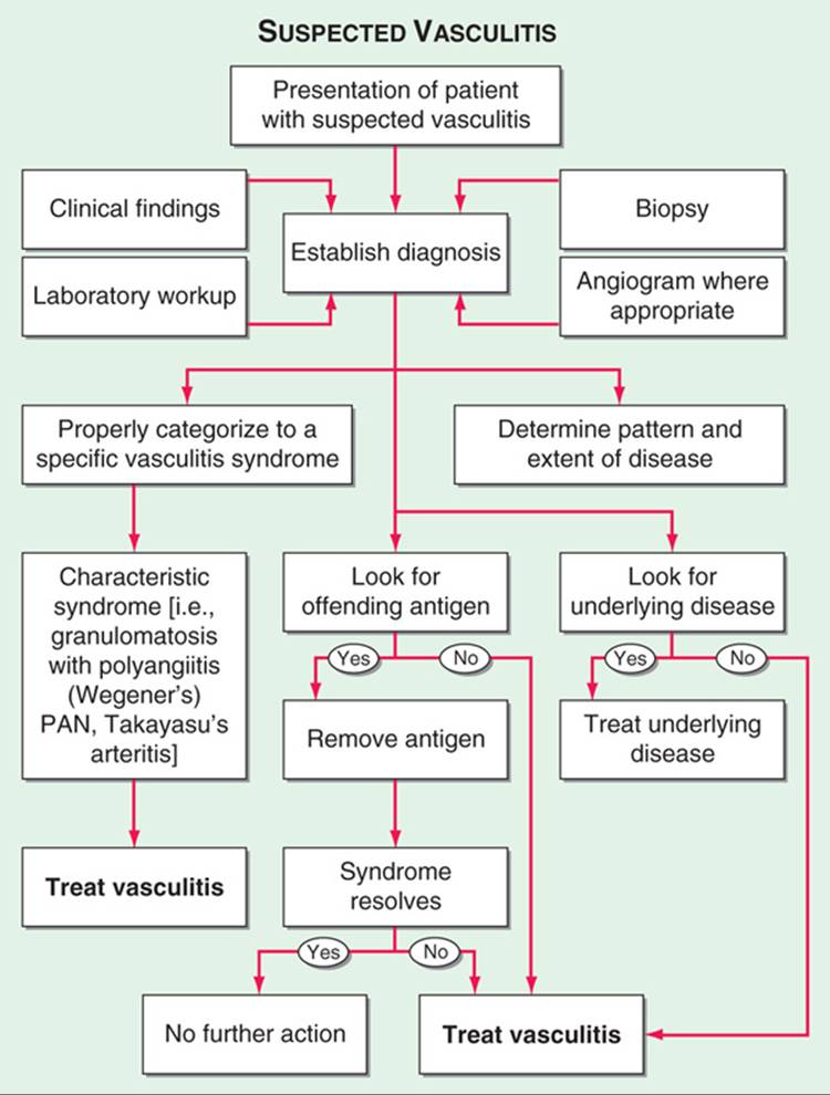
FIGURE 170-1 Algorithm for the approach to a pt with suspected diagnosis of vasculitis. PAN, polyarteritis nodosa.
• Thorough Hx and physical exam—special reference to ischemic manifestations and systemic inflammatory signs/symptoms.
• Laboratories—important in assessing organ involvement: CBC with differential, ESR, renal function tests, UA. Should also be obtained to rule out other diseases: ANA, rheumatoid factor, anti-GBM, hepatitis B/C serologies, HIV.
• Antineutrophil cytoplasmic autoantibodies (ANCA)—associated with granulomatosis with polyangiitis (Wegener’s), microscopic polyangiitis, and some pts with Churg-Strauss syndrome; presence of ANCA is adjunctive and should not be used in place of biopsy as a means of diagnosis or to guide treatment decisions.
• Radiographs—CXR should be performed even in the absence of symptoms.
• Diagnosis—can usually be made only by arteriogram or biopsy of affected organ(s).
DIFFERENTIAL DIAGNOSIS
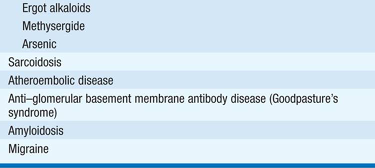
Guided by organ manifestations. In many instances includes infections and neoplasms, which must be ruled out prior to beginning immunosuppressive therapy. Consideration must also be given for diseases that can mimic vasculitis (Table 170-1).
TABLE 170-1 CONDITIONS THAT CAN MIMIC VASCULITIS


TREATMENT Vasculitis
Therapy is based on the specific vasculitic syndrome and the severity of its manifestations. Immunosuppressive therapy should be avoided in disease that rarely results in irreversible organ system dysfunction or that usually does not respond to such agents (e.g., isolated cutaneous vasculitis). Antiviral agents play an important role in treating vasculitis occurring with hepatitis B or C. Glucocorticoids alone may control giant cell arteritis and Takayasu’s arteritis. Therapy that combines glucocorticoids with another immunosuppressive agent is particularly important in syndromes with life-threatening organ system involvement, especially active glomerulonephritis. Frequently used agents:
• Prednisone 1 (mg/kg)/d initially, then tapered.
• Cyclophosphamide 2 (mg/kg)/d, adjusted to avoid severe leukopenia. Morning administration with a large amount of fluid is important in minimizing bladder toxicity. IV cyclophosphamide (15 mg/kg every 2 weeks for 3 doses then every 3 weeks thereafter) can also induce remission but may be associated with a higher relapse rate. Treatment should be limited to 3–6 months followed by transition to maintenance therapy with methotrexate or azathioprine.
• Rituximab 375 (mg/m2)/week for 4 weeks. As effective as cyclophosphamide to induce remission of granulomatosis with polyangiitis (Wegener’s) or microscopic polyangiitis. Relapse rate, long-term safety, repeat dosing frequency are unclear.
• Methotrexate in weekly doses up to 25 mg/week may be used to induce remission in granulomatosis with polyangiitis (Wegener’s) or microscopic polyangiitis pts who do not have immediately life-threatening disease or cannot tolerate cyclophosphamide. It may also be used for maintaining remission after induction with cyclophosphamide. Cannot be used in renal insufficiency or chronic liver disease.
• Azathioprine 2 (mg/kg)/d. Less effective in treating active disease but useful in maintaining remission after induction with cyclophosphamide.
• Mycophenolate mofetil 1000 mg bid. Less effective than azathioprine to maintain remission but an option in pts who cannot take or who have relapsed with methotrexate and azathioprine.
• Plasmapheresis may have an adjunctive role in rapidly progressive glomerulonephritis.

For a more detailed discussion, see Langford CA, Fauci AS: The Vasculitis Syndromes, Chap. 326, p. 2785; in HPIM-18.