
GOUT
DEFINITION
Gout is a metabolic disease most often affecting middle-aged to elderly men and postmenopausal women. Hyperuricemia is the biologic hallmark of gout. When present, plasma and extracellular fluids become supersaturated with uric acid, which, under the right conditions, may crystallize and result in a spectrum of clinical manifestations that may occur singly or in combination.
PATHOGENESIS
Uric acid is the end product of purine nucleotide degradation; its production is closely linked to pathways of purine metabolism, with the intracellular concentration of 5-phosphoribosyl-1-pyrophosphate (PRPP) being the major determinant of the rate of uric acid biosynthesis. Uric acid is excreted primarily by the kidney through mechanisms of glomerular filtration, tubular secretion, and reabsorption. Hyperuricemia may thus arise in a wide range of settings that cause overproduction or reduced excretion of uric acid or a combination of the two (Table 359-2, p. 3183, HPIM-18).
Acute Gouty Arthritis
Monosodium urate (MSU) crystals present in the joint are phagocytosed by leukocytes; release of inflammatory mediators and lysosomal enzymes leads to recruitment of additional phagocytes into the joint and to synovial inflammation.
CLINICAL MANIFESTATIONS
Acute arthritis—most frequent early clinical manifestation of gout. Usually initially affects one joint, but may be polyarticular in later episodes. The first metatarsophalangeal joint (podagra) is often involved. Acute gout frequently begins at night with dramatic pain, swelling, warmth, and tenderness. Attack will generally subside spontaneously after 3–10 days. Although some pts may have a single attack, most pts have recurrent episodes with intervals of varying length with no symptoms between attacks. Acute gout may be precipitated by dietary excess, trauma, surgery, excessive ethanol ingestion, hypouricemic therapy, and serious medical illnesses such as myocardial infarction and stroke.
Chronic arthritis—a proportion of gout pts may have a chronic nonsymmetric synovitis; may rarely be the only manifestation. Can also present with periarticular tophi (aggregates of MSU crystals surrounded by a giant cell inflammatory reaction). Occurs in the setting of long-standing gout.
Extraarticular tophi—often occur in olecranon bursa, helix and anthelix of ears, ulnar surface of forearm, Achilles tendon.
Tenosynovitis
Urate nephropathy—deposition of MSU crystals in renal interstitium and pyramids. Can cause chronic renal insufficiency.
Acute uric acid nephropathy—reversible cause of acute renal failure due to precipitation of urate in the tubules; pts receiving cytotoxic treatment for neoplastic disease are at risk.
Uric acid nephrolithiasis—responsible for 10% of renal stones in the United States.
DIAGNOSIS
• Synovial fluid analysis—should be performed to confirm gout even when clinical appearance is strongly suggestive; joint aspiration and demonstration of both intracellular and extracellular needle-shaped negatively birefringent MSU crystals by polarizing microscopy. Gram stain and culture should be performed on all fluid to rule out infection. MSU crystals can also be demonstrated in chronically involved joints or tophaceous deposits.
• Serum uric acid—normal levels do not rule out gout.
• Urine uric acid—excretion of >800 mg/d on regular diet in the absence of drugs suggests overproduction.
• Screening for risk factors or sequelae—urinalysis; serum creatinine, liver function tests, glucose and lipids; complete blood counts.
• If overproduction is suspected, measurement of erythrocyte hypoxanthine guanine phosphoribosyl transferase (HGPRT) and PRPP levels may be indicated.
• Joint x-rays—may demonstrate cystic changes, erosions with sclerotic margins in advanced chronic arthritis.
• If renal stones suspected, abdominal flat plate (stones often radiolucent), possibly IVP.
• Chemical analysis of renal stones.
Differential Diagnosis
Septic arthritis, reactive arthritis, calcium pyrophosphate dihydrate (CPPD) deposition disease, rheumatoid arthritis.
TREATMENT Gout
ASYMPTOMATIC HYPERURICEMIA As only ~5% of hyperuricemic pts develop gout, treatment of asymptomatic hyperuricemia is not indicated. Exceptions are pts about to receive cytotoxic therapy for neoplasms.
ACUTE GOUTY ARTHRITIS Treatment is given for symptomatic relief only since attacks are self-limited and will resolve spontaneously. Toxicity of therapy must be considered in each pt.
• Analgesia
• NSAIDs—Rx of choice when not contraindicated.
• Colchicine—generally only effective within first 24 h of attack; overdose has potentially life-threatening side effects; use is contraindicated in pts with renal insufficiency, cytopenias, LFTs >2 × normal, sepsis. PO—0.6 mg qh until pt improves, has GI side effects, or maximal dose of 4 mg is reached.
• Intraarticular glucocorticoids—septic arthritis must be ruled out prior to injection.
• Systemic glucocorticoids—brief taper may be considered in pts with a polyarticular gouty attack for whom other modalities are contraindicated and where articular or systemic infection has been ruled out.
URIC ACID–LOWERING AGENTS Indications for initiating uric acid–lowering therapy include recurrent frequent acute gouty arthritis, polyarticular gouty arthritis, tophaceous gout, renal stones, prophylaxis during cytotoxic therapy. Should not start during an acute attack. Initiation of such therapy can precipitate an acute flare; consider concomitant PO colchicine 0.6 mg qd until uric acid <5.0 mg/dL, then discontinue.
1. Xanthine oxidase inhibitors (allopurinol, febuxostat): Decrease uric acid synthesis. Allopurinol must be dose-reduced in renal insufficiency. Both have side effects and drug interactions.
2. Uricosuric drugs (probenecid, sulfinpyrazone): Increases uric acid excretion by inhibiting its tubular reabsorption; ineffective in renal insufficiency; should not be used in these settings: age >60, renal stones, tophi, increased urinary uric acid excretion, prophylaxis during cytotoxic therapy.
3. Pegloticase: Recombinant uricase that lowers uric acid by oxidizing urate to allantoin. Risk of severe infusion reactions. Should be used only in selected pts with chronic tophaceous gout refractory to conventional therapy.
CALCIUM PYROPHOSPHATE DIHYDRATE (CPPD) DEPOSITION DISEASE (PSEUDOGOUT)
DEFINITION AND PATHOGENESIS
CPPD disease is characterized by acute and chronic inflammatory joint disease, usually affecting older individuals. The knee and other large joints are most commonly affected. Calcium deposits in articular cartilage (chondrocalcinosis) may be seen radiographically; these are not always associated with symptoms.
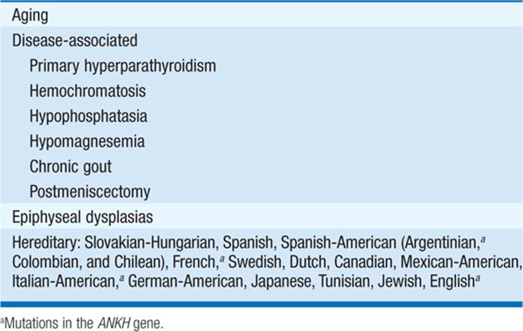
CPPD is most often idiopathic but can be associated with other conditions (Table 175-1).
TABLE 175-1 CONDITIONS ASSOCIATED WITH CPPD DEPOSITION DISEASE
Crystals are thought not to form in synovial fluid but are probably shed from articular cartilage into joint space, where they are phagocytosed by neutrophils and incite an inflammatory response.
CLINICAL MANIFESTATIONS
• Acute CPPD arthritis (“pseudogout”)—knee is most frequently involved, but polyarticular in two-thirds of cases; involved joint is erythematous, swollen, warm, and painful. Most pts have evidence of chondrocalcinosis.
• Chronic arthropathy—progressive degenerative changes in multiple joints; can resemble osteoarthritis (OA). Joint distribution may suggest CPPD with common sites including knee, wrist, MCP, hips, and shoulders.
• Symmetric proliferative synovitis—seen in familial forms with early onset; clinically similar to RA.
• Intervertebral disk and ligament calcification
• Spinal stenosis
DIAGNOSIS
• Synovial fluid analysis—demonstration of CPPD crystals that appear as short blunt rods, rhomboids, and cuboids with weak positive birefringence by polarizing microscopy
• Radiographs may demonstrate chondrocalcinosis and degenerative changes (joint space narrowing, subchondral sclerosis/cysts).
• Secondary causes of CPPD deposition disease should be considered in pts <50 years old.
Differential Diagnosis
OA, RA, gout, septic arthritis.
TREATMENT Pseudogout
• NSAIDs
• Intraarticular injection of glucocorticoids
• Colchicine (variably effective)
CALCIUM APATITE DEPOSITION DISEASE
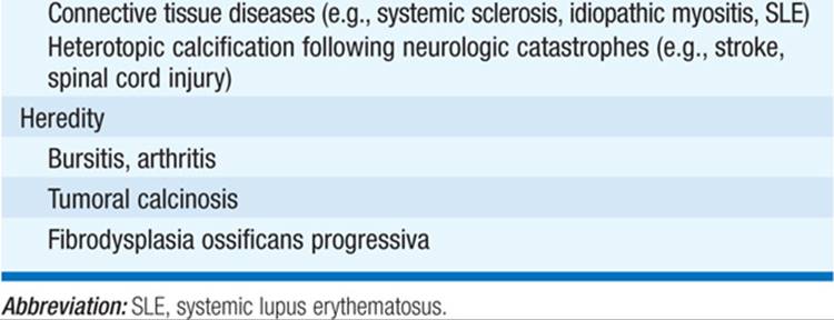
Apatite is the primary mineral of normal bone and teeth. Abnormal accumulation can occur in a wide range of clinical settings (Table 175-2). Apatite is an important factor in Milwaukee shoulder, a destructive arthropathy of the elderly that occurs in the shoulders and knees. Apatite crystals are small; clumps may stain purplish on Wright’s stain and bright red with alizarin red S. Definitive identification requires electron microscopy or x-ray diffraction studies. Radiographic appearance resembles CPPD disease. Treatment: NSAIDs, repeated aspiration, and rest of affected joint.
TABLE 175-2 CONDITIONS ASSOCIATED WITH CALCIUM APATITE DEPOSITION DISEASE

CALCIUM OXALATE DEPOSITION DISEASE
CaOx crystals may be deposited in joints in primary oxalosis (rare) or secondary oxalosis (a complication of end-stage renal disease). Clinical syndrome similar to gout and CPPD disease. Treatment: marginally effective.
For a more detailed discussion, see Burns CM, Wortmann RL: Disorders of Purine and Pyrimidine Metabolism, Chap. 359, p. 3181; and Schumacher HR, Chen LX: Gout and Other Crystal-Associated Arthropathies, Chap. 333, p. 2837, in HPIM-18.