
MENTAL STATUS EXAM
• The bare minimum: During the interview, look for difficulties with communication and determine whether the pt has recall and insight into recent and past events.
The mental status examination is underway as soon as the physician begins observing and talking with the pt. The goal of the mental status exam is to evaluate attention, orientation, memory, insight, judgment, and grasp of general information. Attention is tested by asking the pt to respond every time a specific item recurs in a list. Orientation is evaluated by asking about the day, date, and location. Memory can be tested by asking pt to immediately recall a sequence of numbers and by testing recall of a series of objects after defined times (e.g., 5 and 15 min). More remote memory is evaluated by assessing pt’s ability to provide a cogent chronologic history of the illness or personal life events. Recall of historic events or dates of current events can be used to assess knowledge. Evaluation of language function should include assessment of spontaneous speech, naming, repetition, reading, writing, and comprehension. Additional tests such as ability to draw and copy, perform calculations, interpret proverbs or logic problems, identify right vs. left, name and identify body parts, etc., are also important.
A useful screening examination of cognitive function is the mini-mental status examination (MMSE) (Table 191-1).
TABLE 191-1 THE MINI-MENTAL STATUS EXAMINATION

CRANIAL NERVE (CN) EXAM
• The bare minimum: Check the fundi, visual fields, pupil size and reactivity, extraocular movements, and facial movements.
CN I
Occlude each nostril sequentially and ask pt to gently sniff and identify a mild test stimulus, such as soap, toothpaste, coffee, or lemon oil.
CN II
Check visual acuity with eyeglasses or contact lens correction using a Snellen chart or similar tool. Map visual fields (VFs) by confrontation testing in each quadrant of visual field for each eye individually. The best method is to sit facing pt (2–3 ft apart) and then have pt cover one eye gently and fix uncovered eye on examiner’s nose. A small white object (e.g., a cotton-tipped applicator) is then moved slowly from periphery of field toward center until seen. Pt’s VF should be mapped against examiner’s for comparison. Formal perimetry and tangent screen exam are essential to identify and delineate small defects. Optic fundi should be examined with an ophthalmoscope, and the color, size, and degree of swelling or elevation of the optic disc recorded. The retinal vessels should be checked for size, regularity, AV nicking at crossing points, hemorrhage, exudates, and aneurysms. The retina, including the macula, should be examined for abnormal pigmentation and other lesions.
CN III, IV, VI
Describe size, regularity, and shape of pupils; reaction (direct and consensual) to light; and convergence (pt follows an object as it moves closer). Check for lid drooping, lag, or retraction. Ask pt to follow your finger as you move it horizontally to left and right and vertically with each eye first fully adducted then fully abducted. Check for failure to move fully in particular directions and for presence of regular, rhythmic, involuntary oscillations of eyes (nystagmus). Test quick voluntary eye movements (saccades) as well as pursuit (e.g., follow the finger).
CN V
Feel the masseter and temporalis muscles as pt bites down and test jaw opening, protrusion, and lateral motion against resistance. Examine sensation over entire face. Testing of the corneal reflex is indicated when suggested by the history.
CN VII
Look for asymmetry of face at rest and with spontaneous movements. Test eyebrow elevation, forehead wrinkling, eye closure, smiling, frowning; check puff, whistle, lip pursing, and chin muscle contraction. Observe for differences in strength of lower and upper facial muscles. Taste on the anterior two-thirds of tongue can be affected by lesions of the seventh CN proximal to the chorda tympani.
CN VIII
Check ability to hear tuning fork, finger rub, watch tick, and whispered voice at specified distances with each ear. Check for air vs. mastoid bone conduction (Rinne) and lateralization of a tuning fork placed on center of forehead (Weber). Accurate, quantitative testing of hearing requires formal audiometry. Remember to examine tympanic membranes.
CN IX, X
Check for symmetric elevation of palate-uvula with phonation (“ahh”), as well as position of uvula and palatal arch at rest. Sensation in region of tonsils, posterior pharynx, and tongue may also require testing. Pharyngeal (“gag”) reflex is evaluated by stimulating posterior pharyngeal wall on each side with a blunt object (e.g., tongue blade). Direct examination of vocal cords by laryngoscopy is necessary in some situations.
CN XI
Check shoulder shrug (trapezius muscle) and head rotation to each side (sternocleidomastoid muscle) against resistance.
CN XII
Examine bulk and power of tongue. Look for atrophy, deviation from midline with protrusion, tremor, and small flickering or twitching movements (fibrillations, fasciculations).
MOTOR EXAM
• The bare minimum: Look for muscle atrophy and check limb tone. Assess upper limb strength by checking for pronator drift and strength of wrist or finger reflexes. Test for lower limb strength by asking pt to walk normally and on heels and toes.
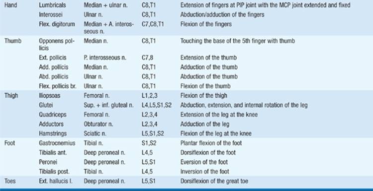
Power should be systematically tested for major movements at each joint (Table 191-2). Strength should be recorded using a reproducible scale (e.g., 0 = no movement, 1 = flicker or trace of contraction with no associated movement at a joint, 2 = movement present but cannot be sustained against gravity, 3 = movement against gravity but not against applied resistance, 4 = movement against some degree of resistance, and 5 = full power; 4 values can be supplemented with + and − signs to provide additional gradations). Speed of movement, ability to relax contractions promptly, and fatigue with repetition should all be noted. Loss in bulk and size of muscle (atrophy) should be noted, as well as the presence of irregular involuntary contraction (twitching) of groups of muscle fibers (fasciculations). Any involuntary movements should be noted at rest, during maintained posture, and with voluntary action.
TABLE 191-2 MUSCLES THAT MOVE JOINTS


REFLEXES
• The bare minimum: Tap the biceps, patellar, and Achilles reflexes.
Important muscle-stretch reflexes to test routinely and the spinal cord segments involved in their reflex arcs include biceps (C5, 6); brachioradialis (C5, 6); triceps (C7, 8); patellar (L3, 4); and Achilles (S1, 2). A common grading scale is 0 = absent, 1 = present but diminished, 2 = normal, 3 = hyperactive, and 4 = hyperactive with clonus (repetitive rhythmic contractions with maintained stretch). The plantar reflex should be tested by using a blunt-ended object such as the point of a key to stroke the outer border of the sole of the foot from the heel toward the base of the great toe. An abnormal response (Babinski sign) is extension (dorsiflexion) of the great toe at the metatarsophalangeal joint. In some cases this may be associated with abduction (fanning) of other toes and variable degrees of flexion at ankle, knee, and hip. Normal response is plantar flexion of the toes. Superficial abdominal and anal reflexes are important in certain situations; unlike muscle stretch reflexes, these cutaneous reflexes disappear with CNS lesions.
SENSORY EXAM
• The bare minimum: Ask whether the pt can feel light touch and the temperature of a cool object in each distal extremity. Check double simultaneous stimulation using light touch on the hands.
For most purposes it is sufficient to test sensation to pinprick, touch, position, and vibration in each of the four extremities (Figs. 191-1 and 191-2). Specific problems often require more thorough evaluation. Pts with cerebral lesions may have abnormalities in “discriminative sensation” such as the ability to perceive double simultaneous stimuli, to localize stimuli accurately, to identify closely approximated stimuli as separate (two-point discrimination), to identify objects by touch alone (stereognosis), or to judge weights, evaluate texture, or identify letters or numbers written on the skin surface (graphesthesia).

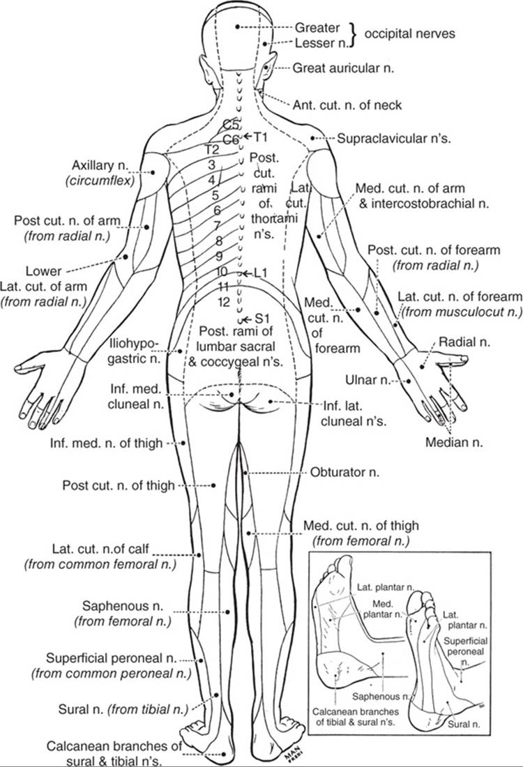
FIGURE 191-1 The cutaneous fields of peripheral nerves. (Reproduced by permission from W Haymaker, B Woodhall: Peripheral Nerve Injuries, 2nd ed. Philadelphia, Saunders, 1953.)
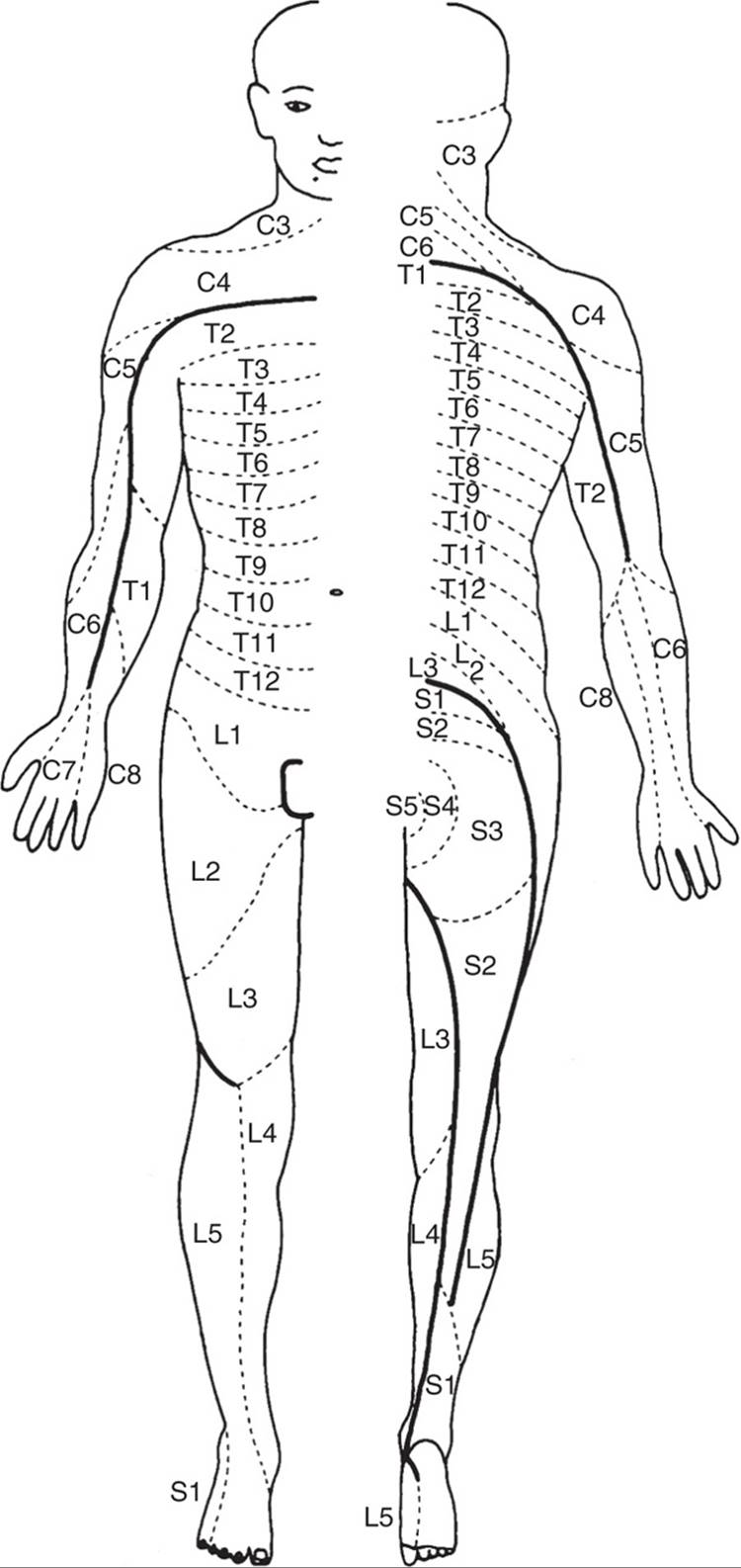
FIGURE 191-2 Distribution of the sensory spinal roots on the surface of the body (dermatomes). (From D Sinclair: Mechanisms of Cutaneous Sensation. Oxford, UK, Oxford University Press, 1981; with permission from Dr. David Sinclair.)
COORDINATION AND GAIT
• The bare minimum: Test rapid alternating movements of the fingers and feet, and the finger-to-nose maneuver. Observe the pt while he or she is walking along a straight line.
The ability to move the index finger accurately from the nose to the examiner’s outstretched finger and the ability to slide the heel of each foot from the knee down the shin are tests of coordination. Additional tests (drawing objects in the air, following a moving finger, tapping with index finger against thumb or alternately against each individual finger) may also be useful. The ability to stand with feet together and eyes closed (Romberg test), to walk a straight line (tandem walk), and to turn should all be observed.
THE NEUROLOGIC METHOD AND LOCALIZATION
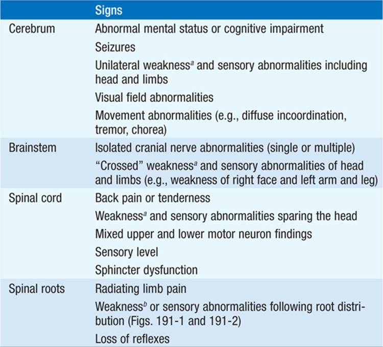
The clinical data obtained from the neurologic examination coupled with a careful history are interpreted to arrive at an anatomic localization that best explains the clinical findings (Table 191-3) and to select the diagnostic tests most likely to be informative in order to define the pathophysiology of the anatomic lesion.
TABLE 191-3 FINDINGS HELPFUL FOR LOCALIZATION WITHIN THE NERVOUS SYSTEM


For a more detailed discussion, see Lowenstein DH, Martin JB, Hauser SL: Approach to the Patient With Neurologic Disease, Chap. 367, p. 3233, in HPIM-18