
SODIUM
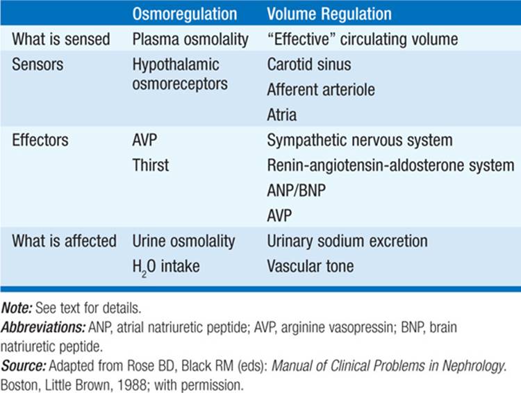
Disturbances of sodium concentration [Na+] result in most cases from abnormalities of H2O homeostasis, which change the relative ratio of Na+ to H2O. Disorders of Na+ balance per se are, in contrast, associated with changes in extracellular fluid volume, either hypo- or hypervolemia. Maintenance of the “effective circulating volume” is achieved in large part by changes in urinary sodium excretion, whereas H2O balance is achieved by changes in both H2O intake and urinary H2O excretion (Table 2-1). Confusion can result from the coexistence of defects in both H2O and Na+ balance. For example, a hypovolemic pt may have an appropriately low urinary Na+ due to increased renal tubular reabsorption of filtered NaCl; a concomitant increase in circulating arginine vasopressin (AVP)—part of the defense of effective circulating volume (Table 2-1)—will cause the renal retention of ingested H2O and the development of hyponatremia.
TABLE 2-1 OSMOREGULATION VERSUS VOLUME REGULATION
HYPONATREMIA
This is defined as a serum [Na+] <135 mmol/L and is among the most common electrolyte abnormalities encountered in hospitalized pts. Symptoms include nausea, vomiting, confusion, lethargy, and disorientation; if severe (<120 mmol/L) and/or abrupt, seizures, central herniation, coma, or death may result (see Acute Symptomatic Hyponatremia, below). Hyponatremia is almost always the result of an increase in circulating AVP and/or increased renal sensitivity to AVP; a notable exception is in the setting of low solute intake (“beer potomania”), wherein a markedly reduced urinary solute excretion is inadequate to support the excretion of sufficient free H2O. The serum [Na+] by itself does not yield diagnostic information regarding total-body Na+ content; hyponatremia is primarily a disorder of H2O homeostasis. Pts with hyponatremia are thus categorized diagnostically into three groups, depending on their clinical volume status: hypovolemic, euvolemic, and hypervolemic hyponatremia (Fig. 2-1). All three forms of hyponatremia share an exaggerated, “nonosmotic” increase in circulating AVP, in the setting of reduced serum osmolality. Notably, hyponatremia is often multifactorial; clinically important nonosmotic stimuli that can cause a release of AVP and increase the risk of hyponatremia include drugs, pain, nausea, and strenuous exercise.

FIGURE 2-1 The diagnostic approach to hyponatremia. See text for details. [From S Kumar, T Berl: Diseases of water metabolism, in Atlas of Diseases of the Kidney, RW Schrier (ed). Philadelphia, Current Medicine, Inc, 1999; with permission.]
Laboratory investigation of a pt with hyponatremia should include a measurement of serum osmolality to exclude “pseudohyponatremia” due to hyperlipidemia or hyperproteinemia. Serum glucose also should be measured; serum Na+ falls by 1.4 mM for every 100-mg/dL increase in glucose, due to glucose-induced H2O efflux from cells. Hyperkalemia may suggest adrenal insufficiency or hypoaldosteronism; increased blood urea nitrogen (BUN) and creatinine may suggest a renal cause. Urine electrolytes and osmolality are also critical tests in the initial evaluation of hyponatremia. In particular, a urine Na+ <20 meq/L is consistent with hypovolemic hyponatremia in the clinical absence of a “hypervolemic,” Na+-avid syndrome such as congestive heart failure (CHF) (Fig. 2-1). Urine osmolality <100 mosmol/kg is suggestive of polydipsia or, in rare cases, of decreased solute intake; urine osmolality >400 mosmol/kg suggests that AVP excess is playing a more dominant role, whereas intermediate values are more consistent with multifactorial pathophysiology (e.g., AVP excess with a component of polydipsia). Finally, in the right clinical setting, thyroid, adrenal, and pituitary function should also be tested.
Hypovolemic Hyponatremia
Hypovolemia from both renal and extrarenal causes is associated with hyponatremia. Renal causes of hypovolemia include primary adrenal insufficiency and hypoaldosteronism, salt-losing nephropathies (e.g., reflux nephropathy, nonoliguric acute tubular necrosis), diuretics, and osmotic diuresis. Random “spot” urine Na+ is typically >20 meq/L in these cases but may be <20 meq/L in diuretic-associated hyponatremia if tested long after administration of the drug. Nonrenal causes of hypovolemic hyponatremia include GI loss (e.g., vomiting, diarrhea, tube drainage) and integumentary loss (sweating, burns); urine Na+ is typically <20 meq/L in these cases.
Hypovolemia causes profound neurohumoral activation, inducing systems that preserve effective circulating volume, such as the renin-angiotensin-aldosterone axis (RAA), the sympathetic nervous system, and AVP (Table 2-1). The increase in circulating AVP serves to increase the retention of ingested free H2O, leading to hyponatremia. The optimal treatment of hypovolemic hyponatremia is volume administration, generally as isotonic crystalloid, i.e., 0.9% NaCl (“normal saline”). If the history suggests that hyponatremia has been “chronic,” i.e., present for 48 h, care should be taken to avoid overcorrection (see below), which can easily occur as AVP levels plummet in response to volume-resuscitation; if necessary, the administration of desmopressin (DDAVP) and free water can reinduce or arrest the correction of hyponatremia (see below).
Hypervolemic Hyponatremia
The edematous disorders (CHF, hepatic cirrhosis, and nephrotic syndrome) are often associated with mild to moderate degrees of hyponatremia ([Na+] = 125–135 mmol/L); occasionally, pts with severe CHF or cirrhosis may present with serum [Na+] <120 mmol/L. The pathophysiology is similar to that in hypovolemic hyponatremia, except that “effective circulating volume” is decreased due to the specific etiologic factors, i.e., cardiac dysfunction, peripheral vasodilation in cirrhosis, and hypoalbuminemia in nephrotic syndrome. The degree of hyponatremia is an indirect index of the associated neurohumoral activation (Table 2-1) and an important prognostic indicator in hypervolemic hyponatremia.
Management consists of treatment of the underlying disorder (e.g., afterload reduction in heart failure, large-volume paracentesis in cirrhosis, immunomodulatory therapy in some forms of nephrotic syndrome), Na+ restriction, diuretic therapy, and, in some pts, H2O restriction. Vasopressin antagonists (e.g., tolvaptan and conivaptan) are also effective in normalizing hyponatremia associated with both cirrhosis and CHF.
Euvolemic Hyponatremia
The syndrome of inappropriate ADH secretion (SIADH) characterizes most cases of euvolemic hyponatremia. Other causes of euvolemic hyponatremia include hypothyroidism and secondary adrenal insufficiency due to pituitary disease; notably, repletion of glucocorticoid levels in the latter may cause a rapid drop in circulating AVP levels and overcorrection of serum [Na+] (see below).
Common causes of SIADH include pulmonary disease (e.g., pneumonia, tuberculosis, pleural effusion) and central nervous system (CNS) diseases (e.g., tumor, subarachnoid hemorrhage, meningitis); SIADH also occurs with malignancies (e.g., small cell carcinoma of the lung) and drugs (e.g., selective serotonin reuptake inhibitors, tricyclic antidepressants, nicotine, vincristine, chlorpropamide, carbamazepine, narcotic analgesics, anti-psychotic drugs, cyclophosphamide, ifosfamide). Optimal treatment of euvolemic hyponatremia includes treatment of the underlying disorder. H2O restriction to <1 L/d is a cornerstone of therapy, but may be ineffective or poorly tolerated. However, vasopressin antagonists are predictably effective in normalizing serum [Na+] in SIADH. Alternatives include the coadministration of loop diuretics to inhibit the countercurrent mechanism and reduce urinary concentration, combined with oral salt tablets to abrogate diuretic-induced salt loss and attendant hypovolemia.
Acute Symptomatic Hyponatremia
Acute symptomatic hyponatremia is a medical emergency; a sudden drop in serum [Na+] can overwhelm the capacity of the brain to regulate cell volume, leading to cerebral edema, seizures, and death. Women, particularly premenopausal women, are particularly prone to such sequelae; neurologic consequences are comparatively rare in male pts. Many of these pts develop hyponatremia from iatrogenic causes, including hypotonic fluids in the postoperative period, prescription of a thiazide diuretic, colonoscopy preparation, or intraoperative use of glycine irrigants. Polydipsia with an associated cause of increased AVP may also cause acute hyponatremia, as can increased H2O intake in the setting of strenuous exercise, e.g., a marathon. The recreational drug Ecstasy [methylenedioxymethamphetamine (MDMA)] can cause acute hyponatremia, rapidly inducing both AVP release and increased thirst.
Severe symptoms may occur at relatively modest levels of serum [Na+], e.g., in the mid-120s. Nausea and vomiting are common premonitory symptoms of more severe sequelae. An important concomitant is respiratory failure, which may be hypercapnic due to CNS depression or normocapnic due to neurogenic, noncardiogenic pulmonary edema; the attendant hypoxia amplifies the impact of hyponatremic encephalopathy.
TREATMENT Hyponatremia
Three considerations are critical in the therapy of hyponatremia. First, the presence, absence, and/or severity of symptoms determine the urgency of therapy (see above for acute symptomatic hyponatremia). Second, pts with hyponatremia that has been present for >48 h (“chronic hyponatremia”) are at risk for osmotic demyelination syndrome, typically central pontine myelinolysis, if serum Na+ is corrected by >10–12 mM within the first 24 h and/or by >18 mM within the first 48 h. Third, the response to interventions, such as hypertonic saline or vasopressin antagonists, can be highly unpredictable, such that frequent monitoring of serum Na+ (every 2–4 h) is imperative.
Treatment of acute symptomatic hyponatremia should include hypertonic saline to acutely increase serum Na+ by 1–2 mM/h to a total increase of 4–6 mM; this increase is typically sufficient to alleviate acute symptoms, after which corrective guidelines for “chronic” hyponatremia are appropriate (see below). A number of equations and algorithms have been developed to estimate the required rate of hypertonic solution; one popular approach is to calculate a “Na+ deficit,” where the Na+ deficit = 0.6 × body weight × (target [Na+] – starting [Na+]). Regardless of the method used to determine the rate of administered hypertonic saline, the increase in serum [Na+] can be highly unpredictable, as the underlying physiology rapidly changes; serum [Na+] should be monitored every 2–4 h during and after treatment with hypertonic saline. The administration of supplemental O2 and ventilatory support can also be critical in acute hyponatremia, if pts develop acute pulmonary edema or hypercapnic respiratory failure. IV loop diuretics will help treat acute pulmonary edema and will also increase free H2O excretion by interfering with the renal countercurrent multiplier system. It is noteworthy that vasopressin antagonists do not have a role in the management of acute hyponatremia.
The rate of correction should be comparatively slow in chronic hyponatremia (<10–12 mM in the first 24 h and <18 mM in the first 48 h), so as to avoid osmotic demyelination syndrome. Vasopressin antagonists are highly effective in SIADH and in hypervolemic hyponatremia due to heart failure or cirrhosis. Should pts overcorrect serum [Na+] in response to vasopressin antagonists, hypertonic saline, or isotonic saline (in chronic hypovolemic hyponatremia), hyponatremia can be safely reinduced or stabilized by the administration of the vasopressin agonist DDAVP and the administration of free H2O, typically IV D5W; again, close monitoring of the response of serum [Na+] is essential to adjust therapy.
HYPERNATREMIA
This is rarely associated with hypervolemia, where the association is typically iatrogenic, e.g., administration of hypertonic sodium bicarbonate. More commonly, hypernatremia is the result of a combined H2O and volume deficit, with losses of H2O in excess of Na+. Elderly individuals with reduced thirst and/or diminished access to fluids are at the highest risk of hypernatremia due to decreased free H2O intake. Common causes of renal H2O loss are osmotic diuresis secondary to hyperglycemia, postobstructive diuresis, or drugs (radiocontrast, mannitol, etc.); H2O diuresis occurs in central or nephrogenic diabetes insipidus (DI) (Chap. 51). In pts with hypernatremia due to renal loss of H2O, it is critical to quantify ongoing daily losses in addition to calculation of the baseline H2O deficit (Table 2-2).
TABLE 2-2 CORRECTION OF HYPERNATREMIA

TREATMENT Hypernatremia
The approach to correction of hypernatremia is outlined in Table 2-2. As with hyponatremia, it is advisable to correct the H2O deficit slowly to avoid neurologic compromise, decreasing the serum [Na+] over 48–72 h. Depending on the blood pressure or clinical volume status, it may be appropriate to initially treat with hypotonic saline solutions (1/4 or 1/2 normal saline); blood glucose should be monitored in pts treated with large volumes of D5W, should hyperglycemia ensue. Calculation of urinary electrolyte-free H2O clearance is helpful to estimate daily, ongoing loss of free H2O in pts with nephrogenic or central DI (Table 2-2). Other forms of therapy may be helpful in selected cases of hypernatremia. Pts with central DI may respond to the administration of intranasal DDAVP. Stable pts with nephrogenic DI due to lithium may reduce their polyuria with amiloride (2.5–10 mg/d) or hydrochlorothiazide (12.5–50 mg/d) or both in combination. These diuretics are thought to increase proximal H2O reabsorption and decrease distal solute delivery, thus reducing polyuria; amiloride may also decrease entry of lithium into principal cells in the distal nephron by inhibiting the amiloride-sensitive epithelial sodium channel (ENaC). Notably, however, most pts with lithium-induced nephrogenic DI can adequately accommodate by increasing their H2O intake. Occasionally, nonsteroidal anti-inflammatory drugs (NSAIDs) have also been used to treat polyuria associated with nephrogenic DI, reducing the negative effect of local prostaglandins on urinary concentration; however, the nephrotoxic potential of NSAIDs typically makes them a less attractive therapeutic option.
POTASSIUM
Since potassium (K+) is the major intracellular cation, discussion of disorders of K+ balance must take into consideration changes in the exchange of intra- and extracellular K+ stores. (Extracellular K+constitutes <2% of total-body K+ content.) Insulin, β2-adrenergic agonists, and alkalosis tend to promote K+ uptake by cells; acidosis, insulinopenia, or acute hyperosmolality (e.g., after treatment with mannitol or D50W) promote the efflux or reduced uptake of K+. A corollary is that tissue necrosis and the attendant release of K+ can cause severe hyperkalemia, particularly in the setting of acute kidney injury. Hyperkalemia due to rhabdomyolysis is thus particularly common, due to the enormous store of K+ in muscle; hyperkalemia may also be prominent in tumor lysis syndrome.
The kidney plays a dominant role in K+ excretion. Although K+ is transported along the entire nephron, it is the principal cells of the connecting segment and cortical collecting duct that play a dominant role in K+ excretion. Apical Na+ entry into principal cells via the amiloride-sensitive epithelial Na+ channel (ENaC) generates a lumen-negative potential difference, which drives passive K+ exit through apical K+channels. This relationship is key to the bedside understanding of potassium disorders. For example, decreased distal delivery of Na+ tends to blunt the ability to excrete K+, leading to hyperkalemia. Abnormalities in the RAA can cause both hypo- and hyperkalemia; aldosterone has a major influence on potassium excretion, increasing the activity of ENaC channels and thus amplifying the driving force for K+ secretion across the luminal membrane of principal cells.
HYPOKALEMIA
Major causes of hypokalemia are outlined in Table 2-3. Atrial and ventricular arrhythmias are the most serious health consequences of hypokalemia. Pts with concurrent Mg deficit and/or digoxin therapy are at a particularly increased risk of arrhythmias. Other clinical manifestations include muscle weakness, which may be profound at serum [K+] <2.5 mmol/L, and, if hypokalemia is sustained, hypertension, ileus, polyuria, renal cysts, and even renal failure.
TABLE 2-3 CAUSES OF HYPOKALEMIA
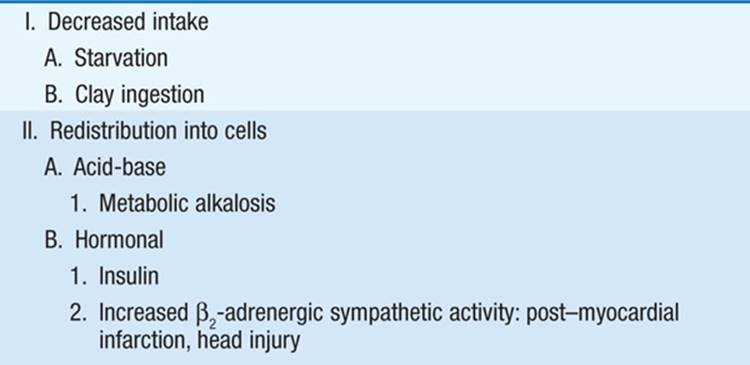

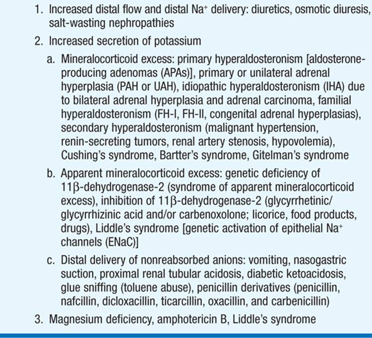
The cause of hypokalemia is usually obvious from history, physical examination, and/or basic laboratory tests. However, persistent hypokalemia may require a more thorough, systematic evaluation (Fig. 2-2). Initial laboratory evaluation should include electrolytes, BUN, creatinine, serum osmolality, Mg2+, and Ca2+, a complete blood count, and urinary pH, osmolality, creatinine, and electrolytes. Serum and urine osmolality are required for calculation of the transtubular K+ gradient (TTKG), which should be <3 in the presence of hypokalemia (see also Hyperkalemia). Further tests such as urinary Mg2+ and Ca2+ and/or plasma renin and aldosterone levels may be necessary in specific cases.

FIGURE 2-2 The diagnostic approach to hypokalemia. See text for details. BP, blood pressure; DKA, diabetic ketoacidosis; FHPP, familial hypokalemic periodic paralysis; FH-I, familial hyperaldosteronism type I; GI, gastrointestinal; HTN, hypertension; PA, primary aldosteronism; RAS, renal artery stenosis; RST, renin-secreting tumor; RTA, renal tubular acidosis; SAME, syndrome of apparent mineralocorticoid excess; TTKG, transtubular potassium gradient. [From Mount DB, Zandi-Nejad K: Disorders of potassium balance, in The Kidney, 8th ed, BM Brenner (ed). Philadelphia, Saunders, 2008; with permission.]
TREATMENT Hypokalemia
Hypokalemia can generally be managed by correction of the underlying disease process (e.g., diarrhea) or withdrawal of an offending medication (e.g., loop or thiazide diuretic), combined with oral KCl supplementation. However, hypokalemia is refractory to correction in the presence of Mg deficiency, which also should be corrected when present; renal wasting of both cations may be particularly prominent after renal tubular injury, e.g., from cisplatin nephrotoxicity. If loop or thiazide diuretic therapy cannot be discontinued, a distal tubular K-sparing agent, such as amiloride or spironolactone, can be added to the regimen. Angiotensin-converting enzyme (ACE) inhibition in pts with CHF attenuates diuretic-induced hypokalemia and protects against cardiac arrhythmia. If hypokalemia is severe (<2.5 mmol/L) and/or if oral supplementation is not feasible or tolerated, IV KCl can be administered through a central vein with cardiac monitoring in an intensive care setting, at rates that should not exceed 20 mmol/h. KCl should always be administered in saline solutions, rather than dextrose; the dextrose-induced increase in insulin can acutely exacerbate hypokalemia.
HYPERKALEMIA
Causes are outlined in Table 2-4; in most cases, hyperkalemia is due to decreased renal K+ excretion. However, increases in dietary K+ intake can have a major effect in susceptible pts, e.g., diabetics with hyporeninemic hypoaldosteronism and chronic kidney disease. Drugs that impact on the renin-angiotensin-aldosterone axis are also a major cause of hyperkalemia, particularly given recent trends to coadminister these agents, e.g., spironolactone or angiotensin receptor blockers with an ACE inhibitor in cardiac and/or renal disease.
TABLE 2-4 CAUSES OF HYPERKALEMIA

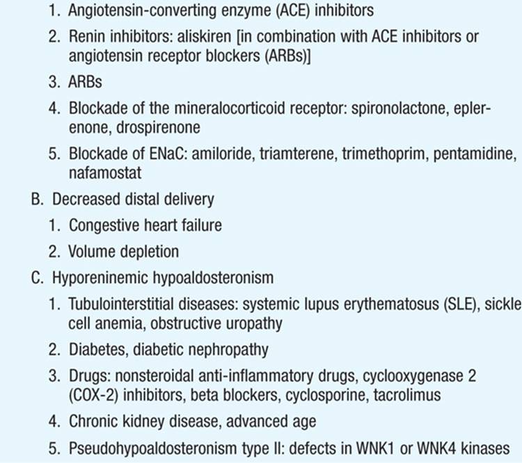
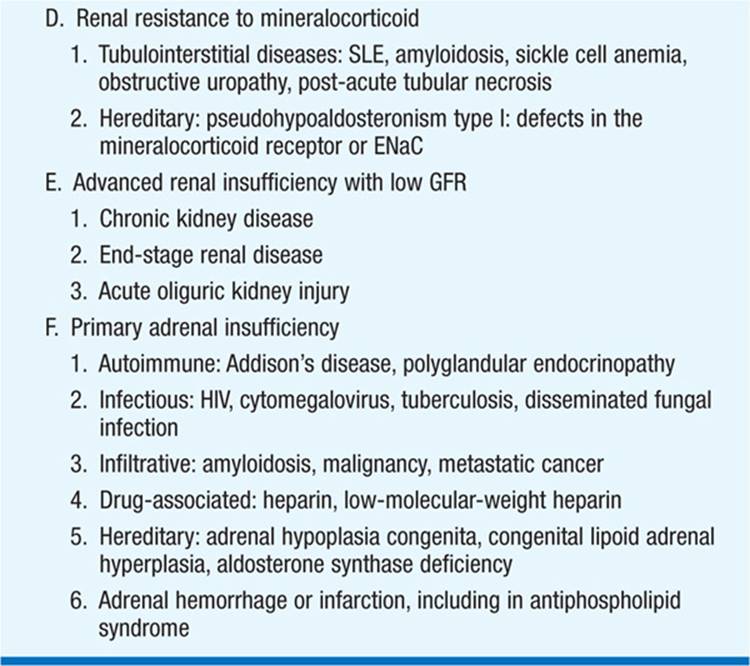
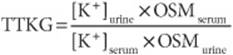
The first priority in the management of hyperkalemia is to assess the need for emergency treatment (ECG changes and/or K+ ≥6.0 mM). This should be followed by a comprehensive workup to determine the cause (Fig. 2-3). History and physical examination should focus on medications (e.g., ACE inhibitors, NSAIDs, trimethoprim/sulfamethoxazole), diet and dietary supplements (e.g., salt substitute), risk factors for acute kidney failure, reduction in urine output, blood pressure, and volume status. Initial laboratory tests should include electrolytes, BUN, creatinine, serum osmolality, Mg2+, and Ca2+, a complete blood count, and urinary pH, osmolality, creatinine, and electrolytes. A urine [Na+] <20 meq/L suggests that distal Na+ delivery is a limiting factor in K+ excretion; volume repletion with 0.9% saline or treatment with furosemide may then be effective in reducing serum [K+] by increasing distal Na+ delivery. Serum and urine osmolality are required for calculation of the TTKG. The expected values of the TTKG are largely based on historic data: <3 in the presence of hypokalemia and >7–8 in the presence of hyperkalemia.
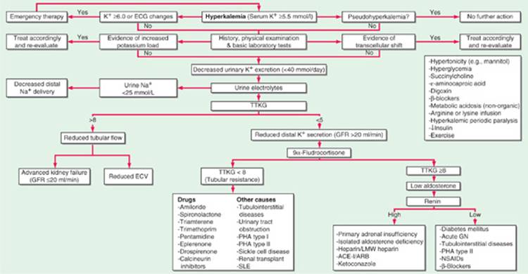
FIGURE 2-3 The diagnostic approach to hyperkalemia. See text for details. ACE-I, angiotensin converting enzyme inhibitor; acute GN, acute glomerulonephritis; ARB, angiotensin II receptor blocker; ECG, electrocardiogram; ECV, effective circulatory volume; GFR, glomerular filtration rate; LMW heparin, low-molecular-weight heparin; NSAIDs, nonsteroidal anti-inflammatory drugs; PHA, pseudohypoaldosteronism; SLE, systemic lupus erythematosus; TTKG, transtubular potassium gradient. [From Mount DB, Zandi-Nejad K: Disorders of potassium balance, in The Kidney, 8th ed, BM Brenner (ed). Philadelphia, Saunders, 2008; with permission.]
TREATMENT Hyperkalemia
The most important consequence of hyperkalemia is altered cardiac conduction, with the risk of bradycardic cardiac arrest. Figure 2-4 shows serial ECG patterns of hyperkalemia; ECG manifestations of hyperkalemia should be considered a true medical emergency and treated urgently. However, ECG changes of hyperkalemia are notoriously insensitive, particularly in pts with chronic kidney disease; given these limitations, pts with significant hyperkalemia (K+ ≥6–6.5 mmol/L) in the absence of ECG changes should also be aggressively managed.

FIGURE 2-4 Diagrammatic ECGs at normal and high serum K. Peaked T waves (precordial leads) are followed by diminished R wave, wide QRS, prolonged P-R, loss of P wave, and ultimately a sine wave.
Urgent management of hyperkalemia constitutes a 12-lead ECG, admission to the hospital, continuous cardiac monitoring, and immediate treatment. Treatment of hyperkalemia is divided into three categories: (1) antagonism of the cardiac effects of hyperkalemia, (2) rapid reduction in [K+] by redistribution into cells, and (3) removal of K+ from the body. Treatment of hyperkalemia is summarized in Table 2-5.
TABLE 2-5 TREATMENT OF HYPERKALEMIA

ACID-BASE DISORDERS (FIG. 2-5)
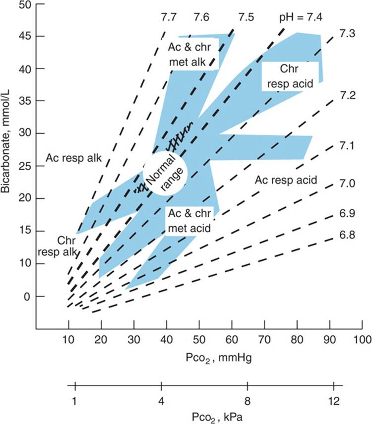
FIGURE 2-5 Nomogram showing bands for uncomplicated respiratory or metabolic acid-base disturbances in intact subjects. Each confidence band represents the mean ±2 SD for the compensatory response of normal subjects or pts to a given primary disorder. Ac, acute; chr, chronic; resp, respiratory; met, metabolic; acid, acidosis; alk, alkalosis. (From Levinsky NG: HPIM-12, p. 290; modified from Arbus GS: Can Med Assoc J 109:291, 1973.)
Regulation of normal pH (7.35–7.45) depends on both the lungs and kidneys. By the Henderson-Hasselbalch equation, pH is a function of the ratio of  (regulated by the kidney) to Pco2 (regulated by the lungs). The HCO3/Pco2 relationship is useful in classifying disorders of acid-base balance. Acidosis is due to gain of acid or loss of alkali; causes may be metabolic (fall in serum ) or respiratory (rise in Pco2). Alkalosis is due to loss of acid or addition of base and is either metabolic (↑ serum
  ) or respiratory (↓ Pco2).
To limit the change in pH, metabolic disorders evoke an immediate compensatory response in ventilation; full renal compensation for respiratory disorders is a slower process, such that “acute” compensations are of lesser magnitude than “chronic” compensations. Simple acid-base disorders consist of one primary disturbance and its compensatory response. In mixed disorders, a combination of primary disturbances is present.
The cause of simple acid-base disorders is usually obvious from history, physical examination, and/or basic laboratory tests. Initial laboratory evaluation depends on the dominant acid-base disorder, but for metabolic acidosis and alkalosis this should include electrolytes, BUN, creatinine, albumin, urinary pH, and urinary electrolytes. An arterial blood gas (ABG) is not always required for pts with a simple acid-base disorder, e.g., mild metabolic acidosis in the context of chronic renal failure. However, concomitant ABG and serum electrolytes are necessary to fully evaluate more complex acid-base disorders. The compensatory response should be estimated from the ABG; Winter’s formula  is particularly useful for assessing the respiratory response to metabolic acidosis. The anion gap should also be calculated; the anion gap =
  = unmeasured anions – unmeasured cations. The anion gap should be adjusted for changes in the concentration of albumin, a dominant unmeasured anion; the “adjusted anion gap” = anion gap + ~2.5 × (4 – albumin mg/dL). Other supportive tests will elucidate the specific form of anion-gap acidosis (see below).
METABOLIC ACIDOSIS
The low in metabolic acidosis results from the addition of acids (organic or inorganic) or from a loss of ; causes of metabolic acidosis are classically categorized by presence or absence of an increase in the anion gap (Table 2-6). Increased anion-gap acidosis (>12 mmol/L) is due to addition of acid (other than HCl) and unmeasured anions to the body. Common causes include ketoacidosis [diabetes mellitus (DKA), starvation, alcohol], lactic acidosis, poisoning (salicylates, ethylene glycol, and methanol), and renal failure.
TABLE 2-6 METABOLIC ACIDOSIS
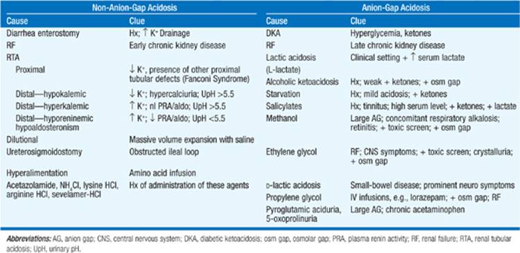
Rare and newly appreciated causes of anion-gap acidosis include D-lactic acidosis, propylene glycol toxicity, and 5-oxoprolinuria (also known as pyroglutamic aciduria). D-Lactic acidosis (an increase in the D-enantiomer of lactate) can occur in pts with removal, disease, or bypass of the short bowel, leading to increased delivery of carbohydrates to colon. Intestinal overgrowth of organisms that metabolize carbohydrate to D-lactate results in D-lactic acidosis; a wide variety of neurologic symptoms can ensue, with resolution following treatment with appropriate antibiotics to change the intestinal flora. Propylene glycol is a common solvent for IV preparations of a number of drugs, most prominently lorazepam. Pts receiving high rates of these drugs may develop a hyperosmolar anion-gap metabolic acidosis, due mostly to increased lactate, often accompanied by acute kidney failure. Pyroglutamic aciduria (5-oxoprolinuria) is a high anion-gap acidosis caused by dysfunction of the γ-glutamyl cycle that replenishes intracellular glutathione; 5-oxoproline is an intermediate product of the cycle. Hereditary defects in the γ-glutamyl cycle are associated with 5-oxoprolinuria; acquired defects occur in the context of acetaminophen therapy, due to derepression of the cycle by reduced glutathione and overproduction of 5-oxoproline. Resolution occurs after withdrawal of acetaminophen; treatment with N-acetyl cysteine to replenish glutathione stores may hasten recovery.
The differentiation of the various anion-gap acidoses depends on the clinical scenario and routine laboratory tests (Table 2-6) in conjunction with measurement of serum lactate, ketones, toxicology screens (if ethylene glycol or methanol ingestion are suspected), and serum osmolality. D-Lactic acidosis can be diagnosed by a specific assay for the D-enantiomer; 5-oxoprolinuria can be diagnosed by the clinical scenario and confirmed by gas chromatographic/mass spectroscopic (GC/MS) analysis of urine, a widely available pediatric screening test for inborn errors of metabolism (typically “urine for organic acids”).
Pts with ethylene glycol, methanol, or propylene glycol toxicity may have an “osmolar gap,” defined as a >10-mosm/kg difference between calculated and measured serum osmolality. Calculated osmolality = 2 × Na+ + glucose/18 + BUN/2.8. Of note, pts with alcoholic ketoacidosis and lactic acidosis may also exhibit a modest elevation in the osmolar gap; pts may alternatively metabolize ethylene glycol or methanol to completion by presentation, with an increased anion gap and no increase in the osmolar gap. However, the rapid availability of a measured serum osmolality may aid in the urgent assessment and management of pts with these medical emergencies.
Normal anion-gap acidosis can result from  loss from the GI tract. Diarrhea is by far the most common cause, but other GI conditions associated with external losses of bicarbonate-rich fluids may lead to large alkali losses—e.g., in ileus secondary to intestinal obstruction, in which liters of alkaline fluid may accumulate within the intestinal lumen. Various forms of kidney disease are associated with non-anion-gap acidosis due to reduced tubular reabsorption of filtered bicarbonate and/or reduced excretion of ammonium
  . The early stages of progressive renal disease are frequently associated with a non-anion-gap acidosis, with development of an anion-gap component in more advanced renal failure. Non-anion-gap acidosis is also seen in renal tubular acidosis or in the context of tubulointerstitial injury, e.g., after acute tubular necrosis, allergic interstitial nephritis, or urinary tract obstruction. Finally, non-anion-gap acidosis due to exogenous acid loads may occur after rapid volume expansion with saline-containing solutions, the administration of NH4Cl (a component of cough syrup), lysine HCl, or treatment with the phosphate binder sevelamer hydrochloride.
Calculation of the urinary anion gap may be helpful in the evaluation of hyperchloremic metabolic acidosis, along with a measurement of urine pH. The urinary anion gap is defined as urinary  = [unmeasured anions] – [unmeasured cations]); the
  ion is the major unmeasured urinary cation in metabolic acidosis, wherein the urinary anion gap should be strongly negative. A negative anion gap thus suggests GI losses of bicarbonate, with appropriate renal response and increased excretion; a positive anion gap suggests altered urinary acidification, as seen in renal failure or distal renal tubular acidoses. An important caveat is that the rapid renal excretion of unmeasured anions in anion-gap acidosis, classically seen in DKA, may reduce the serum anion gap and generate a positive value for the urinary anion gap, despite the adequate excretion of urinary
  ; this may lead to misdiagnosis as a renal tubular acidosis.
TREATMENT Metabolic Acidosis
Treatment of metabolic acidosis depends on the cause and severity. DKA responds to insulin therapy and aggressive hydration; close attention to serum [K+] and administration of KCl is essential, given that the correction of insulinopenia can cause profound hypokalemia. The administration of alkali in anion-gap acidoses is controversial and is rarely appropriate in DKA. It is reasonable to treat severe lactic acidosis with IV  at a rate sufficient to maintain a
  ; treatment of moderate lactic acidosis with is controversial. IV HCO3 is however appropriate to reduce acidosis in D-lactic acidosis, ethylene glycol and methanol toxicity, and 5-oxoprolinuria.
Chronic metabolic acidosis should be treated when is <18–20 mmol/L. In pts with chronic kidney disease, there is some evidence that acidosis promotes protein catabolism and may worsen bone disease. Sodium citrate may be more palatable than oral NaHCO3, although the former should be avoided in pts with advanced renal insufficiency, as it augments aluminum absorption. Oral therapy with NaHCO3 usually begins with 650 mg tid and is titrated upward to maintain serum [].
METABOLIC ALKALOSIS
Metabolic alkalosis is due to a primary increase in serum [], distinguished from chronic respiratory acidosis—with a compensatory increase in renal reabsorption—by the associated increase in arterial pH (normal or decreased in chronic respiratory acidosis). Administered, exogenous alkali (, acetate, citrate, or lactate) may cause alkalosis if the normal capacity to excrete is reduced or if renal reabsorption is enhanced. A recently resurgent problem is “milk alkali syndrome,” a triad of hypercalcemia, metabolic alkalosis, and acute renal failure due to ingested calcium carbonate, typically taken for the treatment or prevention of osteoporosis.
Metabolic alkalosis is primarily caused by renal retention of and is due to a variety of underlying mechanisms. Pts are typically separated into two major subtypes: Cl–-responsive and Cl–-resistant. Measurement of urine Cl– affords this separation in the clinical setting (Fig. 2-6). The quintessential causes of Cl–-responsive alkalosis are GI-induced from vomiting or gastric aspiration through a nasogastric tube, and renal-induced from diuretic therapy. Hypovolemia, chloride deficiency, activation of the renin-angiotensin-aldosterone axis, and hypokalemia play interrelated roles in the maintenance of this hypochloremic or “contraction” alkalosis. The various syndromes of true or apparent mineralocorticoid excess cause Cl–-resistant metabolic alkalosis (Fig. 2-6); most of these pts are hypokalemic, volume-expanded, and/or hypertensive.

FIGURE 2-6 The diagnostic approach to metabolic alkalosis. See text for details. GRA, glucocorticoid-remediable aldosteronism; HTN, hypertension; JGA, juxtaglomerular apparatus. [Modified from Dubose TD: Disorders of acid-base balance, in The Kidney, 8th ed, BM Brenner (ed). Philadelphia, Saunders, 2008; with permission.]
Common forms of metabolic alkalosis are generally diagnosed from the history, physical examination, and/or basic laboratory tests. ABGs will help determine whether an elevated [] is reflective of metabolic alkalosis or chronic respiratory acidosis; ABGs are required for the diagnosis of mixed acid-base disorders. Measurement of urinary electrolytes will aid in separating Cl–-responsive and Cl–-resistant forms. Urinary [Na+] may thus be >20 meq/L in Cl–-responsive alkalosis despite the presence of hypovolemia; however, urinary [Cl–] will be very low. Notably, urinary [Cl–] may be variable in pts with diuretic-associated alkalosis, depending on the temporal relationship to diuretic administration. Other diagnostic tests—e.g., plasma renin, aldosterone, cortisol—may be appropriate in Cl–-resistant forms with high urinary [Cl–] (Fig. 2-6).
TREATMENT Metabolic Alkalosis
The acid-base disorder in Cl–-responsive alkalosis will typically respond to saline infusion; however, the associated hypokalemia should also be corrected. Pts with true or apparent mineralocorticoid excess require specific treatment of the underlying disorder. For example, hyperactive amiloride-sensitive ENaC channels cause Liddle’s syndrome, which can respond to therapy with amiloride and related drugs; pts with hyperaldosteronism may in turn respond to blockade of the mineralocorticoidreceptor with spironolactone or eplerenone. Finally, severe alkalosis in the critical care setting may require treatment with acidifying agents such as acetazolamide or HCl.
RESPIRATORY ACIDOSIS
Respiratory acidosis is characterized by CO2 retention due to ventilatory failure. Causes include sedatives, stroke, chronic pulmonary disease, airway obstruction, severe pulmonary edema, neuromuscular disorders, and cardio-pulmonary arrest. Symptoms include confusion, asterixis, and obtundation.
TREATMENT Respiratory Acidosis
The goal is to improve ventilation through pulmonary toilet and reversal of bronchospasm. Intubation or noninvasive positive pressure ventilation (NPPV) may be required in severe acute cases. Acidosis due to hypercapnia is usually mild; however, combined respiratory and metabolic acidosis may cause a profound reduction in pH. Respiratory acidosis may accompany low tidal volume ventilation in ICU pts and may require metabolic “overcorrection” to maintain a neutral pH.
RESPIRATORY ALKALOSIS
Excessive ventilation causes a primary reduction in CO2 and ↑ pH in pneumonia, pulmonary edema, interstitial lung disease, and asthma. Pain and psychogenic causes are common; other etiologies include fever, hypoxemia, sepsis, delirium tremens, salicylates, hepatic failure, mechanical overventilation, and CNS lesions. Pregnancy is associated with a mild respiratory alkalosis. Severe respiratory alkalosis may acutely cause seizures, tetany, cardiac arrhythmias, or loss of consciousness.
TREATMENT Respiratory Alkalosis
Treatment should be directed at the underlying disorders. In psychogenic cases, sedation or a rebreathing bag may be required.
“MIXED” DISORDERS
In many circumstances, more than a single acid-base disturbance exists. Examples include combined metabolic and respiratory acidosis with cardiogenic shock; metabolic alkalosis and anion-gap acidosis in pts with vomiting and diabetic ketoacidosis; and anion-gap metabolic acidosis with respiratory alkalosis in pts with salicylate toxicity. The diagnosis may be clinically evident and/or suggested by relationships between the Pco2 and [] that diverge from those found in simple disorders. For example, the Pco2 in a pt with metabolic acidosis and respiratory alkalosis will be considerably less than that predicted from the [] and Winter’s formula  .
.
In “simple” anion-gap acidosis, the anion gap increases in proportion to the fall in []. A lesser drop in serum [] than in the anion gap suggests a coexisting metabolic alkalosis. Conversely, a proportionately largerdrop in [] than in the anion gap suggests the presence of a mixed anion-gap and non-anion-gap metabolic acidosis. Notably, however, these interpretations assume 1:1 relationships between unmeasured anions and the fall in [], which are not uniformly present in individual pts or as acidoses evolve. For example, volume resuscitation of pts with DKA will typically increase glomerular filtration and the urinary excretion of ketones, resulting in a decrease in the anion gap in the absence of a supervening non-anion-gap acidosis.

For a more detailed discussion, see Mount DB: Fluid and Electrolyte Disturbances, Chap. 45, p. 341; and DuBose TD Jr: Acidosis and Alkalosis, Chap. 47, p. 363, in HPIM-18. See also Mount DB, Zandi-Nejad K: Disorders of potassium balance, in The Kidney, 9th ed, BM Brenner (ed). Philadelphia, Saunders, 2011; and Ellison DH, Berl T: Clinical practice. The syndrome of inappropriate antidiuresis. N Engl J Med 356:2064, 2007.