
APPROACH TO THE PATIENT Peripheral Neuropathy
Peripheral neuropathy (PN) refers to a peripheral nerve disorder of any cause. Nerve involvement may be single (mononeuropathy) or multiple (polyneuropathy); pathology may be axonal or demyelinating. An approach to pts with suspected neuropathy appears in Fig. 205-1.

FIGURE 205-1 Approach to evaluation of peripheral neuropathies. CIDP, chronic inflammatory demyelinating polyradiculoneuropathy; EDX, electrodiagnostic studies; GBS, Guillain-Barré syndrome; Mg, intravenous immunoglobulin.
Address the following seven initial questions:
1. What systems are involved? It is important to determine if the pt’s symptoms and signs are motor, sensory, autonomic, or a combination of these. If only weakness is present without any evidence of sensory or autonomic dysfunction, consider a motor neuropathy, neuromuscular junction disorder, or myopathy; myopathies usually have a proximal, symmetric pattern of weakness.
2. What is the distribution of weakness? Polyneuropathy involves widespread and symmetric dysfunction of the peripheral nerves that is usually distal more than proximal; mononeuropathy involves a single nerve usually due to trauma or compression; multiple mononeuropathies (mononeuropathy multiplex) can be a result of multiple entrapments, vasculitis, or infiltration.
3. What is the nature of the sensory involvement? Temperature loss or burning/stabbing pain suggests small fiber involvement. Vibratory or proprioceptive loss implicates large fibers.
4. Is there evidence of upper motor neuron involvement? The most common cause is combined system degeneration with B12 deficiency, but should also consider copper deficiency, HIV infection, severe hepatic disease, and adrenomyeloneuropathy.
5. What is the temporal evolution? Most neuropathies are insidious and slowly progressive. Rapidly evolving neuropathies are often inflammatory, including acute inflammatory demyelinating polyneuropathy (AIDP) or Guillain-Barré syndrome (GBS); subacute evolution suggests an inflammatory, toxic, or nutritional cause; chronic neuropathies that are long-standing over years may be hereditary.
6. Is there evidence for a hereditary neuropathy? Consider in pts with a slowly progressive distal weakness over many years with few sensory symptoms but significant sensory deficits on clinical examination. Most common is Charcot-Marie-Tooth disease (CMT; look for foot abnormalities such as high or flat arches, hammer toes).
7. Does the pt have any other medical conditions? Inquire about associated medical conditions (e.g., diabetes, systemic lupus erythematosus); preceding or concurrent infections (e.g., diarrheal illness preceding GBS); surgeries (e.g., gastric bypass and nutritional neuropathies); medications (toxic neuropathy); over-the-counter vitamin preparations (B6); alcohol, dietary habits; and use of dentures (because fixatives contain zinc that can lead to copper deficiency).
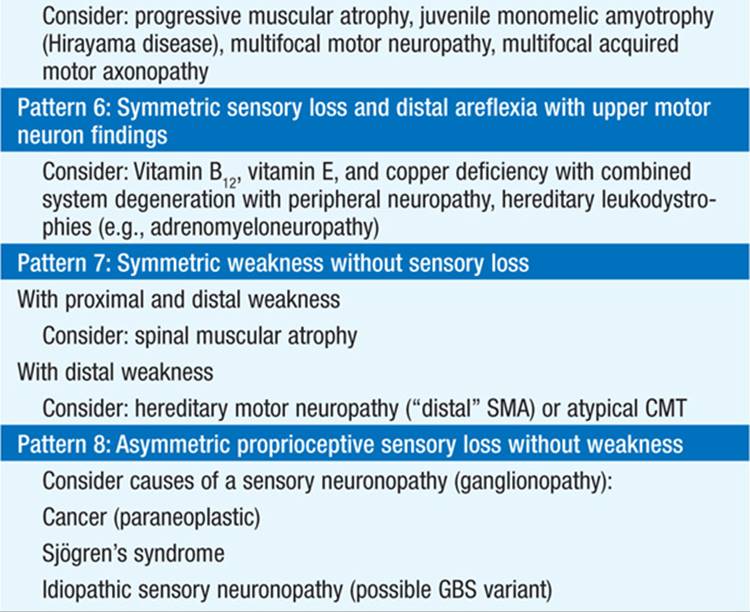
Based on the answers to these seven key questions, neuropathic disorders can be classified into several patterns based on the distribution or pattern of sensory, motor, and autonomic involvement (Table 205-1).
TABLE 205-1 PATTERNS OF NEUROPATHIC DISORDERS


POLYNEUROPATHY
Diagnostic Evaluation
Screening laboratory studies in a distal, symmetric polyneuropathy may include a complete blood count, basic chemistries including serum electrolytes and tests of renal and hepatic function, fasting blood glucose, HbA1c, urinalysis, thyroid function tests, B12, folate, ESR, rheumatoid factor, ASA, SPEP, and urine for Bence Jones protein. An oral glucose tolerance test is indicated in pts with painful sensory neuropathies even if other screens for diabetes are negative.
Diagnostic tests to further characterize the neuropathy include nerve conduction studies (NCS), electromyography (EMG), sural nerve biopsy, muscle biopsy, and quantitative sensory testing. Diagnostic tests are more likely to be informative in pts with asymmetric, motor-predominant, rapid-onset, or demyelinating neuropathies.
Electrodiagnosis
NCS are carried out by stimulating motor or sensory nerves electrically. Demyelination is characterized by slowing of nerve conduction velocity (NCV), dispersion of evoked compound action potentials, conduction block (decreased amplitude of muscle compound action potentials on proximal, as compared to distal, stimulation of the nerve), and prolongation of distal latencies. In contrast, axonal neuropathies exhibit reduced amplitude of evoked compound action potentials with relative preservation of NCV. EMG records electrical potentials from a needle electrode in muscle, at rest and during voluntary contraction; it is most useful for distinguishing myopathic from neuropathic disorders. Myopathic disorders are marked by small, short-duration, polyphasic muscle action potentials; by contrast, neuropathic disorders are characterized by muscle denervation. Denervation decreases the number of motor units (e.g., an anterior horn cell, its axon, and the motor end plates and muscle fibers it innervates). In long-standing denervation, motor unit potentials become large and polyphasic due to collateral reinnervation of denervated muscle fibers by axonal sprouts from surviving motor axons. Other EMG features of denervation include fibrillations (random, unregulated firing of individual muscle fibers) and fasciculations (random, spontaneous firing of motor units).
TREATMENT Polyneuropathy
• Treatment of the underlying disorder, pain management, and supportive care to protect and rehabilitate damaged tissue all need to be considered.
• Examples of specific therapies include tight glycemic control in diabetic neuropathy, vitamin replacement for B12 deficiency, IV immune globulin (IVIg) or plasmapheresis for GBS, and immuno-suppression for vasculitis.
• Painful sensory neuropathies can be difficult to treat. Pain management usually begins with tricyclic antidepressants (TCAs), duloxetine hydrochloride, lidocaine patches, or anticonvulsants such as gabapentin (Table 205-2). Topical anesthetic agents including EMLA (lidocaine/prilocaine) and capsaicin cream can provide additional relief.
TABLE 205-2 TREATMENT OF PAINFUL SENSORY NEUROPATHIES


• Physical and occupational therapy is important. Proper care of denervated areas prevents skin ulceration, which can lead to poor wound healing, tissue resorption, arthropathy, and ultimately amputation.
Specific Polyneuropathies
AIDP or GBS: an ascending, usually demyelinating, motor > sensory polyneuropathy accompanied by areflexia, motor paralysis, and elevated CSF total protein without pleocytosis. Over two-thirds are preceded by an acute respiratory or gastrointestinal infection. Maximum weakness is usually reached within 2 weeks; demyelination by EMG. Most pts are hospitalized; one-third require ventilatory assistance. 85% make a complete or near-complete recovery with supportive care. Variants of GBS include Fisher syndrome (ophthalmoparesis, facial diplegia, ataxia, areflexia; associated with serum antibodies to ganglioside GQ1b) and acute motor axonal neuropathy (more severe course than demyelinating GBS; antibodies to GM1 in some cases).
• IVIg (2 g/kg divided over 5 days) or plasmapheresis (40–50 mL/kg daily for 4–5 days) significantly shortens the course.
• Glucocorticoids are ineffective.
Chronic inflammatory demyelinating polyneuropathy (CIDP): a slowly progressive or relapsing polyneuropathy characterized by diffuse hyporeflexia or areflexia, diffuse weakness, elevated CSF protein without pleocytosis, and demyelination by EMG.
• Begin treatment when progression is rapid or walking is compromised.
• Initial treatment is usually IVIg; most pts require periodic re-treatment at 4- to 6-week intervals.
• Other first-line treatment options include plasmapheresis or glucocorticoids
• Immunosuppressants (azathioprine, methotrexate, cyclosporine, cyclophosphamide) used in refractory cases.
Diabetic neuropathy: typically a distal symmetric, sensorimotor, axonal polyneuropathy. A mixture of demyelination and axonal loss is frequent. Other variants include: isolated sixth or third cranial nerve palsies, asymmetric proximal motor neuropathy in the legs, truncal neuropathy, autonomic neuropathy, and an increased frequency of entrapment neuropathy (see below). Mononeuropathy multiplex (MM): defined as involvement of multiple individual peripheral nerves. When an inflammatory disorder is the cause, mononeuritis multiplex is the term used. Both systemic (67%) and nonsystemic (33%) vasculitis may present as MM. Immunosuppressive treatment of the underlying disease (usually with glucocorticoids and cyclophosphamide) is indicated. A tissue diagnosis of vasculitis should be obtained before initiating treatment; a positive biopsy helps to justify the necessary long-term treatment with immunosuppressive medications, and pathologic confirmation is difficult after treatment has commenced.
MONONEUROPATHY
Clinical Features
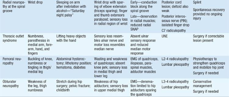
Mononeuropathies are usually caused by trauma, compression, or entrapment. Sensory and motor symptoms are in the distribution of a single nerve—most commonly the ulnar or median nerve in the arm or peroneal nerve in the leg. Intrinsic factors making pts more susceptible to entrapment include arthritis, fluid retention (pregnancy), amyloid, tumors, and diabetes mellitus. Clinical features favoring conservative management of median neuropathy at the wrist (carpal tunnel syndrome) or ulnar neuropathy at the elbow include sudden onset, no motor deficit, few or no sensory findings (pain or paresthesias may be present), and no evidence of axonal loss by EMG. Surgical decompression is considered for chronic mononeuropathies that are unresponsive to conservative treatment, if the site of entrapment is clearly defined. The most frequently encountered mononeuropathies are summarized in Table 205-3.
TABLE 205-3 MONONEUROPATHIES





For a more detailed discussion, see Amato AA, Barohn RJ: Peripheral Neuropathy, Chap. 384, p. 3448; and Hauser SL, Amato AA: Guillain-Barré Syndrome and Other Immune-Mediated Neuropathies, Chap. 385, p. 3473, in HPIM-18.