
Results from lack of delivery of oxygen to the brain because of hypotension or respiratory failure. Most common causes are MI, cardiac arrest, shock, asphyxiation, paralysis of respiration, and carbon monoxide or cyanide poisoning. In some circumstances, hypoxia may predominate. Carbon monoxide and cyanide poisoning are termed histotoxic hypoxia since they cause a direct impairment of the respiratory chain.
CLINICAL MANIFESTATIONS
Mild degrees of pure hypoxia (e.g., at high altitude) cause impaired judgment, inattentiveness, motor incoordination, and, at times, euphoria. However, with hypoxia-ischemia, such as occurs with circulatory arrest, consciousness is lost within seconds. If circulation is restored within 3–5 min, full recovery may occur, but with longer periods permanent cerebral damage usually results. It may be difficult to judge the precise degree of hypoxia-ischemia, and some pts make a relatively full recovery even after 8–10 min of global ischemia. The distinction between pure hypoxia and hypoxia-ischemia is important, since a Pao2as low as 2.7 kPa (20 mmHg) can be well tolerated if it develops gradually and normal blood pressure is maintained, but short durations of very low or absent cerebral circulation may result in permanent impairment.
Clinical examination at different time points after an insult (especially cardiac arrest) helps to assess prognosis (Fig. 22-1). The prognosis is better for pts with intact brainstem function, as indicated by normal pupillary light responses, intact oculocephalic (doll’s eyes) reflexes, and oculovestibular (caloric) and corneal reflexes. Absence of these reflexes and the presence of persistently dilated pupils that do not react to light are grave prognostic signs. A uniformly dismal prognosis is conveyed by the absence of a pupillary light reflex or absence of a motor response to pain on day 3 following the injury. Bilateral absence of the cortical somatosensory evoked potentials (SSEP) in the first several days also conveys a poor prognosis, as does a very elevated serum level (>33 μg/L) of the biochemical marker neuron-specific enolase (NSE). Usefulness of SSEP and NSE often limited: difficult to obtain in a timely fashion; need for expert interpretation (SSEP); and lack of standardization (NSE measurements). Whether administration of mild hypothermia after cardiac arrest will alter the usefulness of these clinical and electrophysiologic predictors is unknown.
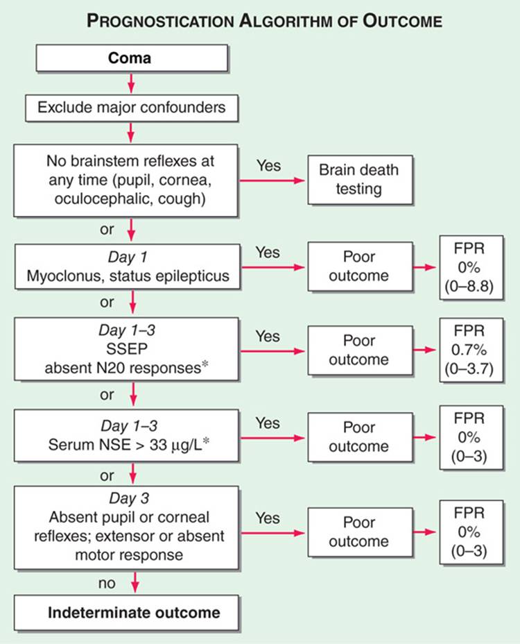
FIGURE 22-1 Prognostication of outcome in comatose survivors of cardiopulmonary resuscitation. Numbers in parentheses are 95% confidence intervals. Confounders could include use of sedatives or neuromuscular blocking agents, hypothermia therapy, organ failure, or shock. Tests denoted with an asterisk (*) may not be available in a timely and standardized manner. SSEP, somatosensory evoked potentials; NSE, neuron-specific enolase; FPR, false-positive rate. [From EFM Wijdicks et al: Practice parameter: Prediction of outcome in comatose survivors after cardiopulmonary resuscitation (an evidence-based review). Neurology 67:203, 2006; with permission.]
Long-term consequences include persistent coma or vegetative state, dementia, visual agnosia, parkinsonism, choreoathetosis, ataxia, myoclonus, seizures, and an amnestic state. Delayed postanoxic encephalopathy is an uncommon phenomenon in which pts appear to make an initial recovery following an insult and then have a relapse with a progressive course often characterized by widespread demeylination on imaging studies.
TREATMENT Hypoxic-Ischemic Encephalopathy
• Initial treatment is directed at restoring normal cardiorespiratory function. This includes securing a clear airway, ensuring adequate oxygenation and ventilation, and restoring cerebral perfusion, whether by cardiopulmonary resuscitation, fluids, pressors, or cardiac pacing.
• Mild hypothermia (33°C), initiated as early as possible and continued for 12–24 h, may improve outcome in pts who remain comatose after cardiac arrest, based on trials in pts whose initial rhythm was primarily ventricular fibrillation or pulseless ventricular tachycardia. Potential complications include coagulopathy and an increased risk of infection.
• Anticonvulsants are not usually given prophylactically but may be used to control seizures.
• Posthypoxic myoclonus can be controlled with clonazepam (1.5–10 mg/d) or valproate (300–1200 mg/d) in divided doses.
• Myoclonic status epilepticus within 24 h after a hypoxic-ischemic insult portends a universally poor prognosis, even if seizures are controlled.
• Severe carbon monoxide intoxication may be treated with hyper-baric oxygen.

For a more detailed discussion, see Hemphill JC III, Smith WS, Gress DR: Neurologic Critical Care, Including Hypoxic-Ischemic Encephalopathy and Subarachnoid Hemorrhage, Chap. 275, p. 2254, in HPIM-18.