
ACUTE MYELOID LEUKEMIA (AML)
AML is a clonal malignancy of myeloid bone marrow precursors in which poorly differentiated cells accumulate in the bone marrow and circulation.
Signs and symptoms occur because of the absence of mature cells normally produced by the bone marrow, including granulocytes (susceptibility to infection) and platelets (susceptibility to bleeding). In addition, if large numbers of immature malignant myeloblasts circulate, they may invade organs and rarely produce dysfunction. There are distinct morphologic subtypes (Table 72-1) that have largely overlapping clinical features. Of note is the propensity of pts with acute promyelocytic leukemia (APL) (FAB M3) to develop bleeding and disseminated intravascular coagulation, especially during induction chemotherapy, because of the release of procoagulants from their cytoplasmic granules.
TABLE 72-1 AML CLASSIFICATION SYSTEMS


Incidence and Etiology
In the United States about 13,780 cases have occurred in 2012. AML accounts for about 80% of acute leukemias in adults. Etiology is unknown for the vast majority. Three environmental exposures increase the risk: chronic benzene exposure, radiation exposure, and prior treatment with alkylating agents (especially in addition to radiation therapy) and topoisomerase II inhibitors (e.g., doxorubicin and etoposide). Chronic myeloid leukemia (CML), myelodysplasia, and myeloproliferative syndromes may all evolve into AML. Certain genetic abnormalities are associated with particular morphologic variants: t(15;17) with APL, inv(16) with eosinophilic leukemia; others occur in a number of types. Chromosome 11q23 abnormalities are often seen in leukemias developing after exposure to topoisomerase II inhibitors. Chromosome 5 or 7 deletions are seen in leukemias following radiation plus chemotherapy. The particular genetic abnormality has a strong influence on treatment outcome. Expression of MDR1 (multidrug resistance efflux pump) is common in older pts and adversely affects prognosis.
Clinical and Laboratory Features
Initial symptoms of acute leukemia have usually been present for <3 months; a preleukemic syndrome may be present in some 25% of pts with AML. Signs of anemia, pallor, fatigue, weakness, palpitations, and dyspnea on exertion are most common. White blood cell count (WBC) may be low, normal, or markedly elevated; circulating blast cells may or may not be present; with  blasts per liter, leukostasis in lungs and brain may occur. Minor pyogenic infections of the skin are common. Thrombocytopenia leads to spontaneous bleeding, epistaxis, petechiae, conjunctival hemorrhage, gingival bleeding, and bruising, especially with platelet count <20,000/μL. Anorexia and weight loss are common; fever may be present.
Bacterial and fungal infection are common; risk is heightened with total neutrophil count <5000/μL, and breakdown of mucosal and cutaneous barriers aggravates susceptibility; infections may be clinically occult in presence of severe leukopenia, and prompt recognition requires a high degree of clinical suspicion.
Hepatosplenomegaly occurs in about one-third of pts; leukemic meningitis may present with headache, nausea, seizures, papilledema, cranial nerve palsies.
Metabolic abnormalities may include hyponatremia, hypokalemia, elevated serum lactate dehydrogenase (LDH), hyperuricemia, and (rarely) lactic acidosis. With very high blast cell count in the blood, spurious hyperkalemia and hypoglycemia may occur (potassium released from and glucose consumed by tumor cells after the blood was drawn).
TREATMENT Acute Myeloid Leukemia
Leukemic cell mass at time of presentation may be 1011–1012 cells; when total leukemic cell numbers fall below ~109, they are no longer detectable in blood or bone marrow and pt appears to be in complete remission (CR). Thus aggressive therapy must continue past the point when initial cell bulk is reduced if leukemia is to be eradicated. Typical phases of chemotherapy include remission induction and postremission therapy, with treatment lasting about 1 year. Figure 72-1 outlines a treatment algorithm.
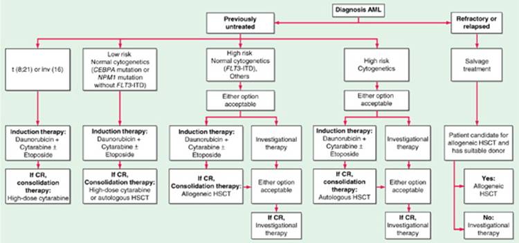
FIGURE 72-1 Flow chart for the therapy of newly diagnosed acute myeloid leukemia. For all forms of AML except acute promyelocytic leukemia (APL), standard therapy includes a 7-day continuous infusion of cytarabine (100–200 mg/m2 per day) and a 3-day course of daunorubicin (60–90 mg/m2 per day) with or without 3 days of etoposide (only with daunorubicin 60 mg/m2 per day) or novel therapies based on their predicted risk of relapse (i.e., risk-stratified therapy). Idarubicin (12–13 mg/m2 per day) could be used in place of daunorubicin (not shown). Patients who achieve complete remission undergo postremission consolidation therapy, including sequential courses of high-dose cytarabine, autologous hematopoietic stem cell transplant (HSCT), allogeneic HSCT, or novel therapies, based on their predicted risk of relapse (i.e., risk-stratified therapy). Patients with APL (see text for treatment) usually receive tretinoin together with anthracycline-based chemotherapy for remission induction and then arsenic trioxide followed by consolidation with anthracycline-based chemotherapy and possibly maintenance with tretinoin. The role of cytarabine in APL induction and consolidation is controversial.
Supportive care with transfusions of red cells and platelets [from cytomegalovirus (CMV)-seronegative donors, if pt is a candidate for bone marrow transplantation] is very important, as are aggressive prevention, diagnosis, and treatment of infections. Colony-stimulating factors offer little or no benefit; some recommend their use in older pts and those with active infections. Febrile neutropenia should be treated with broad-spectrum antibiotics (e.g., ceftazidime 1 g q8h); if febrile neutropenia persists beyond 7 days, amphotericin B should be added.
About 60–80% of pts will achieve initial remission when treated with cytarabine 100–200 (mg/m2)/d by continuous infusion for 7 days, and daunorubicin [45 (mg/m2)/d] or idarubicin [12–13 (mg/m2)/d] for 3 days. Addition of etoposide may improve CR duration. Half of treated pts enter CR with the first cycle of therapy, and another 25% require two cycles. About 10–30% of pts achieve 5-year disease-free survival and probable cure. Pts achieving a CR who have low risk of relapse [cells contain t(8;21) or inv(16)] receive 3–4 cycles of cytarabine. Those at high risk of relapse may be considered for allogeneic bone marrow transplantation.
Response to treatment after relapse is short, and prognosis for pts who have relapsed is poor. In APL, addition of trans-retinoic acid (tretinoin) to chemotherapy induces differentiation of the leukemic cells and may improve outcome. Arsenic trioxide also induces differentiation in APL cells.
Bone marrow transplantation from identical twin or human leukocyte antigen (HLA)-identical sibling is effective treatment for AML. Typical protocol uses high-dose chemotherapy ± total-body irradiation to ablate host marrow, followed by infusion of marrow from donor. Risks are substantial (unless marrow is from identical twin). Complications include graft-versus-host disease, interstitial pneumonitis, opportunistic infections (especially CMV). Comparison between transplantation and high-dose cytarabine as postremission therapy has not produced a clear advantage for either approach. Up to 30% of otherwise end-stage pts with refractory leukemia achieve probable cure from transplantation; results are better when transplant is performed during remission. Results are best for children and young adults.
CHRONIC MYELOID LEUKEMIA (CML)
CML is a clonal malignancy usually characterized by splenomegaly and production of increased numbers of granulocytes; course is initially indolent but eventuates in leukemic phase (blast crisis) that has a poorer prognosis than de novo AML; rate of progression to blast crisis is variable; overall survival averages 4 years from diagnosis.
Incidence and Etiology
In the United States, about 5430 cases have occurred in the United States in 2012. More than 90% of cases have a reciprocal translocation between chromosomes 9 and 22, creating the Philadelphia (Ph) chromosome and a fusion gene product called BCR-ABL. (BCR is from 9, ABL from 22.) The chromosome abnormality appears in all bone marrow–derived cells except T cells. The protein made by the chimeric gene is 210 kDa in chronic phase and 190 kDa in acute blast transformation. In some pts, the chronic phase is clinically silent and pts present with acute leukemia with the Ph chromosome.
Clinical and Laboratory Features
Symptoms develop gradually; easy fatigability, malaise, anorexia, abdominal discomfort and early satiety from the large spleen, excessive sweating. Occasional pts are found incidentally based on elevated leukocyte count. WBC is usually >25,000/μL with the increase accounted for by granulocytes and their precursors back to the myelocyte stage; bands and mature forms predominate. Basophils may account for 10–15% of the cells in the blood. Platelet count is normal or increased. Anemia is often present. Neutrophil alkaline phosphatase score is low. Marrow is hypercellular with granulocytic hyperplasia. Marrow blast cell count is normal or slightly elevated. Serum levels of vitamin B12, B12-binding proteins, and LDH are elevated in proportion to the WBC. With high blood counts, spurious hyperkalemia and hypoglycemia may be seen.
Natural History
Chronic phase lasts 2–4 years. Accelerated phase is marked by anemia disproportionate to the disease activity or treatment. Platelet counts fall. Additional cytogenetic abnormalities appear. Blast cell counts increase. Usually within 6–8 months, overt blast crisis develops in which maturation ceases and blasts predominate. The clinical picture is that of acute leukemia. Half of the cases become AML, one-third have morphologic features of acute lymphoid leukemia, 10% are erythroleukemia, and the rest are undifferentiated. Survival in blast crisis is often <4 months.
TREATMENT Chronic Myeloid Leukemia
Criteria for response are provided in Table 72-2. Allogeneic bone marrow transplantation has the potential to cure the disease in chronic phase. However, the first treatment is imatinib, a molecule that inhibits the chimeric gene product’s tyrosine kinase activity. A daily oral dose of 400 mg produces complete hematologic remission of >90% and cyto-genetic remission in 76%. If a matched donor is available, it is best to transplant pts in complete remission. Several mechanisms of resistance to imatinib have emerged, and it is unlikely that it leads to permanent remissions when used alone; however, follow-up is not sufficient to draw firm conclusions.
TABLE 72-2 RESPONSE CRITERIA IN CHRONIC MYELOID LEUKEMIA

Pts who no longer respond to imatinib may respond to other tyro-sine kinase inhibitors such as dasatinib (100 mg PO qd) or nilotinib (400 mg PO bid). The T315I mutation in the BCR/ABL gene conveys resistance to all three kinase inhibitors. Allopurinol, 300 mg/d, prevents urate nephropathy. The only curative therapy for the disease is HLA-matched allogeneic bone marrow transplantation. The optimal timing of transplantation is unclear, but transplantation in chronic phase is more effective than transplantation in accelerated phase or blast crisis. Transplantation appears most effective in pts treated within a year of diagnosis. Long-term disease-free survival may be obtained in 50–60% of transplanted pts. Infusion of donor lymphocytes can restore remission in relapsing pts. In pts without a matched donor, autologous transplantation may be helpful using peripheral blood stem cells. Treatment of pts in blast crisis with imatinib can obtain responses, but their durability has not been established.
MYELODYSPLASTIC SYNDROMES (MDS)
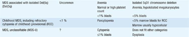
These are clonal abnormalities of marrow cells characterized by varying degrees of cytopenias affecting one or more cell lines. The World Health Organization (WHO) classification of myelodysplastic syndromes is shown in Table 72-3. Other terms that have been used to describe one or more of the entities include preleukemia and oligoblastic leukemia.
TABLE 72-3 WORLD HEALTH ORGANIZATION (WHO) CLASSIFICATION OF MYELODYSPLASTIC SYNDROMES/NEOPLASMS



Incidence and Etiology
About 3000 cases occur each year, mainly in persons >50 years old (median age, 68). As in AML, exposure to benzene, radiation, and chemotherapeutic agents may lead to MDS. Chromosome abnormalities occur in up to 80% of cases, including deletion of part or all of chromosomes 5, 7, and 9 (20 or 21 less commonly) and addition of part or all of chromosome 8.
Clinical and Laboratory Features
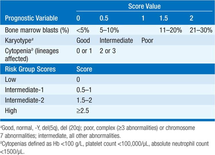
Symptoms depend on the affected lineages. 85% of pts are anemic, 50% have neutropenia, and about one-third have thrombocytopenia. The pathologic features of MDS are a cellular marrow with varying degrees of cytologic atypia including delayed nuclear maturation, abnormal cytoplasmic maturation, accumulation of ringed sideroblasts (iron-laden mitochondria surrounding the nucleus), uni- or bilobed megakaryocytes, micromegakaryocytes, and increased myeloblasts. Table 72-3 lists features used to identify distinct entities. Prognosis is defined by marrow blast %, karyotype, and lineages affected. The International Prognostic Scoring System is shown in Table 72-4.
TABLE 72-4 INTERNATIONAL PROGNOSTIC SCORING SYSTEM (IPSS)
TREATMENT Myelodysplastic Syndromes
Allogeneic bone marrow transplantation is the only curative therapy and may cure 60% of those so treated. However, the majority of pts with MDS are too old to receive transplantation. 5-Azacytidine (75 mg/m2 daily × 7, q 4 weeks) can delay transformation to AML by 8–10 months. Decitabine (15 mg/m2 by continuous IV infusion, q8h daily × 3, q 6 weeks) may induce responses lasting a median of 1 year in 20% of pts. Lenalidomide (10 mg/d), a thalidomide analogue with fewer central nervous system effects, causes a substantial fraction of pts with the 5q– syndrome to become transfusion-independent. Pts with low erythropoietin levels may respond to erythropoietin, and a minority of pts with neutropenia respond to granulocyte colony-stimulating factor. Supportive care is the cornerstone of treatment.
MYELOPROLIFERATIVE SYNDROMES
The three major myeloproliferative syndromes are polycythemia vera, idiopathic myelofibrosis, and essential thrombocytosis. All are clonal disorders of hematopoietic stem cells and all are associated with a mutation in the JAK2kinase (V617F) that results in activation of the kinase. The mutation is seen in 90% of pts with polycythemia vera and ~45% of pts with idiopathic myelofibrosis and essential thrombocytosis.
POLYCYTHEMIA VERA
The most common myeloproliferative syndrome, this is characterized by an increase in red blood cell (RBC) mass, massive splenomegaly, and clinical manifestations related to increased blood viscosity, including neurologic symptoms (vertigo, tinnitus, headache, visual disturbances) and thromboses (myocardial infarction, stroke, peripheral vascular disease; uncommonly, mesenteric and hepatic). It must be distinguished from other causes of increased RBC mass (Chap. 51). This is most readily done by assaying serum erythropoietin levels. Polycythemia vera is associated with very low erythropoietin levels; in other causes of erythrocytosis, erythropoietin levels are high. Pts are effectively managed with phlebotomy. Some pts require splenectomy to control symptoms, and those with severe pruritus may benefit from psoralens and UV light. 20% develop myelofibrosis, <5% acute leukemia.
IDIOPATHIC MYELOFIBROSIS
This rare entity is characterized by marrow fibrosis, myeloid metaplasia with extramedullary hematopoiesis, and splenomegaly. Evaluation of a blood smear reveals teardrop-shaped RBC, nucleated RBC, and some early granulocytic forms, including promyelocytes. However, many entities may lead to marrow fibrosis and extramedullary hematopoiesis, and the diagnosis of primary idiopathic myelofibrosis is made only when the many other potential causes are ruled out. The following diseases are in the differential diagnosis: CML, polycythemia vera, Hodgkin’s disease, cancer metastatic to the marrow (especially from breast and prostate), infection (particularly granulomatous infections), and hairy cell leukemia. Supportive therapy is generally used; novel inhibitors of JAK2 have shown activity in reducing splenomegaly; however, no study has yet shown a particular drug therapy to improve survival.
ESSENTIAL THROMBOCYTOSIS
This is usually noted incidentally upon routine platelet count done in an asymptomatic person. Like myelofibrosis, many conditions can produce elevated platelet counts; thus, the diagnosis is one of exclusion. Platelet count must be >500,000/μL, and known causes of thrombocytosis must be ruled out including CML, iron deficiency, splenectomy, malignancy, infection, hemorrhage, polycythemia vera, myelodysplasia, and recovery from vitamin B12 deficiency. Although usually asymptomatic, pts should be treated if they develop migraine headache, transient ischemic attack, or other bleeding or thrombotic disease manifestations. Interferon α is effective therapy, as are anagrelide and hydroxyurea. Treatment should not be given just because the absolute platelet count is high in the absence of other symptoms.

For a more detailed discussion, see Young NS: Aplastic Anemia, Myelodysplasia, and Related Bone Marrow Failure Syndromes, Chap. 107, p. 887; Spivak JL: Polycythemia Vera and Other Myeloproliferative Diseases, Chap. 108, p. 898; and Wetzler M et al: Acute and Chronic Myeloid Leukemia, Chap. 109, p. 905, in HPIM-18.