
The immunocompromised pt is at increased risk for infection with both common and opportunistic pathogens.
INFECTIONS IN CANCER PTS
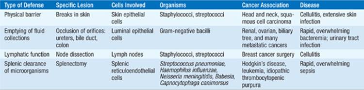
Table 88-1 lists the normal barriers to infection whose disruption may permit infections in immunocompromised pts, with particular relevance for the noted cancers. Infection-associated mortality rates among cancer pts have decreased as a result of an evolving approach entailing early use of empirical broad-spectrum antibiotics; empirical antifungal therapy in neutropenic pts who, after 4–7 days of antibiotic treatment, remain febrile without positive cultures; and use of antibiotics for afebrile neutropenic pts as broad-spectrum prophylaxis against infections.
TABLE 88-1 DISRUPTION OF NORMAL BARRIERS THAT MAY PREDISPOSE TO INFECTIONS IN PATIENTS WITH CANCER

SYSTEM-SPECIFIC SYNDROMES
• Skin infections Skin lesions of various types are common in pts with cancer and may be the first sign of bacterial or fungal sepsis, particularly in neutropenic pts (those with <500 functional neutrophils/μL).
– Cellulitis: most often caused by group A Streptococcus and Staphylococcus aureus. Unusual organisms (e.g., Escherichia coli, Pseudomonas, fungi) may be involved in neutropenic pts.
– Macules or papules: due to bacteria (e.g., Pseudomonas aeruginosa causing ecthyma gangrenosum) or fungi (e.g., Candida)
– Sweet’s syndrome or febrile neutrophilic dermatosis: Most often seen in neutropenic leukemic pts, it presents as red or bluish-red papules or nodules that form sharply bordered plaques; high fever; and an elevated ESR. The skin lesions are most common on the face, neck, and arms.
– Erythema multiforme with mucous membrane involvement: Often due to HSV infection, it is distinct from Stevens-Johnson syndrome, which is associated with drugs and has a more widespread distribution; both conditions are common in pts with cancer.
– Drug rashes: Rashes associated with drugs, particularly cytokines used in cancer therapy, complicate the differential diagnosis of rashes in pts with cancer.
• Catheter-related infections Exit-site infections, often with erythema around the insertion site, are most common.
– Infections caused by coagulase-negative staphylococci can often be treated medically without catheter removal.
– Infections caused by other organisms, including S. aureus, P. aeruginosa, Candida, Stenotrophomonas, or Bacillus, usually require catheter removal.
– If a red streak develops over the SC part of a “tunneled” catheter, the device must be removed to prevent extensive cellulitis and tissue necrosis.
• Upper GI infections Breakdown of mucosal surfaces due to chemotherapy and infection are common.
– Oral mucositis is associated with viridans streptococci and HSV.
– Oral candidal infections (thrush) are common.
– Esophagitis can be caused by Candida albicans and HSV.
• Lower GI infections Transmigration of bowel flora across the intestinal epithelium can lead to severe conditions.
– Chronic disseminated candidiasis: results from seeding of organs (e.g., liver, spleen, kidneys) during neutropenia in pts with hematologic malignancy and generally presents symptomatically when neutropenia resolves. Pts have persistent fever unresponsive to antibiotics, abdominal pain, and increased alkaline phosphatase levels. Although biopsies may reveal granulomas, yeasts, or pseudohyphae, the diagnosis is often made on the basis of radiographic studies (CT, MRI). Treatment should be directed to the causative agent; C. albicans is usually responsible, but C. tropicalis or other Candida species are sometimes involved.
– Typhlitis (necrotizing colitis): more common among children than among adults and among pts with acute myelocytic leukemia or acute lymphocytic leukemia than among pts with other forms of cancer. Pts have fever, RLQ tenderness, and diarrhea that is often bloody. The diagnosis is confirmed by documentation of a thickened cecal wall via imaging. Treatment should include antibiotics directed against bowel flora and surgery (in the case of perforation).
• CNS infections The susceptibility of pts to specific infections depends on whether they have prolonged neutropenia, defects in cellular immunity (e.g., high-dose glucocorticoid therapy, cytotoxic chemotherapy), or defects in humoral immunity [e.g., pts with chronic lymphocytic leukemia, s/p splenectomy, or s/p bone marrow transplantation (BMT)].
– Meningitis: Consider Cryptococcus or Listeria, particularly for pts with defects in cellular immunity. Pts with defects in humoral immunity are also at risk for infection with encapsulated bacteria such as Streptococcus pneumoniae, Haemophilus influenzae, and Neisseria meningitidis.
– Encephalitis: Pts with defects in cellular immunity are particularly prone to infection with VZV, JC virus (progressive multifocal leukoencephalopathy), CMV, Listeria, HSV, and human herpesvirus type 6.
– Brain masses: most often present as headache with or without fever or neurologic abnormalities. Pts with prolonged neutropenia are at increased risk for a brain abscess due to Aspergillus, Nocardia, or Cryptococcus. Pts with defects in cellular immunity are at increased risk for infection with Toxoplasma and EBV (lymphoproliferative disease). A definitive diagnosis may require a biopsy.
• Pulmonary infections Pneumonia can be difficult to diagnose in immunocompromised pts, given that many of the conventional findings (e.g., purulent sputum, physical findings suggestive of chest consolidation) rely on the presence of neutrophils. Radiographic patterns of infiltration can help narrow the differential diagnosis.
– Localized infiltrate: Consider bacterial pneumonia (including Legionella and mycobacteria), local hemorrhage or embolism, and tumor.
– Nodular infiltrate: Consider fungal infection (e.g., Aspergillus, Mucor), Nocardia infection, and recurrent tumor. In pts with Aspergillus infection, hemoptysis may be an ominous sign. A biopsy performed with direct visualization may be required for definitive diagnosis.
– Diffuse infiltrates: Consider infection with viruses (particularly CMV), Chlamydia, Pneumocystis, Toxoplasma, or mycobacteria. Viruses that cause upper respiratory infections in normal hosts (e.g., influenza, respiratory syncytial) may cause fatal pneumonitis in immunocom-promised pts. Noninfectious causes include radiation pneumonitis, CHF, diffuse alveolar hemorrhage (after BMT), and drug-induced lung injury (e.g., bleomycin, alkylating agents).
• Renal and ureteral infections These infections are usually associated with obstructing tumor masses.
– Candida has a predilection for the kidneys, reaching this site via either hematogenous seeding or retrograde spread from the bladder. Persistent funguria should prompt a search for infection in the kidney (e.g., fungus ball).
– BK virus and adenovirus can cause hemorrhagic cystitis.
APPROACH TO THE PATIENT Febrile Neutropenia
Approach to Diagnosis and Treatment of Febrile Neutropenic Pts
Figure 88-1 presents an algorithm for the diagnosis and treatment of pts with febrile neutropenia.

FIGURE 88-1 Algorithm for the diagnosis and treatment of febrile neutropenic pts. Several general guidelines are useful in the initial treatment of these pts: (1) The agents used should reflect both the epidemiology and the antibiotic resistance pattern of the hospital. (2) A single third-generation cephalosporin constitutes an appropriate initial regimen in many hospitals (if the pattern of resistance justifies its use). (3) Most standard regimens are designed for pts who have not previously received prophylactic antibiotics. The development of fever in a pt receiving antibiotics affects the choice of subsequent therapy (which should target resistant organisms and organisms known to cause infections in pts being treated with the antibiotics already administered). (4) Randomized trials have indicated that it is safe to use oral antibiotic regimens to treat “low-risk” pts who have fever and neutropenia. Outpatients who are expected to remain neutropenic for <10 days and who have no concurrent medical problems (such as hypotension, pulmonary compromise, or abdominal pain) can be classified as low-risk and treated with a broad-spectrum oral regimen.
• The initial regimen should be refined on the basis of culture data.
• Adding antibiotics to the initial regimen is not appropriate unless there is a clinical or microbiologic reason to do so. The addition of aminoglycosides to β-lactam therapy does not enhance efficacy (but does increase toxicity), even for infections involving P. aeruginosa.
• For empirical antifungal treatment, amphotericin B is being supplanted by liposomal formulations of amphotericin B, newer azoles (e.g., voriconazole or posaconazole), and echinocandins (e.g., caspofungin). Echinocandins are useful against infections with azole-resistant Candida. Clinical experience related to antiviral therapy is most extensive with acyclovir for HSV and VZV infections. Newer agents (e.g., cidofovir, foscarnet) with a broader spectrum of action have heightened the focus on treatment of viral infections.
– Prophylactic antibiotics (e.g., fluoroquinolones) in pts expected to have prolonged neutropenia or antifungal agents (e.g., fluconazole) in pts with hematopoietic stem cell transplants may prevent infections. Pneumocystisprophylaxis is mandatory for pts with acute lymphocytic leukemia and for those receiving glucocorticoid-containing regimens.
INFECTIONS IN TRANSPLANT RECIPIENTS
Evaluation of infections in transplant recipients must involve consideration of infectious agents harbored by the donor organ and the recipient’s immunosuppressive drug regimen, which increases susceptibility to latent infections (among other infections).
– Pretransplantation evaluation of the donor should include a thorough serologic evaluation for viral pathogens (e.g., HSV-1, HSV-2, VZV, CMV, EBV, HIV, and hepatitis A, B, and C viruses) and mycobacterial disease; other evaluations should be directed by the donor’s history, including diet, exposures, and travel.
– Pretransplantation evaluation of the recipient is generally more comprehensive than that of the donor and should include assessment for respiratory viruses and gastrointestinal pathogens. Given the effects of underlying chronic disease and chemotherapy, serologic testing of the recipient may not be reliable.
HEMATOPOIETIC STEM CELL TRANSPLANTATION (HSCT)
• Pathogenesis The myeloablative processes involved in HSCT result in the complete absence of innate and adaptive immune cells. This transient state of complete immunologic incompetence and the reconstitution that follows make the host extremely susceptible to infections.
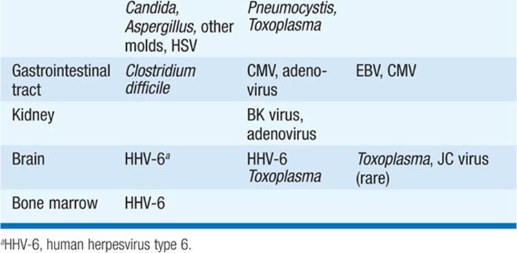
• Etiology Most infections occur in a predictable time frame after HSCT (Table 88-2).
TABLE 88-2 COMMON INFECTIONS AFTER HSCT

– Bacterial infections: Neutropenia-related infectious complications are most common during the first month. Some centers give prophylactic antibiotics (e.g., quinolones) that may decrease the risk of gram-negative bacteremia but increase the risk of Clostridium difficile colitis.
• Skin and bowel flora (e.g., S. aureus, coagulase-negative staphylococci, E. coli) are responsible for most infections in the first few days following HSCT, after which nosocomial pathogens and filamentous bacteria (e.g., vancomycin-resistant enterococci, Acinetobacter and Nocardia species) become more common.
– Fungal infections: Fungal infections are increasingly common beyond the first week after HSCT, particularly among pts who receive broad-spectrum antibiotics. Infections with Candida species are most common, although resistant fungi (e.g., Aspergillus, Fusarium) are becoming more common because of the increased use of prophylactic fluconazole.
• Prolonged treatment with glucocorticoids or other immunosuppressive agents increases the risk of infection with Candida or Aspergillus and of reactivation of endemic fungi even after resolution of neutropenia.
• Maintenance prophylaxis with trimethoprim-sulfamethoxazole (TMP-SMX; 160/800 mg/d starting 1 month after engraftment and continuing for at least 1 year) is recommended to prevent Pneumocystis jiroveci pneumonia.
– Parasitic infections: Prophylaxis with TMP-SMX is also protective against disease caused by Toxoplasma as well as against late infections caused by certain bacteria, including Nocardia, Listeria monocyto-genes, S. pneumoniae, and H. influenzae.
• Given increasing international travel, parasitic diseases (e.g., caused by Strongyloides, Leishmania, Giardia, Cryptosporidium) that are typically restricted to particular environments may be more likely to be reactivated in pts after HSCT.
– Viral infections: Prophylactic acyclovir or valacyclovir for HSV-seropositive pts reduces rates of mucositis and prevents pneumonia and other HSV manifestations.
• Zoster generally occurs several months after HSCT and usually is managed readily with acyclovir.
• Human herpesvirus type 6 delays monocyte and platelet engraftment and may be linked to encephalitis or pneumonitis; the efficacy of antiviral treatment has not been well studied.
• CMV disease (e.g., interstitial pneumonia, bone marrow suppression, colitis, and graft failure) usually occurs 30–90 days after HSCT. Severe disease is more common among allogeneic transplant recipients and is often associated with graft-versus-host disease, with pneumonia as the foremost cause of death. Preemptive therapy (initiation of antiviral therapy only after CMV is detected in blood) has supplanted prophylactic therapy (treatment of all transplant recipients when either the recipient or the donor is seropositive) because of the toxic side effects associated with ganciclovir.
• EBV lymphoproliferative disease as well as infections caused by respiratory viruses (e.g., respiratory syncytial virus, parainfluenza virus, metapneumovirus, influenza virus, adenovirus) can occur. BK virus (a polyomavirus) has been found in the urine of pts after HSCT and may be associated with hemorrhagic cystitis.
SOLID ORGAN TRANSPLANTATION
• Pathogenesis After solid organ transplantation, pts do not go through a stage of neutropenia like that seen after HSCT; thus the infections in these two groups of pts differ. However, solid organ transplant recipients are immunosuppressed for longer periods with agents that chronically impair T cell immunity. Moreover, the persistent HLA mismatch between recipient immune cells (e.g., effector T cells) and the donor organ (allograft) places the organ at permanently increased risk of infection.
• Etiology As in HSCT, the infection risk depends on the interval since transplantation.
– Early infections (<1 month): Infections are most commonly caused by extracellular organisms, which originate in surgical wound or anastomotic sites.
– Middle-period infections (1–6 months): The consequences of suppressing cell-mediated immunity become apparent, and infections result from acquisition—or reactivation—of viruses, mycobacteria, endemic fungi, and parasites.
• CMV can cause severe systemic disease or infection of transplanted organs; the latter increases the risk of organ rejection, prompting increased immunosuppression that, in turn, increases CMV replication.
• Diagnosis, treatment, and prophylaxis of CMV infection are the keys to interrupting this cycle.
– Late infections (>6 months): Infections of this period are similar to those in pts with chronically impaired T cell immunity (e.g., Listeria, Nocardia, Rhodococcus, mycobacteria, various fungi, other intracellular organisms).
• EBV lymphoproliferative disease occurs most commonly in pts who receive a heart or lung transplant (as well as the most intense immunosuppressive regimens); in these cases, immunosuppression should be decreased or discontinued, if possible, and consideration should be given to treatment with anti–B cell antibodies.
• Prophylaxis against Pneumocystis pneumonia for at least 1 year is generally recommended for all solid organ transplant recipients.
• The incidence of tuberculosis within the first 12 months after solid organ transplantation is greater than that after HSCT and reflects the prevalence of tuberculosis in the local population.
• Specific issues While the above information is generally valid for all organ transplants, there are some organ-specific considerations.
– Kidney transplantation: TMP-SMX prophylaxis for the first 4–6 months decreases the incidence of early and middle-period infections, particularly UTIs related to anatomic alterations resulting from surgery. CMV is the predominant pathogen in the middle period; disease is evident in 50% of renal transplant pts presenting with fever 1–4 months after transplantation, prompting many centers to use valacyclovir prophylaxis for high-risk pts. BK viruria and viremia are associated with ureteral strictures, nephropathy, and vasculopathy and require a reduction of immunosuppression to lower rates of graft loss.
– Heart transplantation: Mediastinitis, generally caused by typical skin flora and rarely caused by Mycoplasma hominis, is an early complication of heart transplantation. The overall incidence of toxoplasmosis (a middle-period infection) is so high in the setting of heart transplantation that serologic screening and some prophylaxis (e.g., TMPSMX) are always warranted.
– Lung transplantation: Pts receiving a lung transplant are predisposed to pneumonia and mediastinitis in the early period. The high incidence of CMV disease (75–100% if either the donor or the recipient is seropositive) indicates the importance of antiviral prophylaxis; late disease may occur once prophylaxis is discontinued, although the pt is generally better able to handle it because of reduced immunosuppression.
– Liver transplantation: Bacterial abscesses and peritonitis are common early complications and often result from biliary leaks. Pts receiving a liver transplant have a high incidence of fungal infections correlated with preoperative glucocorticoid use, long-term antimicrobial use, and a high degree of immunosuppression. Recurrent (reactivated) hepatitis B and C infections are problematic; while hepatitis B immunoglobulin administration and prophylaxis with antiviral agents active against hepatitis B virus have been successful in preventing reinfection with hepatitis B virus, reinfection with hepatitis C virus occurs in all pts.
IMMUNIZATIONS IN IMMUNOSUPPRESSED PTS
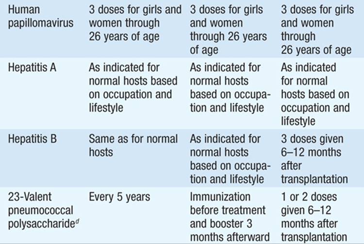
Recommendations for vaccination of cancer pts receiving chemotherapy, pts with Hodgkin’s disease, and hematopoietic stem cell transplant recipients are listed in Table 88-3. In solid organ transplant recipients, the usual vaccines and boosters should be given before immunosuppression. Pts with continued immunosuppression should have pneumococcal vaccination repeated every 5 years and should not receive live vaccines.
TABLE 88-3 VACCINATION OF CANCER PATIENTS RECEIVING CHEMOTHERAPYa




For a more detailed discussion, see Finberg R: Infections in Patients With Cancer, Chap. 86, p. 712; Madoff LC, Kasper DL: Introduction to Infectious Diseases: Host–Pathogen Interactions, Chap. 119, p. 1007; and Finberg R, Fingeroth J: Infections in Transplant Recipients, Chap. 132, p. 1120, in HPIM-18.