
Anthony C. Luke, MD, MPH
C. Benjamin Ma, MD
GENERAL APPROACH TO MUSCULOSKELETAL INJURIES
ESSENTIALS OF DIAGNOSIS
 History is most important in diagnosing musculoskeletal problems.
The mechanism of injury can explain the pathology and symptoms.
Determine whether the injury is traumatic or atraumatic, acute or chronic, high or low velocity (greater velocity suggests more structural damage), or whether any movement aggravates or relieves pain associated with the injury.
 General Considerations
Musculoskeletal problems account for about 10–20% of outpatient primary care clinical visits. Orthopedic problems can be classified as traumatic (ie, injury-related) or atraumatic (ie, degenerative or overuse syndromes) as well as acute or chronic. The mechanism of injury is usually the most helpful part of the history in determining the diagnosis.
The onset of symptoms should be elicited. With acute traumatic injuries, patients typically seek medical attention within 1–6 weeks of onset. The patient should describe the exact location of symptoms, which helps determine anatomic structures that may be damaged. If the patient is vague, the clinician can ask the patient to point with one finger only to the point of maximal tenderness.
Clinical Findings
A. Symptoms and Signs
The chief musculoskeletal complaints are typically pain (most common), instability, or dysfunction around the joints. Since symptoms and signs are often nonspecific, recognizing the expected combination of symptoms and physical examination signs can help facilitate the clinical diagnosis. Patients may describe symptoms of “locking” or “catching,” suggesting internal derangement in joints. Symptoms of “instability” or “giving way” suggest ligamentous injury; however, these symptoms may also be due to pain causing muscular inhibition. Constitutional symptoms of fever or weight loss, swelling with no injury, or systemic illness suggest medical conditions (such as infection, cancer, or rheumatologic disease).
Initial evaluation should follow routine trauma guidelines to rule out serious joint injury. However, typical evaluations in the clinic follow the traditional components of the physical examination and should include inspection, palpation, and assessment of range of motion and neurovascular status.
Inspection includes observation of swelling, erythema, atrophy, deformity, and (surgical) scars (remembered by the mnemonic, “SEADS”). The patient should be asked to move joints of concern (see Table 8–1). If motion is asymmetric, the clinician should assess the passive range of motion for any physical limitation.
Table 8–1. Shoulder examination.
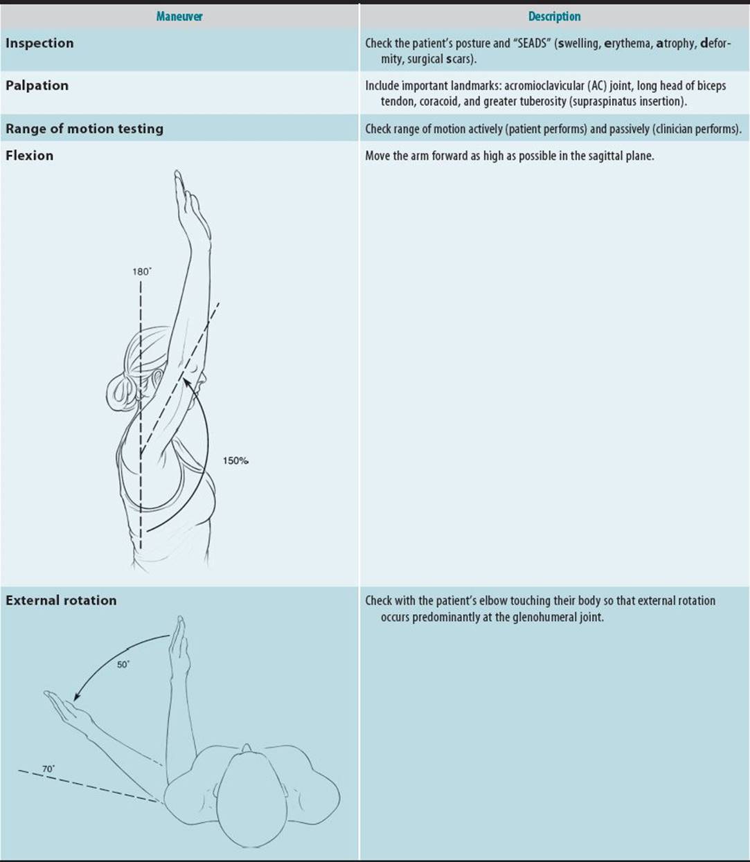

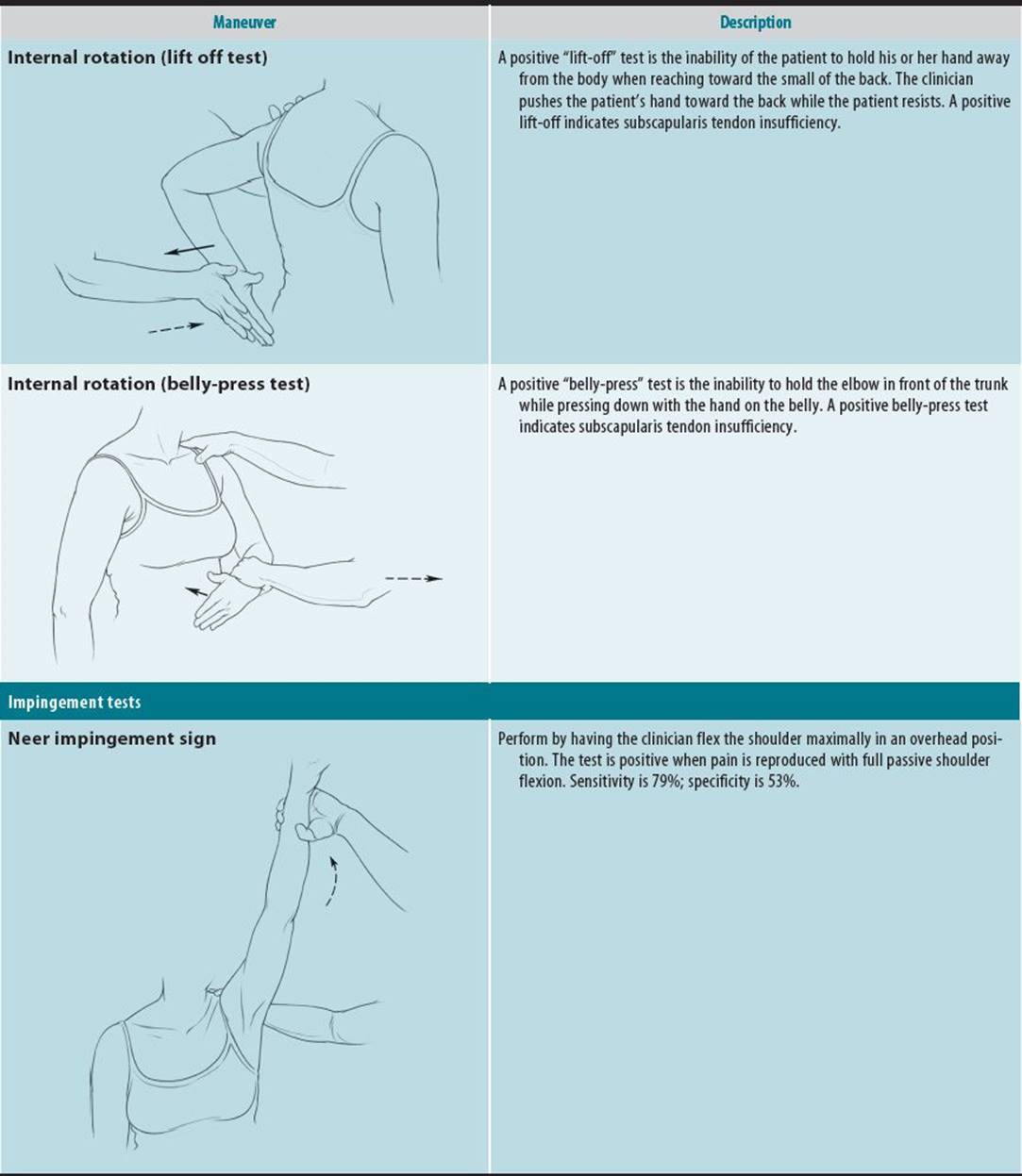
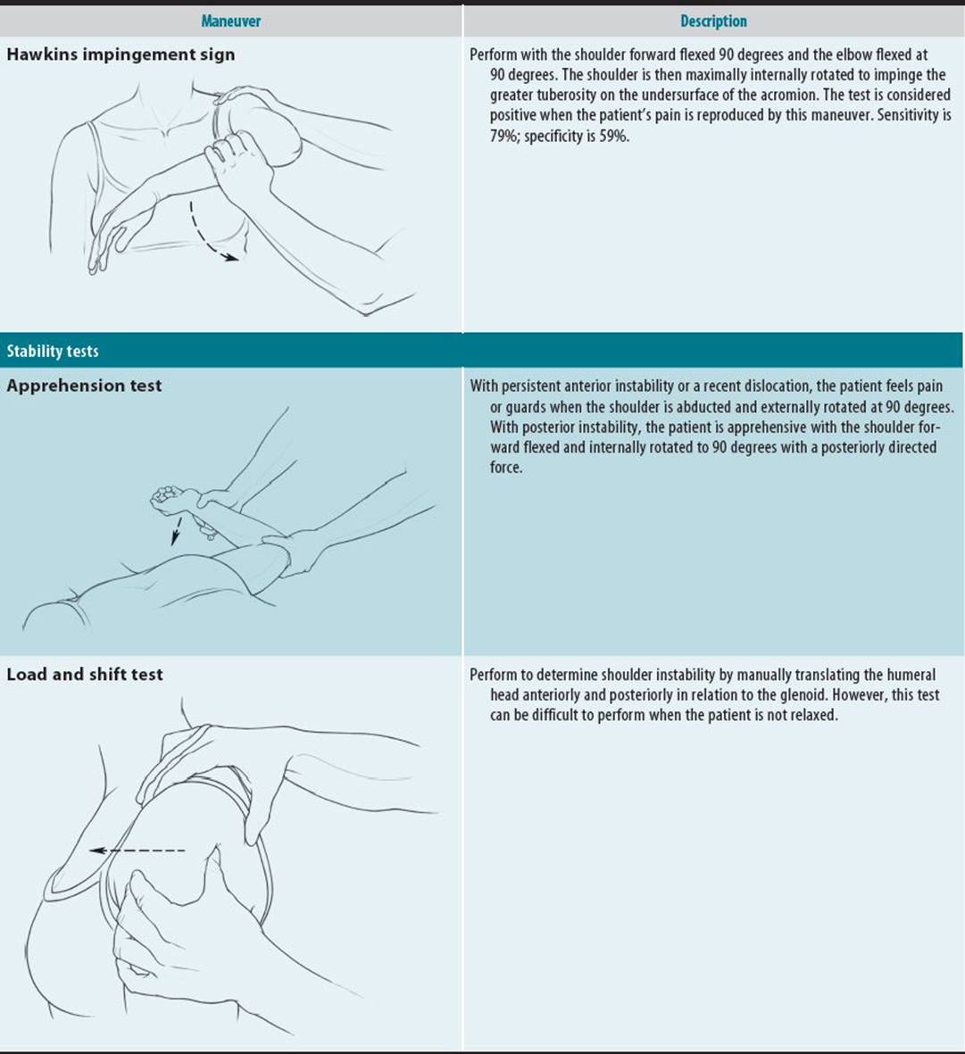
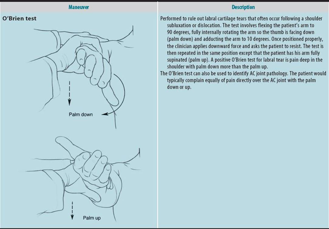
There are special tests to assess each joint. Typically, provocative tests re-create the mechanism of injury with the goal to reproduce the patient’s pain. Stress tests apply load to ligaments of concern. Typically, 10–15 lb of force should be applied when performing stress tests. Functional testing, including simple tasks performed during activities of daily living, is useful to assess injury severity.
B. Imaging
Bony pathology can be assessed using standard radiographs, although there also may be characteristic soft tissue findings. However, CT scans are the most effective method for visualizing any bony pathology, including morphology of fractures. Nuclear bone scans are now less commonly used but are still valuable for identifying stress injuries, infection, malignancy, or multisite pathology. Positron emission tomography (PET) scans are useful in identifying metastatic malignant lesions. MRI provides excellent visualization of ligaments, cartilage, and soft tissues. High-field 3.0 Tesla MRI is more available clinically and allows higher image resolution and decreased examination times compared to the standard 1.5 Tesla machines. Gadolinium contrast can be injected as an MRI arthrogram to increase sensitivity of detecting certain internal derangements in joints such as labral injuries. Musculoskeletal ultrasound, where available, can be useful for identifying superficial tissue problems, including tendinopathies and synovial problems.
C. Special Tests
Arthrocentesis must be performed promptly to rule out an infection when acute knee pain with effusion and inflammation are present and the patient is unable to actively flex the joint. The joint fluid should be sent for cell count, crystal analysis, and culture. Arthrocentesis and joint fluid analysis demonstrating crystals can lead to the diagnosis of gout (negatively birefringent, needle-shaped crystals) or pseudogout (positively birefringent, rectangularshaped crystals). In large, uncomfortable knee joint effusions, removal of excessive joint fluid may improve joint range of motion (flexion) and patient comfort. To avoid infecting the joint, arthrocentesis should not be performed when there is an active cellulitis or abscess overlying the joint. It appears the risk of bleeding after arthrocentesis or joint injection is extremely low even if the patient is taking anticoagulants. Caution should be practiced if the INR is > 3.0; however, even a supratherapeutic INR did not suggest an increased risk of hemarthrosis in one study. Markers of inflammation such as complete blood cell count, erythrocyte sedimentation rate, and C-reactive protein, and rheumatologic tests are useful in evaluating for infectious, oncologic, or rheumatologic processes. Electrodiagnostic studies such as electromyography and nerve conduction studies are useful when there are neurologic concerns; they can also help with prognostication in chronic conditions.
Treatment
While most outpatient musculoskeletal problems are best treated conservatively, the first consideration is whether there is an immediate surgical need. Surgical treatment is chosen when the outcome promises better health, restoration of function, and improved quality of life. During surgery, the musculoskeletal problem is usually repaired, removed, realigned, reconstructed, or replaced (eg, joint replacement).
If surgery is not immediately indicated, conservative treatment in the outpatient setting usually includes modification of activities, ice, compression, and elevation (remembered by the mnemonic, “MICE”). Controlling pain is an early concern for most patients. Commonly prescribed medications are analgesics (nonsteroidal anti-inflammatory drugs [NSAIDs], acetaminophen, or opioids). Other medications that may also be prescribed, albeit less commonly, are muscle relaxants or co-analgesics for neuropathic pain (which include the calcium channel alpha-2-delta ligands [eg, gabapentin] or tricyclic antidepressants). Topical medications, such as capsaicin cream or patch, lidocaine patches, and NSAID patches, can help provide superficial local pain relief.
Immobilization by casting, slings, and braces is helpful to protect an injured limb. Crutches are useful to reduce weight bearing. Rehabilitation and physical therapy are frequently needed. Other modalities commonly used by patients include chiropractic manipulation, massage therapy, acupuncture, and osteopathy.
When to Refer
Indications for emergency referral (immediate)
• Neurovascular injury
• Fractures (open, unstable)
• Unreduced joint dislocation
• Septic arthritis
Indications for urgent referral (within 7 days)
• Fractures (closed, stable)
• Reduced joint dislocation
• “Locked” joint (inability to fully extend a joint due to mechanical derangement, usually a loose body or torn cartilage)
• Tumor
Indications for early orthopedic assessment (2–4 weeks)
• Motor weakness (neurologic)
• Constitutional symptoms (eg, fever not due to septic arthritis, weight loss)
• Multiple joint involvement
Indications for routine orthopedic assessment (for further management)
• Failure of conservative treatment (persistent symptoms > 3 months)
• Persistent numbness and tingling in an extremity
Ahmed I et al: Safety of arthrocentesis and joint injection in patients receiving anticoagulation at therapeutic levels. Am J Med 2012;125:265 [PMID: 22340924].
Shapiro L et al: Advances in musculoskeletal MRI: technical considerations. J Magn Reson Imaging 2012;36:775 [PMID: 22987756].
DEFINITIONS OF COMMON ORTHOPEDIC CONDITIONS
A. Strain
A strained muscle or tendon has been pushed or pulled to its extreme by exposing it to an extreme load. It commonly results from an unexpected external force, such as a fall. The symptoms of strain should resolve within a few days to several weeks.
B. Sprain
A sprain is an injury in which a ligament has been stretched beyond its limit, causing tears or disruption in fibers within the substance of the ligament. Reactive inflammation with associated edema and local venous congestion develops over hours to days. A complete tear of a ligament is sometimes called a third-degree sprain.
C. Tendinosis or Tendinopathy
Tendinosis or tendinopathy is a degenerative change of a tendon with disruption of collagen fibers, the formation of new capillaries, and fibrosis. It may be the result of a primary inflammatory disease, such as rheumatoid arthritis, or it may be secondary to a mechanical injury.
D. Tenosynovitis
Tenosynovitis is inflammation of a tendon sheath.
E. Bursitis
Inflammation of a bursa is known as bursitis. An example is olecranon bursitis caused by inflammation in the thin tissue planes between the skin and olecranon.
F. Arthrosis
Arthrosis indicates an abnormal joint caused by injury, disease, or congenital abnormality. Examples include post-traumatic arthritis or osteoarthritis of the basilar joint of the thumb.
G. Peripheral Neuropathies
The peripheral nerves in the upper extremity can become entrapped in specific locations in the arm, typically in locations where the nerve crosses a joint or is in a tunnel. The entrapments may be due to external compression, exposure to vibrating hand tools, repeated forceful hand exertions, or sustained posture extremes (eg, overhead work). These exposures, if of adequate intensity and duration, can cause edema and fibrosis in adjacent tissues (eg, synovium or tendon) that compresses the nerve leading edema and connective tissue in the nerve and paresthesias and pain. If not addressed, it can progress to denervation and weakness.
H. Repetitive Strain Injures
Repetitive strain injuries are related to cumulative micro-trauma associated with repetitive forceful exertions or exposure to vibrating hand tools. These repeated exposures may lead to edema, microtrauma, acute inflammation, or chronic degenerative changes of the tendon, muscle, ligament, capsule, or nerve with associated pain. The associated edema or tissue swelling can lead to stenosis, which can entrap tendons, nerves, and vascular tissues. Common sites of injury to the upper extremity are the tendon compartments at the wrist, the epicondyles, and the shoulder.
 SELF-ASSESSMENT QUESTIONS
Select the one correct answer to each question.
Question 1: Functional testing
a. re-creates activities of daily living
b. is seldom useful to assess injury severity
c. is synonymous with stress testing
d. replaces the need for stress testing
Question 2: CT scans
a. are the most effective method for visualizing any bony pathology
b. are of limited use in visualizing fractures
c. are superior to MRI in visualizing ligaments, cartilage, and soft tissues
d. have replaced musculoskeletal ultrasound for identifying superficial tissue problems
Question 3: Arthrocentesis
a. must be performed promptly to rule out an infection with any acute knee pain
b. is indicated with knee pain when effusion and inflammation are present
c. should be performed even if there is an active cellulitis or abscess overlying the joint
d. presents a serious risk of bleeding
Question 4: Cumulative trauma
a. is unrelated to repetitive trauma
b. affects bone, but not the tendon, muscle, capsule, or the nerve
c. is seldom painful
d. may involve the extremity
Chapter adapted, with permission, from Luke A, Ma CB. Sports medicine and outpatient orthopedics. In: Papadakis MA, McPhee SJ, Rabow MW, eds. Current Medical Diagnosis and Treatment. 53rd ed. New York, NY: McGraw-Hill; 2014.