
Definition (Mayo Clin Proc 2010;85(6):572)
• Acute pericarditis is a common d/o caused by inflammation of the pericardium that can be isolated or as a manifestation of an underlying systemic dz
• Causes include idiopathic (85–90%), viral (coxsackie, echo), bacterial (staph, strep), TB, or fungal infection, cancer, XRT, connective tissue d/o, myxedema, uremia, Dressler syndrome, recent cardiac surgery or MI, chest wall trauma among others
• Although no formal diagnostic criteria, the Dx of acute pericarditis includes absence of life-threatening causes of CP (ACS, PE, ACS, etc.) & at least 2 of the following:
1. Characteristic CP: Sudden onset, retrosternal, pleuritic, positional (better w/ leaning forward or upright); pain can radiate to neck, arms, shoulders similar to ACS
2. Pericardial friction rub (high pitched, scratch sound heard best at L sternal border)
3. Suggestive ECG findings (see below)
4. New or worsening pericardial effusion
• Can be relapsing (incessant type, where discontinuation of or attempts to wean tx causes relapse in <6 wk; intermittent type, symptom-free intervals >6 wk but recurs)
• Can be a/w pericardial effusion w/ or w/o tamponade, or can be constrictive
Approach (NEJM 2004;351:2195; Mayo Clin Proc 2010;85(6):572)
• Obtain ECG as soon as possible
• Differentiate from other, more serious conditions such as ACS & aortic dissection
History
Pericarditis
• Characteristic CP: Sudden onset, retrosternal, pleuritic, positional (better w/ leaning forward or upright); pain can radiate to neck, arms, shoulders similar to ACS
• ± Fever, SOB, dysphagia
Effusion
• Asymptomatic → tamponade (hypotension, syncope, SOB, etc.)
Findings
Pericarditis
• Friction rub (high pitched, scratch sound heard best at LLSB apex), ↑ HR, ↑ RR, nl BP
Effusion
• Distant heart sounds
• Tamponade: Beck’s triad (hypotension, distended neck veins, distant heart sounds); pulsus paradoxus

Evaluation
• ECG (in pericarditis findings occur in 4 stages, see table below):
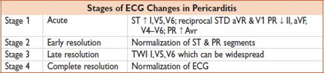
• Low voltage (QRS amplitude <0.5 mV in limb leads & electrical alternans suggest effusion
• Labs: CBC, BUN/Cr (r/o uremia); consider serology (strep, viral, ANA, anti-DNA Ab), TSH, ESR/CRP, CK/cardiac enzymes (30% positive in myopericarditis, JACC 2003;42:2144)
• CXR: >250 cc effusion → cardiomegaly w/ epicardial halo; also r/o other etiologies
• POC cardiac US to assess for: (1) pericardial effusion, (2) tamponade physiology (late diastolic collapse of RA, persistence of RA collapse >1/3 cardiac cycle, early diastolic collapse of RV, collapse of LA, dilated IVC w/ <50% respiratory collapse)
• Comprehensive cardiac echo if indicated to r/o ACS via focal wall motion abnormalities
• CT chest: Visible, often found while looking for other diagnoses (ie, PE, pl effusion)
Treatment (Pericarditis)
• NSAIDs: If viral/idiopathic, give Ibuprofen 600–800 mg TID, ×1–2 wk ± colchicine 0.5 mg BID (COPE, Circulation 2005;112:2012) for 4–6 wk; sx usually resolve w/i 1–3 d (JAMA 2003;289:1150); Colchicine should be used w/ caution in renal insufficiency, hepatobiliary dz blood dyscrasias, & GI motility disorders
• Steroids: For autoimmune or uremic etiologies as well as those who receive no benefit from NSAID or colchicine therapy. May ↑ risk of recurrence (COPE, Circulation 2005;112:2012).
• When used, tx w/ high-dose prednisone (1 mg/kg/d) w/ slow taper after 2–4 wk
• Pericardiocentesis indicated for purulent, Tb, or neoplastic pericarditis
• Abx & drainage: For purulent pericarditis (postsurgical)
• Treat underlying cause, dialysis; eg, for uremia
• Morphine for pain
• Cardiology consult: If tamponade/echo are being considered
• CT surgery consult: For pts w/ recent cardiac surgery or require pericardial window (effusion)
Treatment (Effusion)
• W/u: R/o infection, consider sending BUN, Cr, ANA, RF, malignancy screen
• Tx: Pericardiocentesis/biopsy is diagnostic & therapeutic, but can be done after admission if pt stable
• Acute cardiac tamponade w/ HD compromise requires urgent pericardiocentesis
Disposition
Pericarditis
• Home: If stable & virus is likely etiology
• Admit: Anyone w/ HD abnlty or myocarditis, uremia, ↑ cardiac silhouette on CXR
Effusion
• Admit if new or pt symptomatic
Pearls
• Consider this Dx in any CP pt w/ recent MI
• Can be a/w myocarditis, so check CK/CKMB/Tn
• Always assess for pericardial effusion/cardiac tamponade
• In Dressler syndrome, avoid NSAIDs (↓ healing)/anticoagulation (risks hemorrhagic tamponade)
• If pt is unstable & bedside US is available, obtain subxiphoid/parasternal long views to Dx effusion