
Approach
• Follow ACLS protocols for anyone unstable or severely symptomatic (CP, SOB, AMS)
• Anticipate need for intubation & defibrillator early
• Always obtain ECG & rhythm strip
• Determine (1) rate, (2) wide or narrow QRS, (3) rhythm regular or irregular
Supraventricular Tachycardia
Approach
• Differentiate type based on ECG, rhythm strip, & response to adenosine/vagal maneuvers (see below)
Definition
• Rhythm arises above the ventricles (either atrium or AV junction) w/ narrow QRS unless pre-excitation or aberrant conduction
History
• H/o pulmonary or cardiac dz → AT, MAT, AFL, AF, NPJT; o/w health adult → AVNRT, AVRT
• Gradual onset → ST, AT; abrupt onset → AVNRT, AVRT
Evaluation
• Consider CBC, TSH, tox screen, though in most cases, ECG/rhythm strip is sufficient


Treatment
• Cardiovert any unstable rhythm
• ST: Treat underlying condition
• AT/MAT: Treat underlying condition; consider AV nodal blocker
• AF/AFL: CCB, βB, dig, antiarrhythmic (amiodarone, lidocaine)
• AVNRT/AVRT: Vagal maneuvers, adenosine, CCB preferable to βB → avoid adenosine/nodal agents if e/o pre-excitation (see WPW below)
• NPJT: CCB, βB, amiodarone
Disposition
• Most pts w/ ST, AVNRT, AVRT can be d/c home once rhythm is controlled if → asymptomatic & no acute underlying condition. Admission for other rhythms is variable, but often necessary due to underlying condition.
• Consult cardiology for any pt w/ unstable SVT & those difficult to control w/ standard tx
Pearl
• MAT is often misdiagnosed as AF. Look closely at P wave morphology.
Atrial Fibrillation and Atrial Flutter
Definition (JACC 2006;48:e149)
• AF is a supraventricular tachyarrhythmia characterized by uncoordinated atrial activity w/ consequent deterioration of mechanical function
• Can be 1st episode or recurrent (≥2 episodes) as well as paroxysmal (self-limited), persistent (>7 d), permanent (>1 y) &/or cardioversion has failed
• Valvular → rheumatic heart dz, or postvalve surgery
• Lone AF → <60 y/o & no e/o cardiac dz or HTN
History
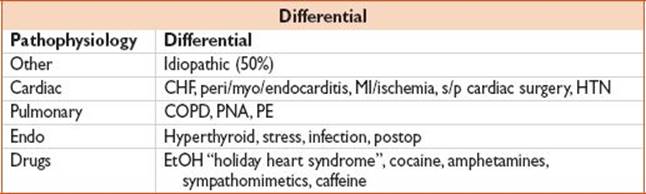
• Abrupt vs. gradual onset (palpitations, DOE, fatigue presyncope/syncope, CP); recent illness, drug & alcohol use
Findings
• Irregularly irregular pulse; may be regular w/ AFL
Evaluation
• ECG, CBC, lytes, Ca, Mg, PO4; CXR
• Consider cardiac markers (if active CAD is suspected); TSH, dig level if appropriate; echo (LA size, ?thrombus, valves, LV fxn)
• Consider outpt holter in pts w/ suggestive hx who arrive in NSR
• ECG in AF: Replacement of consistent P waves by rapid oscillating or fibrillatory waves that vary in amplitude, shape, & timing, a/w irregular, frequently rapid ventricular response rate
• ECG in aflutter: Atrial rate 250–350 bmp w/ ventricular response rate typically 150bpm presence of “sawtooth” flutter (“F”) waves. Can be typical (spiky V1, negative in II, III, aVF, V5–6) or atypical(appearance other than typical). F waves revealed via adenosine or vagal maneuvers. Most commonly 2:1 or 4:1 conduction.
Treatment
• Main objectives: Rate control, prevention of thromboembolism, & correction of rhythm
• When deciding on management strategies in the ED, several things to consider include:
a. Is the pt stable or unstable?
b. Is this 1st episode or recurrent episode, & is this part of a paroxysmal, persistent or permanent duration paradigm?
c. If 1st-episode or paroxysmal, how long have sxs been present (ie, <48 h)?
d. What is the pt’s stroke risk?
e. Does the pt have a cardiologist/PCP w/ whom you can make joint decision or poor f/u?
• Rate control vs. rhythm control: Numerous studies have sought to answer this question, but bottom line is that there appears to be no difference in symptomatic improvement, CHF, thromboembolic cx, severe bleeding or mortality when comparing the 2 strategies (PIAF, Lancet 2000;356:1789; AFFIRM, NEJM 2002;347:1825; STAF, J Am Coll Cardiol 2003;41:1690; HOT CAFÉ, CHEST 2004;126:476); however, rhythm control seems to be a/w increased rates of hospitalization & adverse medication effects (PIAF, Lancet 2000;356:1789; AFFIRM, NEJM 2002;347:1825)
• *Note: majority of these studies included pts w/ persistent AF, thus may not be generalizable to ED pt presenting w/ 1st episode or paroxysmal AF
• Suggested initial tx algorithms (Adapted from guidelines: Can J Cardiol 2011;27(1):27; Can J Cardiol 2011;27(1):38; Can J Cardiol 2011;27(1):47; Can J Cardiol 2011;27:74; Circulation2011;123(10):e269)

Figure 1.5 Unstable patient with afib. (Note: Mean energy level for successful cardioversion 50 J biphasic and 200 J monophasic [Am J Cardiol. 2004;93:1495–1499]. There may be higher first-shock success for DVVC if initial energy used 200 J vs.100 J [BEST-AF, Heart 2008; 94:884–887]).
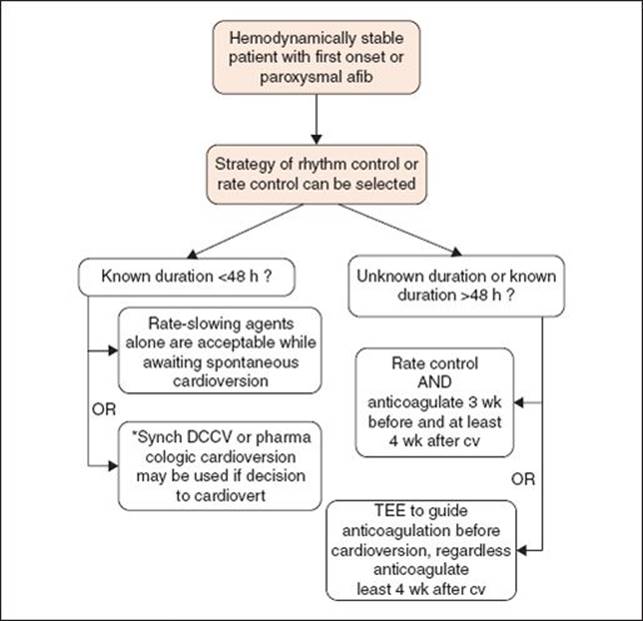
Figure 1.6 Stable patient, first episode or paroxysmal afib. Note: These patients may undergo cardioversion without anticoagulation, however, consider delaying DCCV and anticoagulate for 3 wk if high risk of stroke (i.e., mechanical valve, RHD, recent CVA/TIA).

Figure 1.7 Stable patient, persistent or permanent afib.
• Rate control: βB or nondihydropyridine CCBs recommended as 1st-line therapy for rate control. CCB, however, should be avoided in pts w/ ADHF & AF.
• Digoxin can be added to therapy w/ βB or CCB in pts whose HR is not controlled
• Dronaderone may be added for additional rate control w/ uncontrolled ventricular rate despite above therapy
• IV administration of digoxin or amiodarone is recommended to control HR in pts w/ AF & HF
• Amiodarone for rate control should be reserved for exceptional cases in which other means are not feasible or insufficient
• IV procainamide, disopyramide, ibutilide, or amiodarone may be considered for HD stable pts w/ AF involving conduction over an accessory pathway. In this situation, IV CCB or digoxin should be avoided as they may paradoxically accelerate the ventricular response.

• Direct current cardioversion: Recommended dose 150–200 J biphasic waveform
• Mean energy level for successful cardioversion 50 J biphasic & 200 J monophasic (Am J Cardiol 2004;93:1495). There may be higher 1st-shock success for DVVC if initial energy used 200 J vs.100 J (BEST-AF, Heart2008;94:884).
• Pretreatment w/ amiodarone, flecainide, ibutilide, propafenone or sotalol can be used to enhance the success of DCCV & prevent recurrent AF.
• Pharmacologic cardioversion: Administration of flecainide, dofetilide, propafenone, or ibutilide is recommended for pharmacologic cardioversion
• Procainamide has been shown to be effective in ED population w/ 58.3% cardioversion, w/ 91.7% success rate if followed by DCCV in nonresponders (CMEJ 2010;12(3):181)
• Amiodarone is a reasonable option, but digoxin & sotalol may be harmful for cardioversion & are not recommended
• βB or nondihydropyridine CCBs should be given before administering class I antiarrhythmic agents

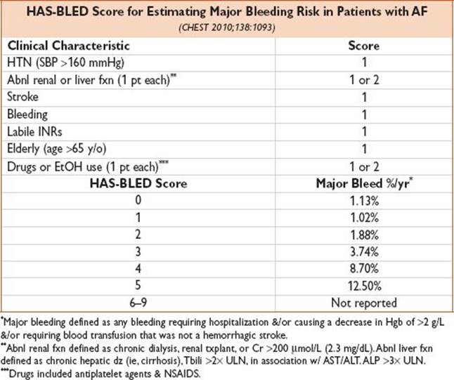
• Anticoagulation: All pts w/ AF or AFL (paroxysmal, persistent, or permanent) should be stratified using a predictive index for stroke (ie, CHADS2 or CHA2DS2-VASc) & for the risk of bleeding (ie, HAS-BLED) & most pts should receive anticoagulation
• Pts w/ very low risk of CVA (CHADS2 = 0) should receive ASA 81–325 mg/d
• Pts w/ low risk of CVA (CHADS2 = 1) should receive oral anticoagulation w/ either warfarin or dabigatran, but ASA is reasonable for some pts
• Pts w/ mod risk of CVA (CHADS2 ≥ 2) should receive oral anticoagulation w/ either warfarin or dabigatran
• Most pts should receive dabigatran 150 mg PO BID preferable to warfarin when anticoagulation indicated
• Newer effective oral anticoagulants include rivaroxaban 20 mg QD (ROCKET-AF, NEJM 2011;365:883) & apixaban 5 mg PO BID (ARISTOTLE, NEJM 2011;365:981)
• Anticoagulation not recommended for pts w/ lone AF

Disposition
• Home: Pts who convert to sinus, or are rate controlled, & anticoagulated if necessary
• All discharged pts should get close PCP or cardiology f/u
• EDOU: Depending on local clinical protocols
• Admit: Pts w/ acute underlying illness, ongoing sxs, or poor rate control
Pearls
• Risk of stroke is similar in all forms of AF/AFL (recurrent paroxysmal, persistent, & permanent AF, & AFL)
• Spontaneous cardioversion occurs w/i 24 h in 50–67% acute AF
• 5–8% elderly have recurrent AF
Guidelines: Gillis AM, Skanes AC, CCS Atrial Fibrillation Guidelines Committee. Canadian Cardiovascular Society atrial fibrillation guidelines 2010: Implementing GRADE and achieving consensus. Can J Cardiol. 2011;27(1):27–30.
Steill IG, Macle L, CCS Atrial Fibrillation Guidelines Committee. Canadian Cardiovascular Society atrial fibrillation guidelines 2010: Management of recent-onset atrial fibrillation and flutter in the emergency department. Can J Cardiol. 2011;27(1):38–46.
Gillis AM, Verma A, Talajic M, et al. Canadian Cardiovascular Society atrial fibrillation guidelines 2010: Rate and rhythm management. Can J Cardiol. 2011;27(1):47–59.
Cairns JA, Connolly S, McMurtry S, et al. Canadian Cardiovascular Society atrial fibrillation guidelines 2010: Prevention of stroke and systemic thromboembolism in atrial fibrillation and flutter. Can J Cardiol. 2011;27:74–90.
Fuster V, Ryden LE, Cannom DS, et al. 2011 ACCF/AHA/HRS focused updates incorporated into the ACC/AHA ESC 2006 guidelines for the management of patients with atrial fibrillation: A report of the American College of Cardiology Foundation/American Heart Association Task Force on Practice Guidelines. Circulation. 2011;123(10):e269–e367.
Pre-excitation
Definition
• Accessory pathway: A bypass tract that conducts impulses b/w atria
• Wolff–Parkinson–White: Accessory conduction pathway evident on resting ECG
• Orthodromic AVRT: Impulse travels down AV node (fast), then conducts retrograde, up the accessory pathway (slowly) → thus narrow-complex QRS
• Antidromic AVRT: Impulse travels down the accessory pathway (slowly), then conducts retrograde, up the AV node (fast) → thus wide-complex QRS
Evaluation
• ECG & rhythm strip
• Orthodromic AVRT: Narrow complex tachycardia
• Antidromic AVRT: WCT
Treatment
• AVRT: Vagal maneuvers, βBs, CCB
• AF/AFL w/ pre-excitation → cardiology consult, DC cardiovert, or use procainamide; βB & CCB are ineffective & can precipitate VF
Ventricular Tachycardia
Approach
• Determine if pt is stable or unstable → use ACLS protocol for any pt w/ unstable VT
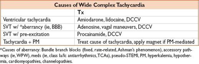
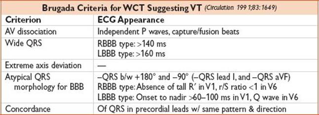
• Differentiate VT from nonsustained VT (NSVT), & other causes of WCT (see above)
• Differentiate monomorphic from polymorphic VT
Definition
• NSVT: VT lasting <30 s
• SVT w/ aberrancy: VT look-alike b/c abnl conduction → WCT. Caused by fixed BBB, rate-related BBB, or accessory pathway
• Torsades de pointes: Polymorphic VT + prolonged QT
Causes
• Monomorphic, structurally abnl heart: Prior MI, CMP, arrhythmogenic RV dysplasia
• Monomorphic, structurally nl heart: Idiopathic VT
• Polymorphic: Ischemia, CMP, torsades de pointes, Brugada syndrome (see below)
History
• Palpitations, lightheadedness, CP, SOB, nausea, syncope, unresponsiveness; PMH: CAD, CMP, multiple CAD RFs, & FHx sudden death all ↑ risk VT
Evaluation
• ECG, rhythm strip, lytes, Ca, Mg, PO4, cardiac markers; CXR; digoxin level if appropriate
Treatment
• Unstable VT: ACLS protocol
• Stable VT: Use either:
• Lidocaine: 100 mg IV load, then 1–4 mg/min
• Amiodarone: 150 mg IV load, then 1 mg/min
• Polymorphic VT: Magnesium 2–4 g IV bolus
• Other: Replete electrolytes (Ca, Mg, PO4); treat coincident ischemia if present
Disposition
• Admit to cardiac step-down unit or cardiac ICU
Pearls
• Assume all WCT to be ventricular unless proven o/w
• Best clinical predictors that WCT is VT → prior MI, CHF, LV dysfxn (Am J Med 1998;84:53)
Brugada Syndrome
Definition
• Incomplete RBBB w/ STE V1–V3 caused by alteration of the myocyte Na channel, a/w VT & sudden cardiac death
History
• Classically young, o/w healthy male, FHx sudden D; sxs: Presyncope, syncope, cardiac arrest
Evaluation
• ECG, electrolytes, Ca, Mg, PO4
Treatment
• Tele; electrophysiology consult
Disposition
• If incidental finding, refer to cardiology for f/u. O/w, admit to Tele bed for EP study, possible ICD placement.