
Hyponatremia
Definition
• Na <135, excess of water relative to sodium, generally from elevated ADH; generally not symptomatic at Na >125
History
• Most sxs are nonspecific & can include fatigue, weakness, muscle cramps, thirst, or postural dizziness. Sxs can range to more severe manifestations including confusion, agitation, delirium, lethargy, somnolence, coma, or szs.
• Other helpful historical features include h/o CHF, cirrhosis, renal dz, cancer, adrenal or pituitary dysfxn, recent GI surgery, thiazide or loop diuretics use, alcoholism
Physical Exam
• Look for signs to assess pt fluid status:
• Hypervolemia: Elevated JVP, peripheral edema, crackles, ascites, anasarca
• Hypovolemia: Tachycardia, hypotension, dry mucous membranes, oliguria, poor skin turgor, IVC collapsibility
• Look for signs of profound hyponatremia: Lethargic, disoriented/abnl sensorium, depressed reflexes, hypothermic, pseudobulbar palsy, Cheyne–Stokes respiration
Diagnostics
• Labs: Chem 7, FSG, urine electrolytes (Na, Cr, Osm), serum Osm, albumin
• VBG w/ stat sodium & Osm may provide more rapid turnaround
• Corrected Naglucose = Serum Na + [0.016 × (serum glucose – 100)] up to 400 mg/dL
• for glucose >400 mg/dL, 4 mEq/L should be added to every additional 100 mg/dL
Step-wise Approach to Hyponatremia
• Step 1: What is the serum osmolality?
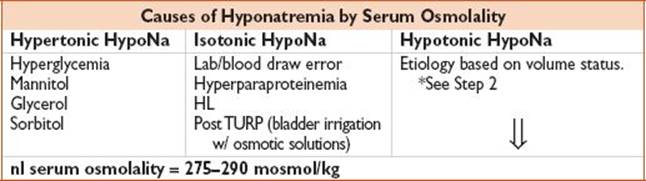
• Step 2: What is the pt’s volume status? Hypervolemic, euvolemic, or hypovolemic?
• Step 3: What are the urine Na, urine Osm, & FeNA values?
• Fractional Excretion of Sodium = FeNa = (Naurine × Crserum)/(Naserum × Crurine)

Treatment
• Asymptomatic or mild sxs of hyponatremia: Correct serum Na at ≤0.5 mEq/L/h
• Severe manifestations of hyponatremia: RAPID correction serum Na at 2 mEq/L/h × 2–3 h OR until sxs resolve

• Euvolemic hyponatremia
• Asymptomatic: Free water restrict (500–1000 mL/d)
• Symptomatic: See above
• SIADH
• Free water restrict + treat underlying cause
• Caution if using hypertonic or nl saline esp if IVF Osm < urine Osm, serum sodium may worsen (higher Osm will draw out fluid)
• May also consider lithium or demeclocycline (NEJM 2007;356:2064)
• Hypovolemic hyponatremia
• Volume replete w/ nl saline, as above (once dehydration resolved, stimulation of ADH will decline & Na will correct)
• Hypervolemic hyponatremia
• Free water restrict (0.5–1.5 L/d)
• Increase arterial volume: W/ vasodilators (Nitro), loop diuretics; consider albumin in cirrhosis
• Severe hyponatremia: Consider diuresis + Na replacement
Disposition
• Home: Mild asymptomatic hyponatremia
• Admit: Symptomatic, comorbidities, elderly. May require ICU admission if severe.
Pearl
• Rapid correction >10–12 mEq/L/d may result in central pontine myelinolysis (dysarthria, szs, quadriparesis due to focal myelin destruction in pons & extrapontine areas)
Hypernatremia
Definition
• Na >145, usually from free water loss or sodium gain (ie, infusion of hypertonic fluid)
• Appropriate response to hypernatremia is increased free water intake stimulated by thirst & renal excretion of a minimal volume of maximally concentrated urine as regulated by ADH
History
• Mild sxs include increased thirst or polyuria
• Severe sxs: AMS (irritability, lethargy, confusion, delirium, coma)
• RFs: Elderly, infants, debilitated. Endocrine pathology; cardiac, renal, liver dzs; psychiatric disorder (see etiology of Central and Nephrogenic Diabetes Insipidus); MEDS (see below chart), living situation (access to free water).
Physical Exam
• Look for signs to assess pt fluid status:
• Hypervolemia: Elevated JVP, peripheral edema, crackles, ascites, anasarca
• Hypovolemia: Tachycardia, hypotension, dry mucous membranes, oliguria, poor skin turgor, IVC collapsibility
• Severe hypernatremia: Lethargy, muscle spasticity, tremor, hyperreflexia, respiratory paralysis, ataxia
Diagnostics
• Labs: Chem 7, FSG, urine electrolytes (Na, Cr, Osm), serum Osm, albumin
• VBG w/ stat sodium & Osm may provide more rapid turnaround
• Corrected Naglucose = Serum Na + [0.016 × (serum glucose – 100)] up to 400 mg/dL
• for glucose >400 mg/dL, 4 mEq/L should be added to every additional 100 mg/dL
Step-wise Approach to Hypernatremia
• Step 1: What is the serum osmolality?
• nl serum osmolality = 275–290 mosmol/kg
• Step 2: What is the pt’s volume status? Hypervolemic, euvolemic, or hypovolemic?
• Step 3: What are the urine Na & urine Osm values?

Treatment

• Hypervolemic hypernatremia
• Treat underlying disorder
• Replace free water deficit (as above)
• Euvolemic hypernatremia
• Replace free water deficit (as above)
• Treat underlying etiology
• Central DI: Vasopressin 10 U SQ
• Hypovolemic hypernatremia
• Restore volume 1st then replace free water deficit (as above); add 40 mEq KCl IV to fluid replacement once pt is urinating
Disposition
• Home: Mild hypernatremia which can be corrected in <24 h
• Admit: Most will be admitted
Hypokalemia
Definition
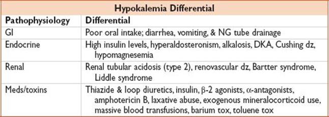
• Kμ+μ <3.5 mEq/L (ie, decreased intake, shift into cells, loss); 98% of potassium is intracellular.
History
• Usually not symptomatic until Kμ+μ <3 mEq/L
• Nausea, vomiting, weakness, fatigue, myalgia, muscle cramps. Meds (see Differential table).
• Pts at highest risk for electrocardiac cx of hypokalemia include those w/ acute ischemia, prolonged QT syndrome, & those taking digoxin
Physical Exam
• Paresthesias, depressed reflexes, proximal muscle weakness, ileus
• Severe hypokalemia: Hypoventilation, paralysis, rhabdomyolysis, myoglobinuria
• ARF, polymorphic VT, asystole
Diagnostics
• Labs: Chem 7, UA, urine electrolytes, urine Osm; consider blood gas, CPK, serum Osm
• Urine Kμ+μ <15 mmol/d suggests extrarenal, while urine Kμ+μ >15 mmol/d suggests renal etiology
• Transtubular Kμ+μ concentration gradient (TTKG) is helpful, but rarely used in the ED: TTKG = (PlasmaOsm × UrineK)/(PlasmaK × UrineOsm)
Note: Hypokalemia w/ TTKG >4 suggests renal Kμ+μ loss due to distal Kμ+μ secretion
• ECG: T-wave flattening/inversion, ST depression, U-waves, prolonged QT/QU interval; may also see PR prolongation, decreased voltage, QRS widening, atrial/ventricular dysrhythmias
Treatment
• ED
• Potassium replacement: Potassium chloride, Potassium bicarbonate, Potassium phosphate

• Treat underlying cause
• Replace Mg as needed (*Note: Concurrent Mg & Kμ+μ deficiency could lead to refractory Kμ+μ repletion)
• Goal Kμ+μ = 4 mEq/L in pts at highest risk
• Home
• Counsel pts to increase dietary intake of Kμ+μ (dried fruits, nuts, avocados, wheat germ lima beans, vegetables [spinach, broccoli, cauliflower, beets, carrots], fruits [banana, kiwi, etc])
• Discuss w/ PCP: Decrease diuretic dose; start/substitute for Kμ+μ-sparing med (βB, ACE, ARB, Kμ+μ-sparing diuretic)
• Potassium replacement: KCl 20 mEq PO QD for prevention; KCl 40–100 mEq PO QD for tx
Disposition
• Home: Mild hypokalemia w/ close f/u to recheck labs
• Admit: Moderate/severe hypokalemia, acid–base abnormalities, arrhythmia
Pearl
• Avoid dextrose solutions (stimulate insulin & inward shift of Kμ+μ)
Guideline: Cohn JN, Kowey PR, Whelton PK, Prisant LM. New guidelines for potassium replacement in clinical practice. Arch Intern Med. 2000;160: 2429–2436.
Hyperkalemia
Definition
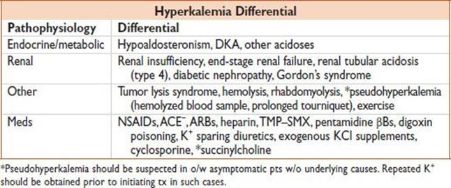
• Kμ+μ >5 mEq/L (ie, Kμ+μ release from cells, decreased renal losses, iatrogenic)
History
• Weakness, muscle cramps, paresthesias, nausea, palpitations. Meds (see Differential table).
Physical Exam
• Paresthesias, tetany; assess fluid status
• Severe hyperkalemia: Flaccid paralysis, hypoventilation, PEA arrest, or asystole
Diagnostics
• Labs: Chem 7; consider blood gas w/ stat Kμ+μ, UA, urine electrolytes, urine Osm, CPK
• ECG: Early: Peaked & symmetric T waves, flattened P waves, PR prolongation, 1° AVB. Late: Widening/slurring of QRS → sinusoidal waveform → VFib or asystole

Treatment
• Continuous cardiac monitoring
• Treating underlying cause
• Check electrolytes every 2–4 h until normalized
Disposition
• Home: Only if mild, stable hyperkalemia
• Admit: Most pts will require admission; may require ICU admission
Pearls
• Think “ABCD” (albuterol, bicarbonate, calcium, dextrose/insulin, dialysis, diuretics)
• Combination therapy is proven more efficacious than any therapy alone
• HD is the most rapid & effective way of lowering plasma Kμ+μ
Hypocalcemia
Definition
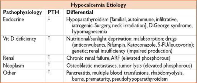
• Ca <8.5 mg/dL (2 mmol/L) OR ionized Ca <4.5 mg/dL (1.1 mmol/L); 50% bound to albumin, 40% is free, 10% complexed to anions
History
• Weakness, muscle cramps, paresthesias, irritability, depression, tetany, AMS. Meds (see Differential table).
Physical Exam
• Paresthesias; Chvostek sign (tap over facial nerve causing facial twitching); +Trousseau sign (inflate a BP cuff to 20 mmHg above systolic BP over bicep × 3 min to cause carpal spasm); may also see psychosis, szs, ↑ ICP, bronchospasm, laryngospasm
Diagnostics
• Labs: Chem 7 w/ Ca/Mg/Phosphorus testing. Check ionized calcium level, albumin, consider PTH for continued inpt w/u:
Corrected Ca = measured serum calcium (mg/dL) + [0.8 × (4-serum albumin (g/dL))]
• ECG: Prolonged QTc, heart blocks, ventricular dysrhythmias, torsade
Treatment
• Asymptomatic: Oral elemental Ca (1–3 g/d in divided doses)
• Symptomatic: [10% Calcium gluconate (1–2 g IV over 20 min) OR 10% Calcium chloride (1–2 g IV diluted in 100 cc D5W to decrease tissue irritation)], ± Vit D, ± Mg (50–100 mEq/d)
Disposition
• Home: Asymptomatic, w/ oral regimen described above & PCP f/u in 5–7 d to recheck electrolytes
• Admit: Severe hypocalcemia, comorbid conditions, HD unstable
Hypercalcemia
Definition
• Ca >10.5 mg/dL; usually asymptomatic at levels up to 11.5 mg/dL

History
• Polyuria, polydipsia, dehydration, nausea, vomiting, depression, confusion, coma, AMS; abdominal pain, anorexia, constipation, bone pain, Meds (see Differential table)
• May cause pancreatitis, nephrolithiasis, pathologic fractures thus suspect hypercalcemia in pts presenting w/ sxs consistent w/ these diagnoses
Physical Exam
• General weakness, epigastric tenderness, depressed deep tendon reflexes, coma
Diagnostics
• Labs: Chem 7 w/ Ca/Mg/Phosphorus testing, ionized Ca, lipase (if considering pancreatitis), urine electrolytes, albumin (see corrected Ca equation above), consider PTH
• ECG: Shortened QTc, PR prolongation, QRS widening; rarely BBB, sinus bradycardia or high-degree AV block
Treatment
• Address/treat underlying causes

Disposition
• Home: Mild stable hypercalcemia
• Admit: Most will need admission until resolution
Pearl
• Hypercalcemia = stones, bones, abdominal groans, & psychiatric overtones
Hypomagnesemia
Definition
• Mg <0.7 mmol/L

History
• Weakness, AMS, muscle cramps. Meds (see Differential table).
Physical Exam
• Tetany, Chvostek/Trousseau signs, papilledema, hyperreflexia
Diagnostics
• Labs: Chem 7 w/ Ca/Mg/Phosphorus testing, ionized Ca, albumin, consider PTH for continued inpt w/u.
• ECG: Similar to hypokalemia & hypocalcemia (prolonged intervals, T-wave flattening, widening of QRS, U waves)
Treatment
• Address underlying cause
• Magnesium replacement: 50% magnesium sulfate 2–4 g (16.6–33 mEq) IV over 30 min. Oral form may cause diarrhea (ie, magnesium citrate, milk of magnesia).
• Alcoholics: Consider thiamine; phosphorous & potassium replacement as needed
Disposition
• Home: Mild hypomagnesemia
• Admit: Severe hypomagnesemia w/ other associated electrolyte abnormalities (potassium, calcium), comorbid conditions
Pearl
• Most exogenously administered Mg will be excreted in urine; full Mg replacement takes days
Hypermagnesemia
Definition
• Mg >3 mEq/L

History
• Nausea, vomiting, lethargy, weakness, AMS; depends on level (renal insufficiency, GI motility disorder, adrenal insufficiency, hyperparathyroidism), Meds (anticholinergic, narcotic, lithium)
Physical Exam
• Depends on level
• Mg >3 mEq/L: N/V cutaneous flushing
• Mg >4 mEq/L: Hyporeflexia
• Mg >5 mEq/L: Hypotension
• Mg >9 mEq/L: Respiratory depression, shock, coma
• Mg >10 mEq/L: Asystole
Diagnostics
• Labs: Chem 7 w/ Ca/Mg/Phosphorus testing, ionized Ca, albumin
• ECG: QRS widening, QT prolongation, prolonged AV conduction → complete block
Treatment
• Calcium:
• Immediate: Calcium gluconate IV or Calcium chloride (see hypocalcemia)
• Continuous: 10% Calcium gluconate 2–4 mg/kg/h if indicated
• Diuretics: Loop diuretics + aggressive hydration (improve excretion)
• Dialysis: Particularly for pts in renal failure
Disposition
• Home: Asymptomatic, stable
• Admit: All need admission until sxs & lab values have normalized
Pearl
• Magnesium abnormalities are often seen w/ Kμ+μ or calcium abnormalities
Hypoglycemia
Definition
• Glucose <60 mg/dL; however, clinical hypoglycemia is any plasma glucose level low enough to cause sxs or signs c/w hypoglycemia (see below). Usually <55 mg/dL causes sx.
• Whipple’s triad: Sign/sxs of hypoglycemia, low plasma glucose, resolution of sx when plasma glucose is raised

History
• Neurogenic/autonomic sxs: Agitation, tremor, diaphoresis, palpitations, pallor, hunger
• Neuroglycopenic sxs: Fatigue, HA, AMS, lethargy, somnolence, coma, sz
• Take detailed med hx (see Differential table); consider new meds, med dose changes, incorrect use, intentional/accidental overdose, OTC/naturopathic meds
• Diabetics: Inquire recent FSG values (if taken), last meal, dietary changes, excess exercise
• ROS of contributing causes: Fever, chills, cough, abdominal pain, diarrhea, urinary sx, etc.
• RFs: Diabetics (esp on insulin), alcoholics, infants, elderly, s/p gastric bypass, critically ill
Diagnostics
• Labs: FSG, Chem 7; consider infectious w/u (CBC, UA, CXR)
*In o/w healthy, nondiabetics, consider LFTs, TSH, insulin, β-hydroxybutyrate, proinsulin, & C-peptide (low in exogenous insulin, high in insulinoma or sulfonylureas) in consultation w/ an endocrine specialist
• Serial glucose assessments may be necessary when prolonged hypoglycemia is expected in pts unable to communicate (ie, dementia, delirium, comatose, infants)
Treatment
• Glucose replacement:
• PO: Glucose paste/tablets (20 g), fruit juice, soft drinks, candy, a meal, etc.
• IV: 1 amp D50; infusion may be needed
• IM: 0.5–1 mg IM or SC glucagon (may cause N/V)
Disposition
• Home: Identifiable cause, does not need further monitoring
• Prompt f/u w/ primary care or endocrinologist should be arranged
• Pts should keep a glucose diary & should become concerned about the possibility of developing hypoglycemia when self-monitored glucose levels fall rapidly or is no greater than 70 mg/dL
• Admit: Long-acting hypoglycemic agents, unable to tolerate POs, HD unstable
Pearls
• βBs can mask adrenergic signs of hypoglycemia
• Efforts should be made to contact pt’s primary physician or endocrinologist
Guideline: Cryer PE, Axelrod L, Grossman AB, Heller SR, Montori VM, Seaquist ER, Service FJ. Evaluation and management of adult hypoglycemic disorders: An Endocrine Society clinical practice guideline. J Clin Endocrinol Metab. 2009;94:709–728.