
RHEUMATOLOGY
VASCULITIS
OVERVIEW
• Inflammation w/in blood vessel walls causing end-organ damage often a/w systemic sx; may be primary or secondary (eg, infection, malignancy) in etiology
• Classified by size of predominant vessel affected (Arthritis Rheum 2013;65:1); overlap of vessel size affected is common
• Clinical manifestations based on size of vessels involved; constitutional sx (low-grade fever, fatigue, weight loss, myalgias, anorexia) common to all

LARGE-VESSEL VASCULITIS
Takayasu’s arteritis (“pulseless disease”)
• Arteritis of aorta and its branches → stenosis/aneurysm → claudication; onset <50 y
• Pattern of involvement: aorta and branches; most often subclavian and innominate arteries (>90%), as well as carotid, coronary, renal, pulmonary (~50%)
• Epidemiology: Most common in Asia;  :
: ~9:1; age <50 y
• Clinical manifestations and physical findings
Systemic inflamm with fever, arthralgias, wt loss
Vessel inflamm w/ pain & tenderness, ↓ & unequal pulses/BPs in extremities, bruits, limb claudication, renovascular HTN (>50%), neurogenic syncope; Ao aneurysm ± AI
“Burnt out” or fibrotic period (eg, vascular stenosis)
• Dx studies: ↑ ESR (75%), CRP; arteriography → occlusion, stenosis, irregularity and aneurysms; carotid U/S Doppler studies; PET-CT; MRA; pathology → focal panarteritis, cellular infiltrate with granulomas and giant cells (bx not required for dx)
• Treatment: steroids ± MTX or AZA; anti-TNF (2nd line, Autoimmun Rev 2012;11:678), ASA, surgical/endovascular revasc (Circ 2008;69:70)
• Monitoring: MRA or PET-CT (Arth Rheum 2012;64:866); ESR/CRP (Ann Rheum Dis 2009;68:318)
Giant cell arteritis (GCA) (Curr Rheumatol Rep 2010;12:436)
• Granulomatous arteritis of aorta/branches w/ predilection for temporal artery, a/w PMR, onset >50 y
• Pattern of involvement: extracranial branches of carotid artery, esp. temporal artery (thus also called temporal arteritis); aorta and/or its branches in 10–80%
• Epidemiology: 90% of Pts >60 y, extremely rare <50 y; :=3:1
• Clinical manifestations (JAMA 2002;287:92)
constitutional sx: fevers, fatigue, wt loss, PMR sx (see below)
temporal artery (TA) → headache, tender TAs and scalp; absent TA pulse
ophthalmic artery (20%) → optic neuritis, diplopia, amaurosis fugax, blindness
facial arteries → jaw claudication
large vessel vasculitis → intermittent claudication of extremities; thoracic Ao aneurysm
• Dx studies: ↑ ESR (ESR <40 in ~5%), ↑ CRP, anemia
(ESR related to fibrinogen & Ig in blood; Ddx for >100: malignancy esp. multiple myeloma, lymphoma; GCA or other vasculitis; ESRD; endocarditis, TB, osteomyelitis)
temporal artery bx whenever GCA suspected (Se ≤85%); 1–2 cm ± bilat to ↑ yield (3–7% discordance) (Ann Rheum Dis 2009;68:318) → vasculitis & granulomas
if suspect aortitis or lg vessel involvement (BP Δ or bruits) → MRI/MRA or PET-CT
• Polymyalgia rheumatica (Lancet 2013;381:63)
seen in 50% of GCA Pts; 15% of Pts w/ PMR develop GCA
age ≥50 y; ESR >40 mm/h (and/or ↑ CRP); bilateral pain & morning stiffness (>30 min × ≥1 mo), involving 2 of 3 areas: neck or torso, shoulders or prox. arms, hips or prox. thighs; nighttime pain; exclude other causes of sx (eg, RA); nl CK
• Rx: steroids (do not await bx/path results to begin steroids, have at least 2 wk to bx)
GCA: 40–60 mg/d w/ slow taper, ASA daily; consider IV pulse if vision threatened
PMR:10–20 mg/d (Semin Arthritis Rheum 2007;37:13)
• Monitoring: follow clinical status & ESR/CRP (Ann Rheum Dis 2009;68:318)
MEDIUM-VESSEL VASCULITIS
Polyarteritis nodosa (“classic” PAN) (Arth Rheum 2010;62:616)
• Necrotizing nongranulomatous vasculitis of medium and small arteries (w/ muscular media) w/o glomerulonephritis or capillary involvement (ie, DAH), not a/w ANCA
• Epidemiology: >; average age of onset ~50 y; primary or HBV-associated (~10%)
• Clinical manifestations
constitutional sx (80%): wt loss, fever, fatigue
neuro (79%): mononeuritis multiplex, peripheral neuropathies, stroke
musculoskeletal (64%): extremity pain, myalgias, arthralgias, arthritis
renal (51%): HTN, hematuria, proteinuria, renal failure, glomerulonephritis unusual
GI (38%): abd pain, GIB/infarction, cholecystitis; GU (25%): ovarian or testicular pain
skin (50%): livedo reticularis, purpura, nodules, ulcers, Raynaud’s
ophthalmic (9%): retinal vasculitis, retinal exudates, conjunctivitis, uveitis
cardiac (22%): coronary arteritis, cardiomyopathy, pericarditis
if lung involvement, suspect other vasculitis
• Dx studies: ↑ ESR/CRP,  ANCA; ✓ HBs Ag; ↓ C3/C4 if HBV-associated
angiogram (mesenteric or renal vessels) → microaneurysms & focal vessel narrowing
CTA may be adequate to make dx, but conventional angiogram is most sensitive
biopsy (sural nerve, skin or affected organ) → vasculitis of small and medium vessel arteries with fibrinoid necrosis without granulomas
• Treatment: steroids ± CYC (if severe or failure to induce remission); antivirals if a/w HBV
ANCA-ASSOCIATED SMALL-VESSEL VASCULITIS
Microvascular vasculitis (eg, capillaries, postcapillary venules, & arterioles)
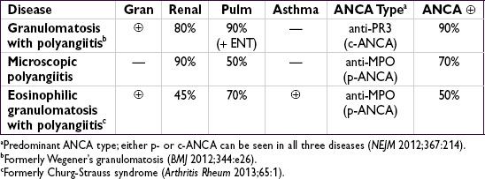
Differential diagnosis of ANCA
• anti-PR3 (c-ANCA): granulomatosis w/ polyangiitis, eosinophilic granulomatosis and polyangiitis, microscopic polyangiitis (rarely)
• anti-MPO (p-ANCA): microscopic polyangiitis, eosinophilic granulomatosis and
polyangiitis, granulomatosis w/ polyangiitis, drug-induced vasculitis, nonvasculitic
rheumatic diseases
• Atypical ANCA patterns: drug-induced vasculitis, nonvasculitic rheumatic diseases,
ulcerative colitis, primary sclerosing cholangitis, endocarditis, cystic fibrosis
Granulomatosis with polyangiitis (GPA, formerly Wegener’s granulomatosis)
• Necrotizing granulomatous systemic vasculitis frequently affecting nose, sinuses and/or upper respiratory tract in addition to kidneys, lungs, etc.
• Epidemiology: any age, but ↑ incidence in young and middle-aged adults; =
• Clinical manifestations
respiratory (90%)
upper: sinusitis, rhinitis, oral/nasal ulcers, saddle-nose deformity, otitis, hearing loss, subglottic stenosis
lower: pulmonary infiltrates, nodules, pulmonary hemorrhage, hemoptysis, pleurisy
renal (80%): RPGN (pauci-immune), RBC casts, dysmorphic RBCs, hematuria
ocular (50%): episcleritis, scleritis, uveitis, orbital granulomas → proptosis, corneal ulcer
neurologic: cranial and peripheral neuropathies, mononeuritis multiplex
skin (50%): palpable purpura, livedo reticularis
hematologic: ↑ incidence DVT/PE (20×) when disease active (Ann Intern Med 2005;142:620)
• Dx studies: 90%  ANCA (80% PR3, 20% MPO), less Se in limited upper airway disease
CXR or CT → nodules, infiltrates, cavities; sinus CT → sinusitis ± bone erosions
↑ BUN & Cr, proteinuria, hematuria; sediment w/ RBC casts, dysmorphic RBCs
Biopsy → necrotizing granulomatous inflammation of arterioles, capillaries, veins
• Treatment (Annals 2009;150:670; NEJM 2010;363:211 & 221)
Induction: RTX (375 mg/m2/wk × 4 wk) + steroids (1 g IV × 3 Δ → 1–2 mg/kg/d) or CYC (2 mg/kg/d × 3–6 mo or pulse 15 mg/kg q2–3wk) + steroids
RPGN: ± plasma exchange to ? ↓ risk of ESRD (Am J Kidney Dis 2011;57:566)
if mild (nonorgan- or life-threatening): MTX + prednisone may be adequate for induction (Arth Rheum 2012;64:3472)
Maintenance: MTX or AZA for ≥2 y after CYC induction (NEJM 2008;359:2790); after RTX induction repeat RTX q6mo (Arth Rheum 2012;64:3760) vs. watchful waiting
Relapse: if severe, reinduce w/ steroids + RTX or CYC; steroids ± MTX or AZA if mild;
↑ ANCA w/o clinical evidence of flare should not prompt Δ Rx (Annals 2007;147:611)
Microscopic polyangiitis (MPA) (Rheum Dis Clin North Am 2010;36:545)
• Similar to GPA, but w/o ENT/airway involvement & nongranulomatous
• Epidemiology: >; avg onset 50–60 y
• Clinical manifestations: similar to GPA w/o upper respiratory involvement; renal (80–100%): glomerulonephritis pulmonary (25–50%): pulmonary capillary alveolitis, pulmonary fibrosis constitutional and neuro sx similar to GPA; skin lesions (eg, palpable purpura) in 30–60%
• Dx studies: 70% ANCA (almost all anti-MPO)
biopsy → necrotizing, nongranulomatous inflammation of small vessels, pauci-immune (minimal deposition of complement or Ig; contrast w/ HSP, cryoglobulinemia, etc.)
urine sediment and CXR findings similar to those seen in GPA
• Treatment: as for GPA; ↓ relapse rate compared to GPA
Eosinophilic granulomatosis with polyangiitis (EGPA, formerly Churg-Strauss)
• Similar to GPA w/ more frequent cardiac involvement, a/w asthma and eosinophilia
• Epidemiology: rare; can present at any age (typically 30–40 y); a/w HLA-DRB4
• Clinical manifestations (Curr Rheumatol Rep 2011;13:489)
initial sx: asthma, sinusitis, allergic rhinitis (new asthma in adult raises suspicion)
eosinophilic infiltrative disease: shifting or transient pulmonary infiltrates, gastroenteritis or esophagitis
systemic small-vessel vasculitis: neuropathy (mononeuritis multiplex), renal (glomerulonephritis), skin (palpable purpura, petechial, nodules)
cardiac: coronary arteritis, myocarditis, CHF, valvular insufficiency (Medicine 2009;88:236)
• Dx studies: 50% ANCA (MPO > PR3), eosinophilia (5–10 k/µL, 80–100%),
biopsy → microgranulomas, fibrinoid necrosis and thrombosis of small arteries and
veins with eosinophilic infiltrates
• Treatment: high-dose corticosteroids + cyclophosphamide if severe
Renal-limited vasculitis
• Small vessel pauci-immune vasculitis causing RPGN w/o other organ involvement
• Dx studies: 80% ANCA (MPO > PR3); biopsy with pauci-immune GN ± granulomas
• Treatment identical to that for GPA/MPA
IMMUNE COMPLEX–ASSOCIATED SMALL-VESSEL VASCULITIS
Henoch-Schönlein purpura (HSP)
• IgA-mediated vasculitis w/ predilection for skin, GI tract and kidneys
• Epidemiology: >, children > adults, onset in winter > summer
• May develop after upper respiratory tract infection (esp. strep) or drug exposure
• Clinical manifestations
palpable purpura on extensor surfaces (lower extremity first) & buttocks
polyarthralgias (nondeforming) esp. involving hips, knees, & ankles
colicky abdominal pain ± GIB or intussusception
nephritis ranging from microscopic hematuria & proteinuria to ESRD
• Dx studies: skin bx w/ immunofluorescence → leukocytoclastic vasculitis w/ IgA
and C3 deposition in vessel wall; renal bx → mesangial IgA deposition
• Treatment: often self-limiting over 4 wk; steroids ± DMARDs for renal or severe disease
Cryoglobulinemic vasculitis (see “Cryoglobulinemia”)
Connective tissue disease–associated vasculitis
• Small vessel vasculitis a/w RA, SLE or Sjögren’s syndrome
• Clinical manifestations
distal arteritis: digital ischemia, livedo reticularis, palpable purpura, cutaneous ulceration
visceral arteritis: pericarditis and mesenteric ischemia
peripheral neuropathy
• Dx studies: skin/sural nerve bx, angiography, EMG; ↓ C′ in SLE; RF or anti-CCP in RA
• Treatment: steroids, cyclophosphamide, MTX (other DMARDs)
Cutaneous leukocytoclastic angiitis
• Heterogeneous group of clinical syndromes due to immune complex deposition in capillaries, venules and arterioles; includes hypersensitivity vasculitis
• Overall the most common type of vasculitis
• Etiologies
drugs: PCN, ASA, amphetamines, levamisole, thiazides, chemicals, immunizations
infections: Strep, Staph, endocarditis, TB, hepatitis
malignancy (paraneoplastic)
• Clinical manifestations: abrupt onset of palpable purpura and transient arthralgias after exposure to the offending agent; visceral involvement rare but can be severe
• Dx studies: ↑ ESR, ↓ complement levels, eosinophilia; ✓ U/A; skin biopsy → leukocytoclastic vasculitis w/o IgA deposition in skin (to distinguish from HSP); if etiology not clear, consider ANCA, cryoglobulins, hepatitis serologies, ANA, RF
• Treatment: withdrawal of offending agent ± rapid prednisone taper
Behçet’s syndrome (Curr Rheum Opin 2010;12:429)
• Systemic vasculitis affecting all vessel sizes, a/w oral and/or genital ulcers
• Epidemiology: usually young adults (25–35 y); a/w HLA-B51 in areas of highest prevalence on the old Silk Road (Turkey, Middle East and other Asian countries)
• Classification criteria (#1 + ≥2 others is 91% Se & 96% Sp; Lancet 1990;335:1078)
1. recurrent oral aphthous ulceration (≥3× in 1 y, usually 1st manifestation)
2. recurrent genital ulceration (labia in females, scrotum in males)
3. eye lesions: uveitis, scleritis, retinal vasculitis, optic neuritis (may threaten vision)
4. skin lesions: pustules, papules, folliculitis, erythema nodosum (scarring)
5. pathergy test (prick forearm w/sterile needle → pustule) (not sensitive in Caucasians)
• Other clinical manifestations: most recur but are not chronic
arthritis: mild, ± symmetric, nondestructive, involving knees and ankles
neurologic: usually involvement of midbrain parenchyma; peripheral neuropathy rare
vascular: superficial or deep vein thrombosis (25%); arterial stenosis, occlusion and aneurysm can also occur; low incidence of thromboembolism
• Dx studies: ↑ ESR/CRP; ulcer swab to r/o HSV; ulcer bx nonspecific; ophtho eval if sx
• Treatment (Rheumatology 2007;46:736; Ann Rheum Dis 2008;67:1656 & 2009;68:1528)
mucocutaneous
mild: topical steroids, colchicine (esp. for erythema nodosum), dapsone
severe: oral steroids, steroid-sparing agents
arthritis: NSAIDs, colchicine, steroids, steroid-sparing agents
ocular: topical and/or systemic steroids ± steroid-sparing agents
steroid-sparing: AZA, anti-TNF, CYC (large vessel and CNS ds), CsA, MTX, IFNɑ-2A
venous thrombosis: steroids and anticoagulation (careful if aneurysm present)