
CARDIOLOGY
VALVULAR HEART DISEASE
AORTIC STENOSIS (AS)
Etiology
• Calcific: predominant cause in Pts >70 y; risk factors include HTN, ↑ chol., ESRD
• Congenital (ie, bicuspid AoV w/ premature calcification): cause in 50% of Pts <70 y
• Rheumatic heart disease (AS usually accompanied by AI and MV disease)
• AS mimickers: subvalvular (HCMP, subAo membrane) or supravalvular stenosis
Clinical manifestations (usually indicates AVA <1 cm2 or concomitant CAD)
• Angina: ↑ O2 demand (hypertrophy) + ↓ O2 supply (↓ cor perfusion pressure) ± CAD
• Syncope (exertional): peripheral vasodil. w/ fixed CO → ↓ MAP → ↓ cerebral perfusion
• Heart failure: outflow obstruct + diastolic dysfxn → pulm. edema; esp. if ↑ HR/AF (↓ LV fill.)
• Acquired vWF disease (~20% of sev. AS): destruction of vWF; GI angiodysplasia
• Natural hx: usually slowly progressive (AVA ↓ ~0.1 cm2/y, but varies; Circ 1997;95:2262), until sx develop; mean survival based on sx: angina = 5 y; syncope = 3 y; CHF = 2 y
Physical exam
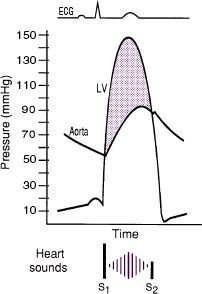
• Midsystolic crescendo-decrescendo murmur at RUSB, harsh, high-pitched, radiates to carotids, apex (holo-systolic = Gallavardin effect), ↑ w/ passive leg raise, ↓ w/ standing & Valsalva. In contrast, dynamic outflow obstruction (HCMP) ↓ w/ leg raise, ↑ w/ standing, Valsalva.
• Ejection click after S1 sometimes heard with bicuspid AoV
• Signs of severity: late-peaking murmur, paradoxically split S2 or inaudible A2, small and delayed carotid pulse (“pulsus parvus et tardus”), LV heave,  S4 (occasionally palpable)
Diagnostic studies
• ECG: may see LVH, LAE, LBBB, AF (in late disease)
• CXR: cardiomegaly, AoV calcification, poststenotic dilation of ascending Ao, pulmonary congestion
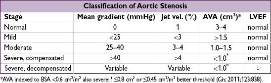
• Echo: valve morphology, estim pressure gradient & calculate AVA, EF
• Cardiac cath: usually to r/o CAD (in ~½ of calcific AS); for hemodyn. if disparity between exam & echo: ✓ pressure gradient (∇) across AoV, calc AVA (underestim. if mod/sev AI)
• Dobutamine challenge during echo or cath if low EF and ∇ <30 to differentiate: afterload mismatch: 20% ↑ SV & ∇, no Δ AVA (implies contractile reserve & ↑ EF post-AVR)
pseudostenosis: 20% ↑ SV, no Δ in ∇, ↑ AVA (implies low AVA artifact of LV dysfxn)
limited contractile reserve: no Δ SV, ∇ or AVA (implies EF prob. will not improve w/ AVR)
Treatment (Circ 2008;118:e523; Lancet 2009;373:956; EHJ 2012;33:2451)
• Management decisions are based on symptoms: once they develop AVR is needed.
If asx, HTN can be cautiously Rx’d; statins have not been proven to ↓ progression.
• AVR: indicated in sx AS (almost invariably severe; if not, look for another cause of sx) & asx severe AS + EF < 50%. May consider if asx but either sx or ↓ BP w/ exercise (can carefully exercise asx AS to uncover sx, do not exercise sx AS) or extremely severe (AVA <0.6 cm2, mean ∇ >60 mmHg, aortic jet >5 m/s). Reasonable if asx mod-severe AS and undergoing CV surgery.
• Medical (if not AVR candidate or to temporize): careful diuresis prn, control HTN, maintain SR; digoxin if ↓ EF & HF or if AF; avoid venodilators (nitrates) &  inotropes (bB/CCB) if severe; avoid vigorous physical exertion once AS mod–severe;
? nitroprusside if p/w CHF w/ sev. AS, EF <35%, CI <2.2, & nl BP (NEJM 2003;348:1756)
• IABP: stabilization, bridge to surgery
• Balloon AoV valvotomy (BAV): 50% ↑ AVA & ↓ peak ∇, but 50% restenosis by 6–12 mo &
↑ risk of peri-PAV stroke/AI (NEJM 1988;319:125), ∴ bridge to AVR or palliation
• Transcatheter AoV replacement (TAVR):  sx, hemodyn, & mortality to surgical AVR, but ↑ (mostly early) risk of vasc complic and stroke/ TIA; paravalvular leaks in ~7% (NEJM 2012;366:1686); in nonoperative Pts, 44% ↓ mortality vs. standard Rx (NEJM 2012;366:1696)
AORTIC INSUFFICIENCY (AI)
Etiology (Circ 2006;114:422)
• Valve disease (43%)
rheumatic heart disease (usually mixed AS/AI and concomitant MV disease)
bicuspid AoV: natural hx: 1⁄3→ normal, 1⁄3 → AS, 1⁄6 → AI, 1⁄6 → endocarditis → AI infective endocarditis
valvulitis: RA, SLE; anorectics (fen/phen) & other serotoninergics (NEJM 2007;356:29,39), XRT
• Root disease (57%)
HTN
aortic aneurysm or dissection, annuloaortic ectasia, Marfan syndrome
aortic inflammation: giant cell, Takayasu’s, ankylosing spond., reactive arthritis, syphilis
Clinical manifestations
• Acute: sudden ↓ forward SV and ↑ LVEDP (noncompliant ventricle) → pulmonary edema ± hypotension and cardiogenic shock
• Chronic: clinically silent while LV dilates (to ↑ compliance to keep LVEDP low) more than it hypertrophies → chronic volume overload → LV decompensation → CHF
• Natural hx: variable progression (unlike AS, can be fast or slow); once decompensation begins, prognosis poor w/o AVR (mortality ~10%/y)
Physical exam
• Early diastolic decrescendo murmur at LUSB (RUSB if dilated Ao root); ↑ w/ sitting forward, expir, handgrip; severity of AI ∝ duration of murmur (except in
acute and severe late); Austin Flint murmur: mid-to-late diastolic
rumble at apex (AI jet interfering w/ mitral inflow)
• Wide pulse pressure due to ↑ stroke volume, hyper- dynamic pulse → many of classic signs (see table); pulse pressure narrows in late AI with ↓ LV fxn; bisferiens (twice-beating) arterial pulse
• PMI diffuse and laterally displaced; soft S1 (early closure of MV); ± S3 (≠ ↓ EF but rather just volume overload in AI)


Diagnostic studies
• ECG: can see LVH, LAD, abnl repol; CXR: cardiomegaly ± ascending Ao dilatation
• Echo: severity of AI (severe = width of regurgitant jet >65% LVOT, vena contracta >0.6 cm, regurg fraction ≥50%, regurg orifice ≥0.3 cm2, flow reversal in descending Ao); LV size & fxn
Treatment (Circ 2008;118:e523; EHJ 2012;33:2451)
• Acute decompensation (consider ischemia and endocarditis as possible precipitants):
surgery usually urgently needed for acute severe AI which is poorly tolerated by LV
IV afterload reduction (nitroprusside) and inotropic support (dobutamine)
± chronotropic support (↑ HR → ↓ diastole → ↓ time for regurgitation)
pure vasoconstrictors and IABP contraindicated
• In chronic AI, management decisions based on LV size and fxn (and before sx occur)
• Surgery (AVR, replacement or repair if possible)
sx (if equivocal, consider stress test) severe AI (if not severe, unlikely to be cause of sx)
asx severe AI and EF ≤ 50% or LV dilation (end syst. diam. >50–55 mm or end diast. diam. >70–75 mm, esp. if progression) or undergoing cardiac surgery
• Transcatheter AoV replacement (TAVR) being explored ( JACC 2013;61:1577)
• Medical therapy: vasodilators (nifedipine, ACEI/ARB, hydralazine) if severe AI w/ sx or LV dysfxn & Pt not operative candidate or to improve hemodynamics before AVR; no clear benefit on clinical outcomes or LV fxn when used to try to prolong compensation in asx severe AI w/ mild LV dilation & nl LV fxn (NEJM 2005;353:1342)
MITRAL REGURGITATION (MR)
Etiology (Lancet 2009;373:1382; NEJM 2010;363:156)
• Leaflet abnormalities: myxomatous degeneration (MVP), endocarditis, calcific
RHD, valvulitis (collagen-vascular disease), congenital, anorectic drugs, XRT
• Functional: inferoapical papillary muscle displacement due to ischemic LV remodeling or other causes of DCMP; LV annular dilation due to LV dilation
• Ruptured chordae tendinae: myxomatous, endocarditis, spontaneous, trauma
• Acute papillary muscle dysfxn b/c of ischemia or rupture during MI [usu. posteromedial papillary m. (supplied by PDA only) vs. anterolateral (suppl. by diags & OMs)]
• HCMP: (see “Cardiomyopathy”)
Clinical manifestations
• Acute: pulmonary edema, hypotension, cardiogenic shock (NEJM 2004;351:1627)
• Chronic: typically asx for yrs, then as LV fails → progressive DOE, fatigue, AF, PHT
• Prognosis: 5-y survival w/ medical therapy is 80% if asx, but only 45% if sx
Physical exam
• High-pitched, blowing, holosystolic murmur at apex; radiates to axilla; ± thrill; ↑ w/ handgrip (Se 68%, Sp 92%),
↓ w/ Valsalva (Se 93%) (NEJM 1988;318:1572)
ant. leaflet abnl → post. jet heard at spine
post. leaflet abnl → ant. jet heard at sternum
• ± diastolic rumble b/c ↑ flow across valve
• Lat. displ. hyperdynamic PMI, obscured S1, widely split S2 (A2 early b/c ↓ LV afterload, P2 late if PHT); ± S3
• Carotid upstroke brisk (vs. diminished and delayed in AS)

Diagnostic studies (NEJM 2005;352:875)
• ECG: may see LAE, LVH, ± atrial fibrillation
• CXR: dilated LA, dilated LV, ± pulmonary congestion
• Echo: MV anatomy (ie, etiol); MR severity: jet area (can underestimate eccentric jets), jet width at origin (vena contracta) or effective regurgitant orifice (ERO; predicts survival); LV fxn (EF should besupranormal if compensated, ∴ EF <60% w/ sev. MR = LV dysfxn); TEE if TTE inconclusive or pre/intraop to guide repair vs. replace
• Cardiac cath: prominent PCWP c-v waves (not spec. for MR), LVgram for MR severity & EF

Treatment (Circ 2008;118:e523; NEJM 2009;361:2261; EHJ 2012;33:2451)
• Acute decompensation (consider ischemia and endocarditis as precipitants)
IV afterload reduction (nitroprusside), ± inotropes (dobuta), IABP, avoid vasoconstrictors
surgery usually needed for acute severe MR as prognosis is poor w/o MVR
• Surgery (repair [preferred if feasible] vs. replacement w/ preservation of mitral apparatus)
sx severe MR, asx severe MR and EF 30–60% or LV sys. diam. >40 mm
consider MV repair for asx severe MR w/ preserved EF, esp. if new AF or PHT
if AF, maze procedure or pulm vein isolation may → NSR and prevent future stroke
• In Pts undergoing CABG w/ mod–sev fxnal MR, consider annuloplasty ring
• Percutaneous MV repair: edge-to-edge clip less effective than surgery, but ? consider for elderly, fxnal MR or low EF (NEJM 2011;364:1395); percutaneous valve under study
• Medical: clinical benefit in asx Pts; bB preserve LV fxn ( JACC 2012;60:833); if sx but not operative candidate ↓ preload (↓ HF and MR by ↓ MV orifice): diuretics, nitrates (esp. if ischemic/fxnal MR); if LV dysfxn: ACEI, bB, ± BiV pacing; maintain SR
MITRAL STENOSIS (MS)
Etiology (Lancet 2012;379:953)
• Rheumatic heart disease (RHD): fusion of commissures → “fish mouth” valve
from autoimmune rxn to b strep infxn; seen largely in developing world today
• Mitral annular calcification (MAC): encroachment upon leaflets → functional MS
• Congenital, infectious endocarditis w/ large lesion, myxoma near MV, thrombus
• Valvulitis (eg, SLE, amyloid, carcinoid) or infiltration (eg, mucopolysaccharidoses)
Clinical manifestations (Lancet 2009;374:1271)
• Dyspnea and pulmonary edema (if due to RHD, sx usually begin in 30s)
precipitants: exercise, fever, anemia, volume overload (incl. pregnancy), tachycardia, AF
• Atrial fibrillation: onset often precipitates heart failure in Pts w/ MS
• Embolic events: commonly cerebral, esp. in AF or endocarditis
• Pulmonary: hemoptysis, frequent bronchitis (due to congestion), PHT, RV failure
• Ortner’s syndrome: hoarseness from LA compression of recurrent laryngeal nerve
Physical exam
• Low-pitched mid-diastolic rumble at apex w/ presystolic accentuation (if not in AF); best heard in L lat decubitus position during expi- ration, ↑ w/ exercise; severity proportional to duration (not intensity) of murmur
• Opening snap (high-pitched early diastolic sound at apex) from fused leaflet tips;
MVA proportional to S2–OS interval (tighter
valve → ↑ LA pressure → shorter interval)
• Loud S1 (unless MV calcified)

Diagnostic studies
• ECG: LAE (“P mitrale”), ± AF, ± RVH
• CXR: dilated LA (straightening of left heart border, double density on right, left mainstem bronchus elevation)
• Echo: estimate pressure gradient (∇), RVSP, valve area, valve echo score (0–16, based on leaflet mobility & thick., subvalvular thick., Ca++); exer. TTE (to assess ∆ RVSP and ∇) if sx & severity of MS at rest discrepant; TEE to assess for LA thrombus before PMV
• Cardiac cath: ∇ from simultaneous PCWP & LV pressures, calculated MVA; LA pressure tall a wave and blunted y descent; ↑ PA pressures

Treatment (NEJM 1994;331:961; Circ 2002;105:1465 & 2008;118:e523; EHJ 2012;33:2451)
• Medical: Na restriction, cautious diuresis, bB, sx-limited physical stress
• Antibiotic Ppx recommended if h/o RHD w/ valvular disease for 10 y or until age 40
• Anticoag if: AF, prior embolism, LA thrombus; ? LA >55 mm or lg LA w/ spont contrast
• Mechanical intervention if: heart failure sx w/ MVA ≤1.5, or
heart failure sx w/ MVA >1.5 but ↑ PASP, PCWP, or MV ∇ w/ exercise, or
asx Pts w/ MVA ≤1.5 and PHT (PASP >50 or >60 mmHg w/ exercise) or new-onset AF
• Percutaneous mitral valvotomy (PMV): preferred Rx if RHD; MVA doubles, ∇↓ by 50%; MVR if valve score <8, ≤ mild MR, AF or LA clot
• Surgical (MV repair if possible, o/w replacement): consider in sx Pts w/ MVA ≤1.5
if PMV unavailable/contraindicated (mod. MR, LA clot), or valve morphology unsuitable
• Pregnancy: if NYHA class III/IV → PMV, o/w medical Rx w/ low-dose diuretic & bB
MITRAL VALVE PROLAPSE (MVP)
Definition and Etiology
• Billowing of MV leaflet ≥2 mm above mitral annulus in parasternal long axis echo view
• Leaflet redundancy from myxomatous proliferation of spongiosa of MV apparatus
• Idiopathic, familial and a/w connective tissue diseases (eg, Marfan’s, Ehlers-Danlos)
• Prevalence 1–2.5% of gen. population,  >
>  (NEJM 1999;341:1), most common cause of MR
Clinical manifestations (usually asymptomatic)
• MR (from leaflet prolapse or ruptured chordae); infective endocarditis; embolic events
• Arrhythmias, rarely sudden cardiac death
Physical exam
• High-pitched, midsystolic click ± mid-to-late systolic murmur
• ↓ LV volume (standing) → click earlier; ↑ LV volume or afterload → click later, softer
Treatment
• Endocarditis prophylaxis no longer recommended (Circ 2007:116:1736)
• Aspirin or anticoagulation if prior neurologic event or atrial fibrillation
TRICUSPID REGURGITATION
• Primary etiol: rheumatic, CTD, radiation, IE, Ebstein’s anomaly, carcinoid, tumors
• Fxnal etiol: RV and/or pulm HTN (may be 2° to L-sided dis.), RV dilation and/or infarct
• Consider repair, annuoplasty or replacement for sx and severe TR (eg, ERO ≥0.40 cm2)
PROSTHETIC HEART VALVES
Mechanical (60%)
• Bileaflet (eg, St. Jude Medical); tilting disk; caged-ball
• Very durable (20–30 y), but thrombogenic and ∴ require anticoagulation
consider if age <~65 y or if anticoagulation already indicated ( JACC 2010;55:2413)
Bioprosthetic (40%)
• Bovine pericardial or porcine heterograft (eg, Carpentier-Edwards), homograft
• Less durable, but min. thrombogenic; consider if >~65 y, lifespan <20 y or anticoag
Physical exam
• Normal: crisp sounds, ± soft murmur during forward flow (normal to have small ∇)
• Abnormal: regurgitant murmurs, absent mechanical valve closure sounds
Anticoagulation & antiplatelet therapy (Circ 2008;118:e523; JAMA 2012;308:2118)
• Assess for high-risk features: prior thromboembolism, AF, EF<30–35%, hypercoagulable
• Warfarin: low-risk mech AVR: INR 2–3 (consider 2.5–3.5 for 1st 3 mo)
mech MVR or high-risk mech AVR: INR 2.5–3.5
high-risk bioprosthetic: INR 2–3 (and consider in low-risk for 1st 3 or even ? 6 mo)
• ASA (75–100 mg) for all prosthetic valves; avoid adding to warfarin if h/o GIB, uncontrolled HTN, erratic INR or >80 y; ASA + clopidogrel (or warfarin) × 3–6 mo after TAVR
• If thrombosis, ↑ intensity (eg, INR 2–3 → 2.5–3.5; 2.5–3.5 → 3.5–4.5; add ASA if not on)

Correction of overanticoagulation (Circ 2008;118:e626)
• Risk from major bleeding must be weighed against risk of valve thrombosis
• Not bleeding: withhold warfarin, give vit K 1–2.5 mg PO only if INR 5–10, ✓ serial INRs
• Bleeding or INR >10: FFP ± low-dose (1 mg) vit K IV
Endocarditis prophylaxis: for all prosthetic valves (see “Endocarditis”)
Complications
• Structural failure (r/o endocarditis); mechanical valves: rare except for Bjork-Shiley; bioprosthetic valves: up to 30% fail rate w/in 10–15 y, mitral > aortic
• Paravalvular leak (r/o endocarditis); small central jet of regurg is normal in mech. valves
• Obstruction from thrombosis or pannus ingrowth: ✓ TTE, TEE and/or fluoroscopy if ? clot significantly sx pannus ingrowth: remove w/ surgery
thrombosis: surgery if L-sided valve & either severe sx or lg (? >1 cm) clot burden; lytic often ineffective for L-sided thrombosis & 12–15% risk of stroke; consider UFH ± lytic (? low-dose tPA via slow infusion, JACC CV Imaging 2013;6:206) if mild sx & small clot burden or poor surg candidate; lytic reasonable for R-sided
• Infective endocarditis ± valvular abscess and conduction system dis. (see “Endocarditis”)
• Embolization (r/o endocarditis); risk ~1%/y w/ warfarin (vs. 2% w/ ASA, or 4% w/o meds)
mech MVR 2 × risk of embolic events vs. mech AVR (Circ 1994;89:635)
• Bleeding (from anticoag), hemolysis (esp. w/ caged-ball valves or paravalvular leak)
HEART VALVES (superior view, JAMA 1976;235:1603)
