
CONSULTS
SURGICAL ISSUES
ABDOMINAL PAIN

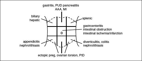
Figure 10-1 Etiologies of abdominal pain based on location
Initial evaluation
• History: onset of pain, location, exacerbating/relieving factors
• Assoc. sx: fevers/chills, N/V, Δ in bowel habits (diarrhea/constipation, stool diam. or color, hematochezia, melena), jaundice, Δ in urine color, Δ in wt, menstrual hx in women
• PMHx: previous incisions or abdominal surgeries; Ob/Gyn hx
• Exam: VS; general posture of Pt; comprehensive abdominal exam specifically looking for signs of peritonitis, which include rebound tenderness and involuntary guarding, abdominal wall rigidity, pain w/ percussion/minimal palpation; presence of hernias; rectal/pelvic
• Labs: CBC, electrolytes, LFTs, amylase/lipase, pregnancy test
• Imaging: depends on suspected etiology, may include RUQ U/S for biliary/hepatic disease, KUB for intestinal obstruction, CT for pancreatitis or intestinal disease. Do not delay resucitation or surgical consultation for ill Pt while waiting for imaging.
ACUTE ABDOMEN
Definition
• Acute onset abdominal pain that portends need for urgent surgery
Etiologies
• Perforated viscous → peritonitis (perforated ulcer, complicated diverticulitis, trauma)
• Intraperitoneal bleed
• Bowel obstruction (adhesions from previous surgeries, malignancies, hernias)
• Mimics: severe pancreatitis can resemble peritonitis; renal colic causes severe abdominal pain but not abdominal rigidity
Initial evaluation
• H&P as above
• Labs as above plus: PT/INR, PTT, type & screen
• Imaging: KUB (upright) or if stable, CT abomen/pelvis w/ IV contrast (IV/PO if suspect obstruction)
Initial management
• Immediate surgical consultation for suspected acute abdomen
• NPO, start IV fluids (NS or LR)
• Broad spectrum abx if perforation suspected
EXTREMITY EMERGENCIES
Acute limb ischemia (see “Peripheral Artery Disease” for details)
• Definition: sudden ↓ in perfusion causing threat to limb viability
• Evaluation: detailed vascular exam; CT angiography or arteriography
• Initial management: anticoag for embolism/thrombosis; immediate surgical consultation
Compartment syndrome (Clin Orthop Relat Res 2010;468:940)
• Definition: ↑ intracompartmental pressure w/ compressive closure of venules → ↑ hydrostatic force resulting in further increases in compartment pressure
• Etiologies: orthopedic (fracture), vascular (ischemia-reperfusion), iatrogenic (eg, vascular injury in anticoagulated Pt), soft tissue injury (eg, prolonged limb compression)
• Clinical manifestations: pain esp. on passive movement, swollen/tense compartment, paraesthesia, pallor, pulselessness, paralysis (late)
• Evaluation: ✓ compartment pressures (needle manometry), ICP >30 or difference between diastolic pressure and ICP of >10–30 is diagnostic
• Treatment: fasciotomy
SURGICAL TUBES, DRAINS, WOUNDS
Tracheostomy (Otolaryngol Head Neck Surg 2013;148:6)
• Inserted either percutaneously or surgically
• Monitor for secretions and suction frequently
• Typically a cuffed tube, which creates a tight seal to facilitate ventilation throught tube
• Speaking valve (eg, Passy-Muir): 1-way valve that allows inhalation through tube, but exhalation around tube through vocal cords (nb, cuff should not be inflated)
• 1st routine tube change for percutaneously placed tubes should be ~10 d postop, whereas surgically placed tubes can be changed >5 d postop and should be overseen by experienced personnel
• Accidental dislodgement of tube:
intubate from above (if airway/vent necessary & anatomically possible)
w/in 7 d of placement: emergent surgical consultation
>7 d after placement: replace with a similar size tube or smaller
Chest tubes (Eur J Cardiothorac Surg 2011;40:291)
• Inserted for PTX, chest trauma or after thoracic surgery for drainage of air/ fluid from thoracic cavity. Tubes range from small 10-Fr catheters placed for spontaneous PTX to large bore tubes (28–32 Fr) placed after pulmonary resections.
• Connected to 3-chamber chest drainage system:
1st: collection chamber for pleural fluid
2nd: water seal chamber used to allow air to exit pleural space on exhalation and prevent air from entering on inhalation
3rd: suction control chamber which regulates suction transmitted to pleural space
• Monitor for ouput and presence of air leak (indicated by bubbling in water seal chamber)
• Removal determined by overall daily outputs and presence of air leak
• If accidentally removed or dislodged so not functional, tube should be completely removed and an occlusive dressing (eg, 4 × 4 covered w/ Tegederm or silk tape) should be placed rapidly over site. CXR STAT; new tube should be placed if persistent PTX.
Gastrostomy/jejunostomy tubes (Paediatr Child Health 2011;16:281)
• Placed for tube feedings, hydration and delivery of medications
• Securely anchor to skin to prevent inadvertent removal
• Surrounding skin should be kept dry to prevent breakdown
• Should not be removed for ≥6–8 wk to allow establishment of mature gastrocutaneous tract
• Obstructed tubes can be cleared by flushing with agents such as carbonated water, meat tenderizer, pancreatic enzymes. ↓ obstruction by flushing before & after meds and flushing q4–6h when receiving continuous feeds.
• If becomes inadvertently removed a foley catheter of similar size or smaller should be placed in the tract immediately to prevent stoma from closing. Tube then replaced and confirmed via fluoro study w/ gastrograffin.
Suture/staple removal
• Should be done in consultation w/ surgical team
• Timing of removal depends on location of wound: wait 3–4 d before removal from face, 6 d for scalp, 7 d for chest, abdomen & arms, 10 d for back & legs, 14 d for hands
• Should not be removed if there is evidence of wound separation during removal!
• After removal, wound should be reapproximated w/ steri-strips
MAXIMIZING A SURGICAL CONSULT
• For ill Pt, call surgical consult early, do not wait for labs & imaging results
• If potential surgical emergency, make Pt NPO, start IVF, ✓ coags, type & screen
• Have appropriate-level MD who knows & has examined Pt call consult