
CARDIOLOGY
ARRHYTHMIAS
BRADYCARDIAS, AV BLOCK AND AV DISSOCIATION
Sinus bradycardia (SB) (NEJM 2000;342:703)
• Etiologies: meds (incl bB, CCB, amio, Li, dig), ↑ vagal tone (incl. athletes, sleep, IMI), metabolic (hypoxia, sepsis, myxedema, hypothermia, ↓ glc), OSA, ↑ ICP
• Treatment: usually none required; atropine, b1 agonists or temp. pacing if symptomatic
• Most common cause of sinus pause is blocked premature atrial beat
Sick sinus syndrome (SSS)
• Features may include: periods of unprovoked SB, SA arrest, paroxysms of SB and atrial
tachyarrhythmias (“tachy-brady” syndrome), chronotropic incompetence w/ ETT
• Treatment: meds alone usually fail (adeq. control tachy → unacceptable brady); usually need combination of meds (bB, CCB, dig) for tachy & PPM for brady

AV dissociation
• Default: slowing of SA node allows subsidiary pacemaker (eg, AV junction) to take over
• Usurpation: acceleration of subsidiary pacemaker (eg, AV junctional tach, VT)
• 3° AV block: atrial pacemaker unable to capture ventricles, subsidiary pacemaker emerges distinguish from isorhythmic dissociation (A  V rate, some P waves nonconducting)
Temporary pacing wires
• Consider w/ bradycardia with hemodyn instability or unstable escape rhythm when perm pacer not readily available. Risks: RV perf, VT, PTX, CHB if existing LBBB, etc.
• Consider instead of PPM for sx bradycardia due to reversible cause (bB/CCB O/D, Lyme, myocarditis, SBE, s/p cardiac surgery/trauma), TdP, acute MI (sx brady, high grade AVB)
SUPRAVENTRICULAR TACHYCARDIAS (SVTS)
Arise above the ventricles, ∴ narrow QRS unless aberrant conduction or pre-excitation.
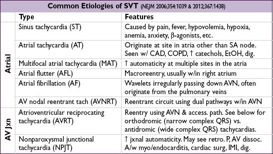

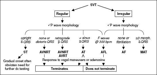
Figure 1-4 Approach to SVT (adapted from NEJM 2012;367:1438)
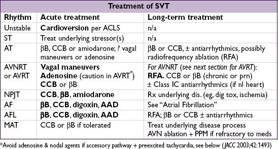
• Catheter ablation: high overall success rate (AFL/AVNRT ~95%, AVRT ~90%, AF ~80%)
Complications: stroke, MI, bleeding, perforation, conduction block (JAMA 2007;290:2768)
ACCESSORY PATHWAYS (WOLFF-PARKINSON-WHITE)
Definitions
• Accessory pathway (bypass tract) of conducting myocardium connecting atria & ventricles, allowing impulses to bypass normal AVN delay
• Preexcitation (WPW) pattern: ↓ PR interval, ↑ QRS width w/ Δ wave (slurred onset, can be subtle), ST & Tw abnl (can mimic old IMI); only seen w/ pathways that conduct antegrade (if pathway only conducts retrograde then
ECG will be normal during SR; “concealed” bypass tract)
PAC can exaggerate preexcitation if AV node conduction slowed
• WPW syndrome: accessory pathway + paroxysmal tachycardia

Classic tachycardias of WPW
• Orthodromic AVRT: narrow-complex SVT (typically), conducting ↓ AVN & ↑ accessory pathway; requires retrograde conduction and ∴ can occur w/ concealed bypass tracts
• Antidromic AVRT (rare): wide-complex SVT, conducting ↓ accessory pathway & ↑ AVN;
requires antegrade conduction and ∴ should see WPW pattern during SR
• AF w/ rapid conduction down accessory pathway; ∴ wide-complex irregular SVT; requires antegrade conduction; ∴ should see WPW pattern in SR. Rarely can degenerate into VF.
Treatment
• AVRT: vagal, bB, ? CCB; caution w/ adenosine (can precip. AF); have defibrillator ready
• AF/AFL w/ conduction down accessory pathway: need to Rx arrhythmia and ↑ pathway
refractoriness; use procainamide, ibutilide, amio, flecainide or DCCV; avoid CCB & bB (ineffective), dig/adenosine (can ↓ refractoriness of pathway → ↑ vent. rate → VF)
• Long term: Rx sx tachycardias w/ RFA, antiarrhythmics (IA, IC) if not candidate for RFA;
consider RFA if asx but AVRT or AF inducible on EPS (NEJM 2003;349:1803) of if rapid conduction possible (✓ w/ EPS if preexcitation persists despite exercise testing)
risk of SCD related to how short RR interval is in AF and if SVT inducible w/ exercise
WIDE-COMPLEX TACHYCARDIAS (WCTS)
Etiologies (Lancet 2012;380:1520)
• Ventricular tachycardia (VT)
• SVT conducted with aberrancy: either fixed BBB, rate-dependent BBB (usually RBBB), conduction via an accessory pathway or atrially triggered ventricular pacing
Monomorphic ventricular tachycardia (MMVT)
• All beats look similar; predominantly upward in V1 = RBBB-type vs. downward = LBBB-type

• In structurally abnormal heart: prior MI (scar); CMP; myocarditis;
arrhythmogenic RV CMP (ARVC): incomplete RBBB,
ε wave (terminal notch in QRS) & TWI in V1–V3 on resting ECG, LBBB-type VT, dx w/ MRI (Lancet 2009;373:1289)
• In structurally normal heart (w/ normal resting ECG):
RVOT VT: LBBB-type VT w/ inferior axis; typically ablate
idiopathic LV VT: RBBB-type VT w/ superior axis; responds to verapamil
Polymorphic ventricular tachycardia (PMVT)
• QRS morphology changes from beat to beat
• Etiologies: ischemia; CMP; catecholaminergic;
torsades de pointes (TdP = “twisting of the points,” PMVT + ↑ QT): ↑ QT acquired (meds, lytes, stroke, see "ECG") w/ risk ↑ w/ ↓ HR, freq PVCs (pause dependent) or congenital (K/Na channelopathies) w/ resting Tw abnl & TdP triggered by sympathetic stimulation (eg, exercise, emotion, sudden loud noises) (Lancet 2008;372:750).
Brugada syndrome (Na channelopathy):  >
>  ; pseudo-RBBB w/ STE in V1–V3 (provoked w/ class IA or IC) on resting ECG

Diagnostic clues that favor VT (assume until proven o/w)
• Prior MI, CHF or LV dysfunction best predictors that WCT is VT (Am J Med 1998;84:53)
• Hemodynamics and rate do not reliably distinguish VT from SVT
• MMVT is regular, but initially it may be slightly irregular, mimicking AF w/ aberrancy; grossly irregularly irregular rhythm suggests AF w/ aberrancy
• ECG features that favor VT (Circ 1991;83:1649)
AV dissociation (independent P waves, capture or fusion beats) proves VT
very wide QRS (>140 ms in RBBB-type or >160 in LBBB-type); extreme axis deviation
QRS morphology atypical for BBB RBBB-type: absence of tall R′ (or presence of monophasic R) in V1, r/S ratio <1 in V6 LBBB-type: onset to nadir >60–100 ms in V1, q wave in V6
concordance (QRS in all precordial leads w/ same pattern/direction)
Long-term management ( JACC 2006;48:1064)
• Workup: echo to ✓ LV fxn, cath or stress test to r/o ischemia, ? MRI and/or RV bx to
look for infiltrative CMP or ARVC, ? EP study to assess inducibility
• ICD: 2° prevention after documented VT/VF arrest (unless due to reversible cause)
1° prev. if high risk, eg, EF <30–35%, ARVC, Brugada, certain LQTS, severe HCMP. See “Intracardiac Devices.” ? Wearable vest if revers. etiol. waiting for ICD (Circ 2013;127:854).
Antitachycardia pacing (ATP = burst pacing faster than VT) can terminate VT w/o shock
• Meds: bB, antiarrhythmics (eg, amio, mexiletine) to suppress VT which could trigger shock
• If med a/w TdP → QT >500 ± VPBs: d/c med, replete K, give Mg, ± pacing (JACC 2010;55:934)
• Radiofrequency ablation if isolated VT focus or if recurrent VT triggering ICD firing; ablation before ICD implantation ↓ discharge rate by 40% (Lancet 2010;375:31)