
CARDIOLOGY
SYNCOPE
Definition
• Symptom of sudden transient loss of consciousness due to global cerebral hypoperfusion
• If CPR or cardioversion required, then SCD and not syncope (different prognosis)
Etiologies (NEJM 2002;347:878; JACC 2006;47:473; Eur Heart J 2009;30:2631)
• Neurocardiogenic (a.k.a. vasovagal, ~20%; NEJM 2005;352:1004): ↑ sympathetic tone → vigorous contraction of LV → mechanoreceptors in LV trigger ↑ vagal tone (hyperactive Bezold-Jarisch reflex) → ↓ HR (cardioinhibitory) and/or ↓ BP (vasodepressor)
cough, deglutition, defecation, & micturition → ↑ vagal tone and thus can be precipitants
related disorder: carotid sinus hypersensitivity (exagg vagal resp to carotid massage)
• Orthostatic hypotension (10%)
hypovolemia/diuretics, deconditioning; vasodilat. (esp. if combined w/  chronotropes)
autonomic neuropathy [1° = Parkinson’s, Shy-Drager, Lewy body dementia, POTS (dysautonomia in the young); 2° = DM, EtOH, amyloidosis, CKD] (NEJM 2008;358:615)
• Cardiovascular
Arrhythmia (15%)
Bradyarrhythmias: SSS, high-grade AV block, chronotropes, PPM malfunction
Tachyarrhythmias: VT, SVT (syncope rare unless structural heart disease or WPW)
Mechanical (5%)
Endocardial/Valvular: AS, MS, PS, prosthetic valve thrombosis, myxoma
Myocardial: pump dysfxn from MI or outflow obstruction from HCMP (but usually VT)
Pericardial: tamponade
Vascular: PE, PHT, aortic dissection, ruptured AAA, subclavian steal
• Neurologic (10%): seizure (technically not syncope), TIA/CVA, vertebrobasilar
insufficiency, dissection of cerebral arteries, migraine, narcolepsy
• Misc. causes of LOC (but not syncope): hypoglycemia, hypoxia, anemia, psychogenic
Workup (etiology cannot be determined in ~40% of cases)
• H&P incl. orthostatic VS have highest yield and most cost effective (Archives 2009;169:1299)
• History (from Pt and witnesses if available)
activity and posture before the incident
precipitating factors: exertion (AS, HCMP, PHT), positional Δ (orthostatic hypotension), stressors such as sight of blood, pain, emotional distress, fatigue, prolonged standing, warm environment, N/V, cough/micturition/defecation/swallowing (neurocardiogenic), head turning or shaving (carotid sinus hypersens.); arm exercise (subclavian steal)
prodrome (eg, diaphoresis, nausea, blurry vision): cardiac <~5 sec, vasovagal >~5 sec
associated sx: chest pain, palp., neurologic, postictal, bowel or bladder incontinence (convulsive activity for <10 sec may occur w/ transient cerebral HoTN & mimic seizure)
• PMH: prior syncope, previous cardiac or neurologic dis.; no CV disease at baseline → 5% cardiac, 25% vasovagal; CV disease → 20% cardiac, 10% vasovagal (NEJM 2002;347:878)
• Medications that may act as precipitants
vasodilators: a-blockers, nitrates, ACEI/ARB, CCB, hydralazine, phenothiazines, antidep.
diuretics; chronotropes (eg, bB and CCB)
proarrhythmic or QT prolonging: class IA, IC or III antiarrhythmics (see “ECG”)
psychoactive drugs: antipsychotics, TCA, barbiturates, benzodiazepines, EtOH
• Family history: CMP, SCD, syncope (vasovagal may have genetic component)
• Physical exam
VS including orthostatics ( if supine → standing results in >20 mmHg ↓ SBP, >10 mmHg ↓ DBP, or >10–20 bpm ↑ HR), BP in both arms
cardiac: HF (↑ JVP, displ. PMI, S3), murmurs, LVH (S4, LV heave), PHT (RV heave, ↑ P2)
vascular: ✓ for asymmetric pulses, carotid/vertebral/subclavian bruits; carotid sinus massage to assess for carotid hypersensitivity (if no bruits)
neurologic exam: focal findings, evidence of tongue biting; FOBT
• ECG (abnormal in ~50%, but only definitively identifies cause of syncope in ~10%)
Conduction: SB, sinus pauses/sinus arrhythmia, AVB, BBB/IVCD
Arrhythmia: ectopy, ↓ QT, preexcitation (WPW), Brugada, e wave (ARVC), SVT/VT
Ischemic changes (new or old): atrial or ventricular hypertrophy
Other diagnostic studies (consider based on results of H&P and ECG)
• Ambulatory ECG monitoring: if suspect arrhythmogenic syncope
Holter monitoring (continuous ECG 24–48 h): useful if frequent events arrhythmia + sx (4%); asx but signif. arrhythmia (13%); sx but no arrhythmia (17%)
Event recorder (activated by Pt to record rhythm strip): limited role as only useful if established prodrome (because must be Pt activated)
Loop recorders (continuously saves rhythm, ∴ can be activated after an event): useful for episodes (including w/o prodrome) likely to occur w/in month.
Implantable loop recorders (inserted SC; can record up to 3 y): useful for infrequent episodes (<1/mo); recommended for recurrent syncope w/o prodrome
• Echo: consider to r/o structural heart disease (eg, CMP [incl HCMP & ARVC], valvular disease [incl AS, MS, MVP], myxoma, amyloid, PHT, ± anomalous coronaries)
• ETT: esp. w/ exertional syncope; r/o ischemia- or catecholamine-induced arrhythmias
• Cardiac catheterization: consider if noninvasive tests suggest ischemia
• Electrophysiologic studies (EPS): consider in high-risk Pts in whom tachy or brady etiology is strongly suspected, but cannot be confirmed;
50% abnl (inducible VT, conduction abnormalities) if heart disease, but ? significance
3–20% abnl if abnl ECG; <1% abnl if normal heart and normal ECG (Annals 1997;127:76)
• ? Tilt table testing: utility is debated due to poor Se/Sp/reproducibility; consider only if vasovagal dx suspected but can’t be confirmed by hx
• Cardiac MRI: helpful to dx ARVC if suggestive ECG, echo (RV dysfxn) or FHx of SCD
• Neurologic studies (cerebrovascular studies, CT, MRI, EEG): if H&P suggestive; low yield
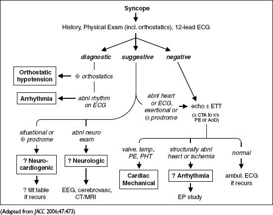
Figure 1-6 Approach to syncope
High-risk features (usually admit w/ telemetry & testing; J Emerg Med 2012;42:345)
• Age >60 y, h/o CAD, HF/CMP, valvular or congenital heart dis., arrhythmias, FHx SCD
• Syncope c/w cardiac cause (lack of prodrome, exertional, resultant trauma) or recurrent
• Complaint of chest pain or dyspnea; abnl VS or cardiac exam
• ECG suggesting tachy or brady-induced syncope; Pt w/ PPM/ICD
Treatment
• Arrhythmia, cardiac mechanical or neurologic syncope: treat underlying disorder
• Vasovagal syncope: no proven benefit for midodrine, fludrocortisone, disopyramide, SSRI ? 16 oz of H2O before at-risk situations (Circ 2003;108:2660)
no proven benefit w/ bB (Circ 2006;113:1164)
? benefit w/ PPM if ≥3 episodes/2y & loop recorder w/ asystole >3 sec (Circ 2012;125:2566)
• Orthostatic syncope: volume replete (eg, 500 mL PO q a.m.); if chronic → rise from supine to standing slowly, compressive stockings, midodrine, fludrocortisone, high Na diet
Prognosis (Ann Emerg Med 1997;29:459; NEJM 2002;347:878)
• 22% overall recurrence rate if idiopathic, else 3% recurrence
• Cardiac syncope: 2-fold ↑ in mort., 20–40% 1-y SCD rate, median survival ~6 y
• Unexplained syncope w/ 1.3-fold ↑ in mort., but noncardiac or unexplained syncope w/ nl
ECG, no h/o VT, no HF, age <45 → low recurrence rate and <5% 1-y SCD rate
• Vasovagal syncope: Pts not at increased risk for death, MI or stroke
• ✓ state driving laws and MD reporting requirements. Consider appropriateness of Pt involvement in exercise/sport, operating machinery, high-risk occupation (eg, pilot).