
GASTROENTEROLOGY
ABNORMAL LIVER TESTS
Tests of hepatocellular injury or cholestasis
• Aminotransferases (AST, ALT): intracellular enzymes released 2° necrosis/inflammation
ALT more specific for liver than is AST (heart, skeletal muscle, kidney, brain, RBC/WBC)
ALT > AST → viral hepatitis or fatty liver/nonalcoholic steatohepatitis (precirrhotic)
AST: ALT >2:1 → alcoholic hepatitis, cirrhosis; nonhepatic source
ALT/AST >15× ULN → etiologies of acute liver failure (↑↑↑ LDH → ischemia/toxic)
• Alkaline phosphatase (AΦ): enzyme bound in hepatic canicular membrane
besides liver, also found in bone, intestines, kidney and placenta
confirm liver origin with: ↑ 5′-NT, ↑ GGT or AΦ heat fractionation
↑ levels seen with biliary obstruction or intrahepatic cholestasis (eg, hepatic infiltration)
Tests of hepatic function
• Albumin: marker for liver protein synthesis, ↓ slowly in liver failure (t1/2 ~20 d)
• Prothrombin time (PT): depends on synthesis of coag factors by liver (except FVIII); b/c t1/2 of some factors (eg, V, VII) is short, ↑ PT can occur w/in hrs of liver dysfxn
• Bilirubin: product of heme metab (unconjugated, “indirect”) carried by alb to liver where taken up for conjugation (“direct”) to make soluble, then excreted into bile; most sensitive test to detect parenchymal disease; in those w/ normal LFTs, high nl Tbili (? marker of ↑ heme oxygenase) a/w ↓ resp disease & death (JAMA 2011;305:691)
Patterns of liver injury
• Hepatocellular: ↑↑ aminotransferases, ± ↑ bilirubin or AΦ
↑↑↑ ALT & AST (>1000): severe viral hepatitis, drugs, ischemia, Wilson’s, AIH
• Cholestasis: ↑↑ AΦ and bilirubin, ± ↑ aminotransferases
• Isolated hyperbilirubinemia: ↑↑ bilirubin (direct or indirect), nl AΦ and aminotransferases
• Infiltrative: ↑ AΦ, ± ↑ bilirubin or aminotransferases
• Jaundice is a clinical sign seen when bilirubin >2.5 mg/dL (esp. in sclera or under
tongue); if hyperbilirubinemia conjugated → ↑ urine bilirubin
Figure 3-3 Approach to abnormal liver tests with hepatocellular pattern
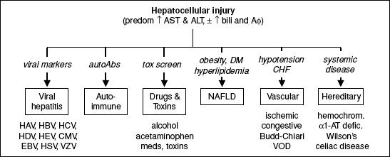
• Acute workup: toxins (EtOH, acetaminophen); vascular abnl (U/S w/ Doppler); viral tests: IgM anti-HAV, HBsAg, IgM anti-HBc, HBV DNA, HCV RNA, anti-HEV, ± EBV, CMV, HSV, VZV;autoimmune (ANA, ASMA, ALKM); ceruloplasmin
• Chronic workup: HBsAg, anti-HCV; Fe, TIBC; glc, HbA1c, TG; ANA, ASMA, ALKM; anti-tissue transglutaminase; ceruloplasmin & ɑ1-AT; TSH; vascular abnl (U/S w/ Doppler)
Figure 3-4 Approach to abnormal liver tests with cholestatic pattern

Figure 3-5 Approach to abnormal liver tests with isolated hyperbilirubinemia

Figure 3-6 Approach to abnormal liver tests with infiltrative pattern

Abnormal liver tests in asymptomatic patients (Clin Liver Dis 2009;13:167)
• Careful review of history (meds, EtOH/drug use, exposures, risk factors for liver disease) and physical exam. Evaluate for any clues to etiology 1st (eg, d/c med and repeat LFTs).
• Confirm hepatic source: if primarily ↑ AΦ (✓ GGT) or AST > ALT (✓ CK, aldolase, TFT)
• Hepatocellular
Evaluate for most common causes: hepatitis A/B/C, hemochromatosis; screen for evidence of chronic liver disease (platelets, PT/INR, albumin)
If  evaluation → lifestyle modification (wt loss, DM control) + repeat test 6 mo
If evidence of chronic liver disease or persistent lab abnl, screen for less common causes: AIH, Wilson’s, celiac, ɑ1-AT; ✓ U/S & consider liver bx
If still → liver bx if ALT or AST >2× ULN for >6 mo; o/w observe
• Cholestatic: ✓ RUQ U/S, AMA
if biliary dilatation or obstruction → MRCP
if AMA  and U/S , or AMA and U/S w/ abnl parenchyma → liver bx
if AMA & U/S : AΦ >1.5× ULN → consider bx; AΦ <1.5× ULN → observe
• Isolated hyperbilirubinemia: ✓ conjugated vs. unconjugated
conjugated → perform abdominal U/S → MRCP if dilatation or obstruction; if nl ultrasound ✓ AMA and consider MRCP or liver bx
unconjugated → ✓ Hct, retic count, smear, LDH, haptoglobin
Common medications that cause abnormal liver tests (http://livertox.nlm.nih.gov)
