
NEPHROLOGY
POTASSIUM HOMEOSTASIS
Overview (Annals 2009;150:619)
• Renal: potassium excretion regulated at distal nephron (collecting tubule) distal Na delivery & urine flow: Na absorption → lumen electronegative → K secretion metabolic alkalemia and aldosterone: increase Na absorption and K secretion
• Transcellular shifts: most common cause of acute change in serum potassium Acid-base disturbance: K+/H+ exchange across cell membranes Insulin → stimulates Na-K ATPase → hypokalemia (mitigates postprandial ↑ K) Catecholamines → stimulate Na-K ATPase → hypokalemia; reversed by b-blockers Digoxin → blocks Na-K ATPase → hyperkalemia Massive necrosis (eg, tumor lysis, rhabdo, ischemic bowel) → release of intracellular K Hypo- or hyperkalemic periodic paralysis: rare disorders due to channel mutations
• Diet: alone rarely causes ↑ or ↓ K (total body store ~3500 mEq, daily intake ~100 mEq)
HYPOKALEMIA
Transcellular shifts
• Alkalemia, insulin, catecholamines, hypokalemic/thyrotoxic periodic paralysis, acute ↑ in hematopoiesis (megaloblastic anemia Rx w/ B12, AML crisis), hypothermia, chloroquine, barium/cesium intoxication, antipsychotic overdose (risperidone, quetiapine)
GI potassium losses (UK<25 mEq/d or <5 mEq/L or TTKG <3)
• GI losses plus metabolic acidosis: diarrhea, laxative abuse, villous adenoma
• Vomiting & NGT drainage usually manifest as renal losses due to 2° hyperaldo & met. alk.
Renal potassium losses (UK>30 mEq/d or >15 mEq/L or TTKG >7)
• Hypotensive or normotensive acidosis: DKA, RTA [proximal RTA (type II) and some distal RTAs (type I)] alkalosis: diuretics, vomiting/NGT drainage (via 2° hyperaldosteronism) Bartter’s syndrome (loop of Henle dysfxn → furosemide-like effect; NEJM 1999;340:1177)
Gitelman’s syndrome (distal convoluted tubule dysfxn → thiazide-like effect)
↓ Mg: ? release Mg-mediated inhib. of ROMK channel ∴ ↑ K secretion (JASN 2007;18:2649)
• Hypertensive: mineralocorticoid excess
1° hyperaldosteronism (eg, Conn’s syndrome, glucocorticoid-remediable aldosteronism)
2° hyperaldosteronism (eg, renovascular disease, renin-secreting tumor)
nonaldosterone mineralocorticoid (eg, Cushing’s, Liddle’s, exogenous mineralocort., licorice, congenital adrenal hyperplasia)
Clinical manifestations
• Nausea, vomiting, ileus, weakness, muscle cramps, rhabdomyolysis, polyuria
• ECG: U waves, ± ↑ QT interval, ventricular ectopy (PVCs, VT, VF)
Workup (NEJM 1998;339:451)
• Rule-out transcellular shifts
• ✓ 24-h UK and transtubular potassium gradient (TTKG) = (UK/PK) / (Uosm/Posm)
UK >30 mEq/d or >15 mEq/L or TTKG >7 → renal loss
UK <25 mEq/d or <15 mEq/L or TTKG <3 → extrarenal loss
• If renal losses, ✓ BP, acid-base, UCl (UNa unreliable for volume status w/ alkalemia)
Figure 4-7 Approach to hypokalemia

Treatment
• If true potassium deficit: potassium repletion (↓ 1 mEq/L  200 mEq total body loss) KCl 40 mEq PO q4–6h if nonurgent, KCl 10 mEq/h IV if urgent, recheck K freq
• Beware of excessive potassium repletion if transcellular shift cause of hypokalemia
• Treat underlying cause (if hydration needed, avoid dextrose-containing solutions as dextrose → ↑ insulin → intracellular potassium shifts)
• Replete low Mg: IV Mg-SO4 1–2 g q2h (oral Mg-oxide poorly tolerated b/c diarrhea) Causes of low Mg: GI loss (diarrhea, bypass, pancreatitis, malnutrition, PPI); renal loss (diuretics, nephrotoxic drugs, EtOH, ↑ Ca, 1° wasting syndromes, volume expansion)
HYPERKALEMIA
Transcellular shifts (BMJ 2009;339:1019)
• Acidemia, insulin defic. (DM), b-blockers, dig intox., massive cellular necrosis (tumorlysis, rhabdo, ischem. bowel, hemolysis), hyperkalemic periodic paralysis, succinylcholine
Decreased GFR
• Any cause of oliguric or anuric AKI or any cause of end stage renal disease
Normal GFR but with Ø renal K excretion
• Normal aldosterone function
↓ EAV (K excretion limited by ↓ distal Na delivery & urine flow): CHF, cirrhosis excessive K intake: in conjunction with impairment in K excretion or transcellular shift ureterojejunostomy (absorption of urinary K in jejunum)
• Hypoaldosteronism: same as etiologies of hypoaldo RTA (type IV)
↓ renin: diabetic nephropathy, NSAIDs, chronic interstitial nephritis, HIV normal renin, ↓ aldo synthesis: 1° adrenal disorders, ACEI, ARBs, heparin
↓ response to aldosterone meds: K-sparing diuretics, TMP-SMX, pentamidine, calcineurin inhibitors tubulointerstitial disease: sickle cell, SLE, amyloid, diabetes
Clinical manifestations
• Weakness, nausea, paresthesias, palpitations
• ECG: peaked T waves, ↑ PR interval, ↑ QRS width, loss of P wave, sine wave pattern, PEA/VF (ECG: low sensitivity, cardiac arrest can be first electrical manifestation!)
Workup (Crit Care Med 2008;36:3246)
• Rule out pseudohyperkalemia (IVF with K, hemolysis during venipuncture, ↑ plt or WBC)
• Rule out transcellular shift
• Assess GFR, if normal: Consider ↓ distal Na delivery and urine flow
✓ transtubular K gradient (TTKG) = (UK/PK)/(Uosm/Posm) <6 c/w hypoaldo (JASN 2008;19:424)
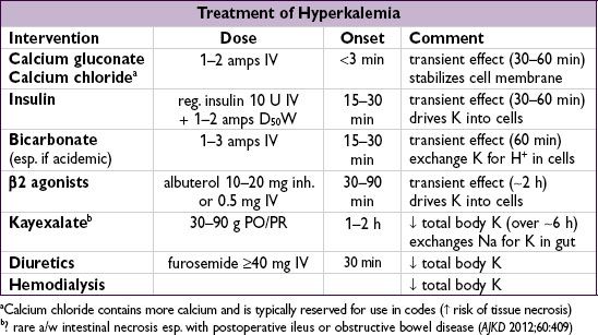
• Rate of onset important to note when establishing a treatment plan
• Calcium helps prevent/treat cardiac complications; ∴ should be initial Rx, esp. if ECG Ds
• Insulin, bicarbonate (esp. if acidemic), and b2 agonists should follow to ↓ plasma K
• Treatments that eliminate total body K essential as other Rxs will wear off with time; Kayexalate ± diuretics may be effective in many cases, but emergent hemodialysis should be considered in life-threatening situations
• Patient information for diet education: http://www.kidney.org/atoz/content/potassium.cfm