HEMATOLOGY-ONCOLOGY
ANEMIA
↓ in RBC mass: Hct <41% or Hb <13.5 g/dL (men); Hct <36% or Hb <12 g/dL (women)
Clinical manifestations
• Symptoms: ↓ O2 delivery → fatigue, exertional dyspnea, angina (if CAD)
• Signs: pallor (mucous membranes, palmar creases), tachycardia, orthostatic hypotension
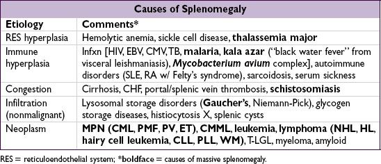
• Other findings: jaundice (hemolysis), splenomegaly (thalassemia, neoplasm, chronic hemolysis), petechiae/purpura (bleeding disorder), glossitis (iron, folate, vitamin B12 defic.), koilonychia (iron defic.), neurologic abnormalities (B12 defic.)
Diagnostic evaluation
• History: bleeding, systemic illness, drugs, exposures, alcohol, diet (including pica), FHx
• CBC w/ diff.; RBC params incl. retics, MCV (nb, mixed disorder can → nl MCV), RDW
• Reticulocyte index (RI) = [reticulocyte count × (Pt’s Hct/nl Hct)]/maturation factor maturation factors for a given Hct: 45% = 1, 35% = 1.5, 25% = 2, 20% = 2.5
RI >2% → adequate marrow response; RI <2% → hypoproliferation
• Peripheral smear: select area where RBCs evenly spaced and very few touch each other; ✓ RBC size, shape, inclusions (see Appendix & Peripheral Smear inserts), WBC morphology, plt count
• Additional labs as indicated: hemolysis labs (if RI >2%), iron/TIBC, ferritin, folate, B12, LFTs, BUN and Cr, TFTs, Hb electrophoresis, enzyme analyses, gene mutation screens
• Bone marrow (BM) aspirate and biopsy (bx) with cytogenetics as indicated
Figure 5-1 Approach to anemia

MICROCYTIC ANEMIAS
Figure 5-2 Approach to microcytic anemias

Iron deficiency (NEJM 1999;341:1986; Gut 2011;60:1309)
• ↓ marrow iron & depleted body iron stores → ↓ heme synthesis → microcytosis → anemia
• Special clinical manifestations: angular cheilosis, atrophic glossitis, pica (consumption of nonnutritive substances such as ice, clay), koilonychia (nail spooning) Plummer-Vinson syndrome (iron deficiency anemia, esophageal web & atrophic glossitis)
• Etiologies: chronic bleeding (GI—incl. cancer, menstrual, parasites, etc.), ↓ supply (malnutrition; ↓ absorp. due to celiac sprue, Crohn’s, ↑ gastric pH, subtotal gastrectomy), ↑ demand (preg., Epo). Rare Fe refractory genetic disorder due to hepcidin dysregulation (Nat Genet 2008;40:569).
• Diagnosis: ↓ Fe, ↑ TIBC, ↓ ferritin (esp. <15), ↓ transferrin sat (Fe/TIBC; esp. <15%), ↑ soluble transferrin receptor; ↑ plt; unless hx c/w other etiology, initiate workup for GIB; incl. H. pylori serology, ? celiac sprue labs (anti-TTG, antigliadin, antiendomysial Ab)
• Treatment (Fe supplementation): oral Fe tid (~6 wk to correct anemia; ~6 mo to replete Fe stores); in cases of excessive/persistent GI losses or for dialysis or cancer Pts prior to Epo Rx, IV iron (Fe-sucrose, -gluconate, -dextran) should be considered
Thalassemias (Lancet 2013;379:373)
• ↓ synthesis of ɑ- or β-globin chains of Hb → ≠ subunits → destruction of RBCs and erythroid precursors; ∴ anemia from hemolysis and ineffective erythropoiesis
• ɑ-thalassemia: deletions in ɑ-globin gene complex on chr. 16 (nl 4 ɑ genes)
3 ɑ → ɑ-thal-2 trait = silent carrier; 2 ɑ → ɑ-thal-1 trait or ɑ-thal minor = mild anemia
1 ɑ → HbH (β4) disease = severe anemia, hemolysis and splenomegaly
0 ɑ genes → Hb Barts (γ4) = intrauterine hypoxia and hydrops fetalis
• β-thalassemia: mutations in β-globin gene on chr. 11 → absent or ↓ gene product
1 mutated β gene → thal minor (or trait) = mild anemia (no transfusions)
2 mutated β genes → thal intermedia (occasional transfusions) or thal major ( = Cooley’s anemia; transfusion dependent) depending on severity of mutations
• Special clinical manifestations (in severe cases): chipmunk facies, pathologic fractures, hepatosplenomegaly (due to extramedullary hematopoiesis), high-output CHF, bilirubin gallstones, iron overload syndromes (from chronic transfusions)
• Diagnosis: MCV <70, normal Fe, MCV/RBC count<13 [Mentzer Index, 60% Se, 98% Sp; (Ann Hem 2007;86:486)], ± ↑ retics, basophilic stippling; Hb electrophoresis: ↑ HbA2 (ɑ2δ2) in β-thal;normal pattern in ɑ-thal trait
• Treatment: folate; transfusions + deferoxamine, deferasirox (oral iron chelator); splen-ectomy if ≥50% ↑ transfusions; consider allo-HSCT in children w/ severe β-thal major
Anemia of chronic inflammation (see below)
Sideroblastic anemia
• Defective heme biosynthesis within RBC precursors
• Etiologies: hereditary/X-linked (ALAS2 mutations), idiopathic, MDS-RARS, reversible (alcohol, lead, isoniazid, chloramphenicol, copper deficiency, hypothermia)
• Special clinical manifestations: hepatosplenomegaly, iron overload syndromes
• Dx: review social, work & TB hx; can be microcytic, normocytic or macrocytic; variable pop of hypochromic RBCs; ↑ Fe, nl TIBC, ↑ ferritin, basophilic stippling, RBC Pappenheimer bodies (Fe-containing inclusions), ring sideroblasts (w/ iron-laden mitochondria) in BM
• Treatment: treat reversible causes; trial of pyridoxine, supportive transfusions for severe anemia; high-dose pyridoxine for some hereditary cases
NORMOCYTIC ANEMIAS
Pancytopenia (see below)
Anemia of chronic inflammation (ACI; NEJM 2005;352:1011; 2009;361:1904)
• ↓ RBC production due to impaired iron utilization and functional iron deficiency from ↑ hepcidin; cytokines (IL-6, TNF-a) cause ↓ Epo responsiveness/production
• Etiologies: autoimmune disorders, chronic infection, inflammation, HIV, malignancy
• Dx: ↓ Fe, ↓ TIBC (usually normal or low transferrin sat), ± ↑ ferritin; usually normochromic, normocytic (~70% of cases) but can be microcytic if prolonged
• Coexisting iron deficiency common. Dx clues include ↓ serum ferritin levels, absence of iron staining on BM bx,  response to a trial of oral iron and/or ↑ soluble transferrin receptor/ferritin index (Blood1997;89:1052).
• Treatment: treat underlying disease ± iron and/or erythropoiesis-stimulating agent (ESA, eg, Epo). Iron if ferritin <100 or Fe/TIBC <20%. Consider ESA if Epo <500. Avoid ESA in cancer if treatment goal is cure (Leuk Res 2012;36:939). Unclear if one should treat highly sx Pts w/ goal Hb 10–12 g/dL; weigh risk of thrombosis.
Anemias of chronic disorders
• Anemia of chronic inflammation (see above)
• Anemia of chronic kidney disease: ↓ Epo; treat w/ Epo (see “Chronic Kidney Disease”)
• Endocrine deficiencies: hypometabolism and ↓ O2 demand with thyroid, pituitary, adrenal, or parathyroid disease → ↓ Epo; can be normocytic or macrocytic
Sideroblastic anemia (see above)
Pure red cell aplasia
• Destructive antibodies or lymphocytes → ineffective erythropoiesis
• Associated with thymoma, CLL and parvovirus infection
• Diagnostic studies: lack of erythroid precursors on BM bx, other lines normal
• Treatment: thymectomy if thymus enlarged; IVIg if parvovirus infection; immunosuppression if CLL or idiopathic; supportive care with PRBC transfusions; ? erythropoietin receptor agonist if due to antierythropoietin Ab (NEJM 2009;361:1848)
MACROCYTIC ANEMIAS
includes megaloblastic and nonmegaloblastic causes
Megaloblastic anemia
• Impaired DNA synthesis → cytoplasm matures faster than nucleus → ineffective erythropoiesis and macrocytosis; due to folate or B12 deficiency; MDS
• ✓folate and vitamin B12; ↑ LDH & indirect bilirubin (due to ineffective erythropoiesis)
• Smear: neutrophil hypersegmentation, macro-ovalocytes, anisocytosis, poikilocytosis
Folate deficiency
• Folate present in leafy green vegetables and fruit; total body stores sufficient for 2–3 mo
• Etiologies: malnutrition (alcoholics, anorectics, elderly), ↓ absorption (sprue), impaired metabolism (methotrexate, pyrimethamine, trimethoprim), ↑ requirement (chronic hemolytic anemia, pregnancy, malignancy, dialysis)
• Diagnosis: ↓ folate; ↓ RBC folate, ↑ homocyst. but nl methylmalonic acid (unlike B12 defic.)
• Treatment: folate 1–5 mg PO qd for 1–4 mo or until complete hematologic recovery; critical to r/o B12 deficiency first (see below)
Vitamin B12 deficiency (NEJM 2013;368:149)
• B12 present only in foods of animal origin; total body stores sufficient for 2–3 y
• Binds to intrinsic factor (IF) secreted by gastric parietal cells; absorbed in terminal ileum
• Etiologies: malnutrition (alcoholics, vegans), pernicious anemia (PA, autoimmune dis- ease against gastric parietal cells, a/w polyglandular endocrine insufficiency and ↑ risk of gastric carcinoma), other causes of ↓ absorption (gastrectomy, sprue, Crohn’s disease), ↑ competition (intestinal bacterial overgrowth, fish tapeworm)
• Clinical manifestations: neurologic changes (subacute combined degeneration) affecting peripheral nerves, posterior and lateral columns of the spinal cord and cortex → numbness, paresthesias, ↓ vibratory and positional sense, ataxia, dementia
• Dx: ↓ B12; ↑ homocysteine and methylmalonic acid; anti-IF Ab; Schilling test; ↑ gastrin in PA
• Treatment: 1 mg B12 IM qd × 7 d → q wk × 4–8 wk → q month for life neurologic abnormalities are reversible if treated w/in 6 mo folate can reverse hematologic abnormalities of B12 deficiency but notneurologic changes (and can lead to “steal” of B12 stores → worsening of neuro complications) oral supplementation (2 mg qd) appears feasible as well (Blood 1998;92:1191) even w/o IF
Nonmegaloblastic macrocytic anemias
• Liver disease: often macrocytic, may see target cells
• Alcoholism: BM suppression & macrocytosis independent of folate/B12 defic. or cirrhosis
• Reticulocytosis
• Other causes: hypothyroidism; MDS; meds that impair DNA synthesis (zidovudine, 5-FU, hydroxyurea, Ara-C); hereditary orotic aciduria; Lesch-Nyhan syndrome.
PANCYTOPENIA
Etiologies
• Hypocellular bone marrow (nl cellularity ~100 – age): aplastic anemia, hypoplastic MDS
• Cellular bone marrow: MDS, aleukemic leukemia, PNH, severe megaloblastic anemia
• Marrow replacement (myelophthisis): myelofibrosis, metastatic solid tumors, granulomas
• Systemic diseases: hypersplenism, sepsis, alcohol, toxins
Clinical manifestations
• Anemia → fatigue
• Neutropenia → recurrent infections
• Thrombocytopenia → mucosal bleeding & easy bruisability
Aplastic anemia = stem cell failure (Lancet 2005;365:1647; Blood 2012;120:1185)
• Epidemiology: 2–5 cases/106/y; biphasic (major peak in adolescents, 2nd peak in elderly)
• Diagnosis: pancytopenia w/ ↓ retics, BM bx w/ cytogenetics showing hypocellularity
• Etiologies: idiopathic (1/2 –1/3 of cases)
stem cell destruction: radiation, chemotherapy, chemicals (eg, benzene) idiosyncratic med rxn (eg, chloramphenicol, NSAIDs, sulfa drugs, gold, carbamazepine, antithyroid)
viruses (HHV-6, HIV, EBV, parvovirus B19); also post-hepatitis (non A, B or C)
immune disorders (SLE, GVHD post-HSCT, thymoma)
PNH (see below); Fanconi’s anemia (congenital disorder w/ pancytopenia, macrocytic anemia, ↑ risk of MDS, AML, & SCC of head & neck, and multiple physical anomalies);
shortened telomeres: seen w/ telomerase (TERT, TERC) mut (10% of aplastic anemia), dyskeratosis congenita/DKC1 mut; a/w IPF, cirrhosis (NEJM 2009;361:2353)
• Treatment and prognosis
allogeneic HSCT: for young Pts → ~80% long-term survival and significantly ↓ risk of malignant evolution, but has risk of transplant-related morbidity & mortality; if possible avoid transfusions (and alloimmunization) pretransplant
immunosuppression (CsA/tacrolimus, ATG): 70–80% respond, with 80–90% 5-y survival in responders (96% vs. 76% w/ horse vs. rabbit ATG; NEJM 2011;365:430); 15–20% 10-y incidence of clonal disorders (mostly MDS, AML, PNH)
TPO mimetics (eg, eltrombopag) may be option in refractory disease (NEJM 2012;367:11)
supportive care: transfusions, antibiotics, possible utility of G-CSF and Epo
Myelodysplastic syndromes (MDS) (qv)
Paroxysmal nocturnal hemoglobinuria (PNH) (Blood 2009;113:6522)
• Acquired clonal stem cell disorder = inactivating somatic mutation of PIG-A gene → deficiency of GPI-anchor for CD55 & CD59 (inhib of complement) → complement-mediated RBC lysis, plt aggreg., & hypercoagulability
• Clinical: intravascular hemolytic anemia, hypercoagulability (venous > arterial; esp. intraabdominal, cerebral), smooth muscle dystonias, deficient hematopoiesis (cytopenias); a/w aplastic anemia, MDS and evolution to AML
• Dx: flow cytometry (↓ CD55 & CD59) on RBCs and granulocytes; urine hemosiderosis
• Treatment: supportive care (iron, folate, transfusions); consider anti-coagulation allogeneic HSCT for hypoplasia or severe thrombosis eculizumab (Ab inactivates terminal complement C5s): ↓ hemolysis, improves QoL & stabilizes Hb levels (NEJM 2004;350:552 & 2006;355:1233; Lancet 2009;373:759); must have meningococcal vaccination
Myelophthisic anemia (see also “Primary Myelofibrosis”)
• Infiltration of bone marrow by cancer, leukemia, infection, fibrosis (primary myelofi- brosis), granulomas, lysosomal storage disorders
HEMOLYTIC ANEMIAS

Diagnostic evaluation
• ↑ reticulocyte count (RI >2%), ↑ LDH, ↓ haptoglobin (83% Se, 96% Sp), ↑ indirect bili
• Autoimmune hemolysis: Coombs’ test = direct antiglobulin test (DAT) → if agglutination occurs when antisera against Ig or C3 are applied to patient RBCs
• Intravascular: ↑↑ LDH, ↓↓ haptoglobin; hemoglobinemia, hemoglobinuria, hemosiderinuria
• Extravascular: splenomegaly
• Family h/o anemia; personal or family h/o cholelithiasis
Glucose-6-phosphate dehydrogenase (G6PD) deficiency (Lancet 2008;371:64)
• X-linked defect of metabolism (G6PD mutations) w/ ↑ susceptibility to oxidative damage
• Most common in  of African or Mediterranean descent (malaria-endemic areas)
• Hemolysis precipitated by drugs (sulfonamides, dapsone, primaquine, doxorubicin, methylene blue), infection, DKA or foods (fava beans in children)
• Diagnosis: smear may show RBC Heinz bodies (oxidized Hb) that result in bite cells once removed by spleen; ↓ G6PD levels (may be normal after acute hemolysis as older RBCs have already lysed and young RBCs may still have near normal levels)
Sickle cell anemia (Lancet 2010;376:2018)
• Recessive β-globin mutation → structurally abnl hemoglobin (HbS). ~8% African Americans heterozygotes (“sickle trait”; usually w/o sx); ~1/400 homozygotes (sickle cell disease).
• ↓ O2 → HbS polymerizes → RBC sickles, ↓ RBC deformability → hemolysis & microvascular occlusion
• Anemia: chronic hemolysis ± acute aplastic (parvo. B19) or splenic sequestration crises
• Vaso-occlusion and infarction: painful crises, acute chest syndrome, CVA, splenic sequestration, hand-foot syndrome, renal papillary necrosis, aseptic necrosis, priapism
• Infection: splenic infarction → overwhelming infection by encapsulated organisms; infarcted bone → osteomyelitis (Salmonella, Staph. aureus)
• Diagnosis: sickle-shaped RBCs and Howell-Jolly bodies on smear; Hb electrophoresis
• Treatment: hydroxyurea causes ↑ HbF → ↓ painful crises, acute chest episodes and may ↓ mortality (NEJM 2008;358:1362); allogeneic HSCT may have a role in young Pts w/ severe disease (Blood2000;95:1918) and adults (NEJM 2009;361:2309)
• Supportive care: folic acid qd; pneumococcal, meningococcal, H. flu & HBV vaccination; pain crises treated with hydration, oxygen and analgesia; simple or exchange transfusion for TIA or stroke, severe acute chest syndrome, or preop (goal Hb 10 g/dL)
Hereditary spherocytosis (HS) (Br J Hematol 2004;126:455)
• Defect in a cytoskeletal protein of RBC membrane → membrane loss mutations in ankyrin, a- and β-spectrin, band 3 and pallidin have been identified
• Most common in N. European populations (1/5000 births); FHx (75% of Pts)
• Anemia, jaundice (mostly neonates), splenomegaly, pigmented gallstones
• Diagnosis: spherocytes on smear, osmotic fragility test (~80% Se), ↓ eosin-5-maleimide (EMA) binding (92% Se; 99% Sp)
• Treatment: folate, transfusions, splenectomy for moderate and severe HS (balance w/ ↑ risk of future thrombosis and infection (J Thromb Haemost 2008;6:1289)
Paroxysmal nocturnal hemoglobinuria (see above)
Autoimmune hemolytic anemia (AIHA)
• Acquired, antibody-mediated RBC destruction
• Warm AIHA: IgG Abs opsonize RBCs at body temp → removal by spleen Etiologies: idiopathic, lymphoproliferative (CLL, NHL), autoimmune (SLE), drugs
• Cold AIHA: IgM Ab binds to RBCs at temp <37°C → complement fixation → intravascular hemolysis and acrocyanosis on exposure to cold
Etiologies: idiopathic, lymphoprolif. disorders (eg, Waldenström’s; monoclonal), Mycoplasma pneumoniae infxn and infectious mononucleosis (polyclonal)
• Diagnosis: spherocytes on smear, Coombs’; ✓ cold agglutinin titer, splenomegaly
• Treatment: treat underlying disease
warm AIHA: corticosteroids ± splenectomy, IVIg, cytotoxic agents, rituximab
cold AIHA: avoid cold; steroids ineffective; rituximab (Blood 2004;103:2925)
Drug-induced hemolytic anemia
• Acquired, antibody-mediated, RBC destruction precipitated by a medication:
abx: cephalosporins, sulfa drugs, rifampin, ribavirin
CV: methyldopa, procainamide, quinidine, thiazides
TCAs, phenothiazines, NSAIDs, sulfonylureas, MTX, 5-FU, rasburicase (G6PD defic.)
• Diagnosis: Coombs’ usually negative, ↑ LDH
• Treatment: discontinue offending agent
Microangiopathic hemolytic anemia (MAHA)
• Intra-arteriolar fibrin damages RBCs → acquired intravascular hemolysis
• Etiologies: hemolytic-uremic syndrome (HUS), thrombotic thrombocytopenic purpura (TTP), disseminated intravascular coagulation (DIC), malignancy, malignant HTN, eclampsia/HELLP, mech. cardiac valves, infected vascular prostheses
• Diagnosis: schistocytes ± thrombocytopenia ± abnormalities a/w specific disorders (eg, ↑ PT in DIC, ↑ Cr in HUS, ↑ LFTs in HELLP)
• Treatment: treat underlying abnormality; urgent plasma exchange for TTP
Hypersplenism
• Stasis/trapping in spleen → mf attack & remodeling of RBC → spherocytosis → hemolysis